[ Ana Sayfa | Editörler | Danışma Kurulu | Dergi Hakkında | İçindekiler | Arşiv | Yayın Arama | Yazarlara Bilgi | E-Posta ]
Fırat Üniversitesi Sağlık Bilimleri Veteriner Dergisi
2022, Cilt 36, Sayı 2, Sayfa(lar) 101-107
[ Özet ] [ PDF ] [ Benzer Makaleler ] [ Yazara E-Posta ] [ Editöre E-Posta ]
Sıçanlarda Sisplatin ile Oluşturulan Deneysel Testis Hasarı Modelinde Boldin ve Dekspantenolün Koruyucu Etkilerinin Araştırılması
Zeynep TUNA1, Haki KARA2, Mahmut ŞAHİN2, Hüseyin GÜNGÖR2, Alper Serhat KUMRU2
1Ministry of Agriculture and Forestry, Ankara, TÜRKİYE
2Sivas Cumhuriyet University, Faculty of Veterinary Medicine, Department of Pharmacology and Toxicology, Sivas, TÜRKİYE
Anahtar Kelimeler: Sisplatin, testis, oksidatif stres, boldin, dekspantenol
Özet
Bu çalışma, sisplatinin neden olduğu olası testis hasarına karşı dekspantenol ve boldin'in koruyucu etkilerini belirlemek amacıyla yapılmıştır. Çalışmada 48 adet 4-5 aylık erkek Wistar rat (250-270 g) kullanıldı. Her grupta 6 hayvan olacak şekilde 8 grup oluşturuldu. Ratlara sisplatin (7 mg/kg) ile birlikte boldin (20 ve 40 mg/kg) ve dekspantenol (250 ve 500 mg/kg) uygulandı. 14. günün sonunda spermatolojik ve histopatolojik incelemelerle testis dokusu malondialdehit (MDA), süperoksit dismutaz (SOD), glutatyon peroksidaz (GSH-Px) ve katalaz (CAT) düzeyleri ölçüldü. Sisplatin grubunda sperm kalitesi ve motilitesinde anlamlı azalma, MDA'da anlamlı artış, SOD, GSH-Px, CAT düzeylerinde de anlamlı azalma gözlendi. MDA seviyeleri diğer gruplarda sisplatin grubuna göre anlamlı derecede azalmıştır. Tedavi gruplarında SOD, GSH-Px ve CAT enzim seviyeleri sisplatin grubuna göre arttı ve kontrol grubu düzeyine yaklaştı. Her iki maddenin yüksek dozlarının sisplatinle kombine uygulaması histopatolojik lezyonları azalttı. Dekspantenol ve boldinin her iki dozunun sisplatinle kombine uygulandığı gruplarda sisplatin grubuna göre anlamlı artışlar tespit edildi. Sonuç olarak, ratlarda sisplatinin indüklediği testis hasarına karşı dekspantenol ve boldin uygulamalarının koruyucu etkisinin olabileceği kanaatine varıldı.
  • Başa Dön
  • Özet
  • Giriş
  • Materyal ve Metot
  • Bulgular
  • Tartışma
  • Kaynaklar
  • Giriş
    Cisplatin is a DNA alkylating antineoplastic agent that kills cells by several mechanisms including production of reactive oxygen species (ROS), DNA damage, and induction of apoptosis. As a result of oxidative stress, deterioration in spermatogenesis, gonadal function, androgenesis and Leydig cell functions may occur. Cisplatin induced reproductive toxicity is reported to be closely related to increased ROS production 1.

    ROS causes damage to cellular macromolecules such as DNA, lipids, and proteins, causing lipid peroxidation and protein denaturation. Malondialdehyde (MDA) is considered as lipid peroxidation index and cisplatin has been reported to significantly increase serum MDA levels 2. It has been reported that cisplatin inhibits antioxidant enzymes such as glutathione peroxidase (GSH-Px) catalase (CAT) and superoxide dismutase (SOD) 3.

    Alkaloids are powerful antioxidants that inhibit lipid peroxidation and platelet aggregation, scavenge tissue from ROS and reactive nitrogen species (RNS), and protect from free radicals by activating antioxidant enzymes 4. Boldine is rich in alkaloid, which is isolated from obtained Peumus boldus 5.

    Boldine is a natural substance that has been reported to have antioxidant, anti-inflammatory, antipyretic, antiplatelet properties in addition to its cell protective properties 6. Previous studies stated that antioxidant substances such as lycopene 7, grape seed extracts 8, hesperidin 9, eugenol 10 could be effective in reducing testicular damage caused by cisplatin and increasing sperm quality.

    Dexpanthenol is a derivative of provitamin B5 and is required for the biosynthesis of co-enzyme A 11. Dexpanthenol is enzymatically oxydized to pantothenic acid (PA). PA has been reported to protect against cell damage caused by ROS. It has been reported that PA supports enzymatic reactions against physiopathological conditions mediated by ROS with cellular antioxidant systems including SOD, CAT, GSH and GPx 12.

    This study was conducted to determine the protective effects of boldin and dexpanthenol on testicular damage and spermatological changes caused by cisplatin.

  • Başa Dön
  • Özet
  • Giriş
  • Materyal ve Metot
  • Bulgular
  • Tartışma
  • Kaynaklar
  • Materyal ve Metot
    Animals and Experimental Groups: In this study, a total of 48 male rats (250-270 g) were used. This study was carried out in the experimental animals unit under optimum conditions and attention to animal welfare. This study was performed with the permission of the local ethics committee.

    Forty-eight rats were randomly divided into eight groups, each group included 6 rats. The administration route was intraperitoneal (i.p.) injection for all substances. Cisplatin was administered as a single dose on the 10th day in the treatment groups.

    • Group 1 (Control): 0.5 mL physiological saline (%0.9) for 14 days.

    • Group 2 (Cis): A single dose of cisplatin (7 mg/kg b.w.) was administered 13.

    • Group 3 (Cis + Dex250): Cisplatin + dexpanthenol (250 mg/kg b.w.) for 14 days.

    • Group 4 (Cis + Dex500): Cisplatin + dexpanthenol (500 mg/kg b.w.) for 14 days.

    • Group 5 (Dex500): Dexpanthenol was given for 14 days (500 mg/kg b.w., i.p.) 14.

    • Group 6 (Cis + Bold20): Cisplatin + boldine (20 mg/kg b.w.) for 14 days.

    • Group 7 (Cis + Bold40): Cisplatin + boldine (40 mg/kg b.w.) for 14 days.

    • Group 8 (Bold40): Boldine was given for 14 days (40 mg/kg b.w., i.p.) 15,16.

    At the end of the experiment, the rats were euthanized by cervical dislocation under ketamine + xylazine anesthesia. Spermatological analysis was performed after the testicular and epididymal tissues were quickly removed. spermatological examination following one testis was put into the Bouin’s solution form for pathological examination, the other testis was stored at –80 ⁰C for further analysis.

    Sperm Collection: Aspiration technique was used to remove sperm from rats according to the method described by Linder et al. 17. Briefly, vas deferens were compressed with the help of a hemostat, pressure was created on the cauda epididymis, a few incisions were made distal to the right cauda epididymis and collected liquid with a pasteur pipette.

    Determination of Sperm Motility: Determination of the total motility rates of the collected sperm samples were performed on the heating table and using phase contrast microscope (Axioscope A1, Zeiss, Germany). For this procedure, a drop of semen was aspirated with a glass pipette and diluted with an equal volume of tris buffer solution and distilled water. The coverslip was closed and the motility rate was evaluated at 400x magnification 18.

    Determination of Sperm Concentration: Hemocytometric method was used to determine the concentration of sperm samples. Sperm samples diluted 1/200 with Eosin-Nigrosin were taken into Thoma slide. Sperm concentration was determined by counting spermatozoa at 400 small squares in Thomas slide using counting on phase contrast microscope 18.

    Determination of Abnormal Sperm Rate: To determine the abnormal sperm rate, the smears were prepared using the mixture of 5 μL sperm and 5 μL eosin-nigrozin dye. Approximately 400 spermatozoa were examined in each smear, and the rate of head and total abnormal sperm was expressed as a percentage 7.

    Histopathological Examination: Testicular tissues were fixed in Bouin’s solution for 24 hours and dehydrated through a graded ethanol series. Then, 5μm sections were taken from the paraffin-blocked tissues. Tissue sections taken were stained with hematoxylin eosin (HE). Testicular tissues were examined under a light microscope (Zeis ERc5s camera) and photographs were taken.

    Determination of Lipid Peroxidation (MDA): Testicular tissue MDA level was determined according to the method of Ohkawa et al. 19. The complex formed as a result of the reaction of animal tissues with thiobarbituric acid was measured in the spectrophotometer device (Perkin Elmer Lambda 25 USA).

    Antioxidant Enzyme Activities: CAT, SOD and GSH-Px levels were determined with the help of commercial kits (Cayman Chemical Company, USA). Tissue homogenates were prepared in accordance with the instructions of the relevant company and spectrophotometric measurements were made. Enzyme activities were expressed as ng/mg protein for CAT, U/mg protein for SOD and nm/min/mg protein for GSH-Px.

    Protein Assay: The total amount of protein was measured by the Bradford method 20.

    Statistical Analysis: The data were analyzed using the SPSS 25 for Windows (SPSS Inc., Chicago, IL, USA). The results were expressed as means ± standard deviation (SD). The normal distribution of data was evaluated. Then, the data were analyzed using one-way analysis of variance (ANOVA), followed by Kolmogorov Smirnov test. Post hoc multiple comparisons were performed using the Tukey test. P values less than 0.05 were deemed to indicate significance 17.

  • Başa Dön
  • Özet
  • Giriş
  • Materyal ve Metot
  • Bulgular
  • Tartışma
  • Kaynaklar
  • Bulgular
    Spermatological Findings: Cisplatin administration significantly decreased sperm motility and density compared to the control group (P<0.05), it increased the rate of abnormal spermatozoa head and total abnormality in spermatozoa (P<0.05). Sperm density increased in cis+bold40 group compared to Cis group (P<0.05). Compared to the control group, sperm density decreased in the dex 500 and bold40 groups, while percentage of spermatozoa with abnormal head increased (P<0.05). Sperm motility increased in the treatment groups (Cis+Dex250, Cis+Dex500, Cis+Bold20, and Cis+Bold40) compared to the cisplatin group (P<0.05, Table 1)


    Büyütmek İçin Tıklayın
    Tablo 1: Spermatological values in control and treatment groups

    Findings of MDA, SOD, GSH-Px and CAT: It was observed that the MDA level increased in the cis group compared to the control group. MDA value decreased in other groups compared to cis group (P<0.05). Looking at SOD, GSH-Px, CAT levels; while these enzymes decreased significantly in the cis group, those were increased in the other groups in Figure 1-4 (P<0.05).


    Büyütmek İçin Tıklayın
    Figure 1: Malondialdehyde (MDA) levels in testis tissue of control and treatment groups. Different letters (a-c) indicate differences among the groups (P<0.05).


    Büyütmek İçin Tıklayın
    Figure 2: Superoxide dismutase (SOD) levels in testis tissue of control and treatment groups. Different letters (a-b) indicate differences among the groups (P<0.05).


    Büyütmek İçin Tıklayın
    Figure 3: Glutathione peroxidase (GSH-Px) levels in testis tissue of control and treatment groups. Different letters (a, b, c) indicate differences among the groups (P<0.05)


    Büyütmek İçin Tıklayın
    Figure 4: Catalase (CAT) levels in testis tissue of control and treatment groups. Different letters (a, b, c) indicate differences among the groups (P<0.05).

    Histopathological Findings: Results obtained from the histopathological examination of the testicles were scored in Table 2, and histopathological images are given in Figure 5.


    Büyütmek İçin Tıklayın
    Figure 5: Histopathological findings of the control and treatment groups. a. Control, b Cis group, severe vacuolar degeneration (arrows) and severe desguamation (arrowheads) in seminiferous epitheliu m. c. Cis group, severe vacuolar degeneration (arrows) and severe necrosis (arrowheads) in seminiferous epithelium d. Cis + Dex500 group, moderate vacuolar degeneration (arrow) and moderate desguamation (arrowhead) in seminiferous epithelium

    Severe vacuolar degeneration and severe desquamation were observed in the testicular tissues of the cis group (Figure 5.b) and severe necrosis was observed in the epithelium and seminiferous tubular epithelium (Figure 5.c). No lesions were found in Dex500 and Bold40 groups, the tissues in this group were similar to the control groups. 250 mg dexpanthenol and 20 mg boldine administrations to cisplatin-treated animals had no effect on severe necrosis, degeneration and desguamation lesions caused by cisplatin. In the groups treated with Cis + Dex500 and Cis + Bold40, it was observed that the lesions in the testicular seminiferous tubular epithelium regressed from severe to moderate only when compared with the cisplatin-treated group (Figure 5d).

  • Başa Dön
  • Özet
  • Giriş
  • Materyal ve Metot
  • Bulgular
  • Tartışma
  • Kaynaklar
  • Tartışma
    Cisplatin is a widely used antineoplastic agent with its proven efficacy and long duration of action in many types of cancer. However, it is known that there are restrictions in its use due to damage in organs such as testicles, liver and kidneys 21,22.

    Oxidative stress is a very common mechanism in cisplatin toxicity, with the formation of free oxygen radicals depending on the dose and exposure time. These free radicals can initiate chain reactions in microseconds and programmed apoptotic cell deaths can be observed. The places where oxidative stress is most effective are the plasma membrane, mitochondria, nucleus, golgi complex and lysosomes. Free oxygen radicals cause a decrease in spermatogenesis and damage most organs and tissues along with the male reproductive organs. Spermatozoa membranes are particularly sensitive to oxidative stress damage due to their high level of poly-unsaturated faty acids. In addition, most DNA damage in spermatozoa is a result of oxidative stress. Also it has been reported that sperm quality decrease, sertoli, Leydig and germ cells, sperm count and motility decreases with cisplatin administration 23,24.

    According to the sperm analysis results (Table 1), there was a significant decrease in sperm motility and density (P>0.05) and a significant increase in the number of abnormal head spermatozoa and abnormal total spermatozoa in the cis group (P<0.05). It is seen that these results support the literature information. Our opinion that these spermatological changes are caused by cell damage caused by the increase in ROS caused by cisplatin.

    Chemotherapy and radiotherapy are the treatments that limit the antioxidant capacities of the cells and antioxidants and prooxidants are used considering the cell type, exposure time and environmental conditions. In studies conducted in rats, it was stated that MDA level increased due to oxidative damage with cisplatin application and GSH and CAT levels decreased. Various researchers have stated that natural and synthetic antioxidant substances such as Vitamin A, Vitamin C, melatonin, quercetin and royal jelly are effective against oxidative damage caused by cisplatin 22,23,25.

    Yadav et al. 26 reported that lipid peroxidation increased, GSH, SOD and CAT levels decreased, in addition that degeneration in the seminiferous tubules, loss of germ cells and free radical-induced oxidative tissue necrosis occurred in the testicular tissue in histopathological examinations after 10 mg/kg cisplatin administration to rats. According to Simsek et al. 27 in their study investigating the effect of selenium against the damage caused by cisplatin in the testicles, they stated that selenium showed protective properties. Observed that the tubules had more regular areas, MDA levels were significantly lower than the cisplatin group.

    Ilbey et al. 28, investigated the effect of melatonin against testicular damage caused by cisplatin. At the end of the study, they found that sperm count and motility increased, oxidative parameters improved, and Sertoli cells returned to normal as a result of melatonin application.

    Figure 5 shows the degeneration, necrosis and desquamation that occurred in the seminiferous epithelium of the testes of rats given cisplatin in the histopathological examination of the testes. It has been observed that these findings are similar to the findings of the researchers mentioned above.

    Ekinci Akdemir et al. 10, in their studies on the antiapoptotic and antioxidant effects of eugenol against cisplatin-induced testicular damage, it was observed that cisplatin increased testicular MDA levels and decreased GSH-Px, SOD, CAT values. In addition, dense spermatocytes in the walls of the seminiferous tubules decreased, severe degeneration and necrotic changes were observed in rats treated with cisplatin. In addition, edema in the intertubular interstitial areas, dilated veins in these areas and hyperemia were observed.

    In our study, when the damage-relieving effects of the groups given boldine and dexpanthenol were taken into consideration, it was seen that there were serious improvements especially in the groups given 500 mg/kg dexpanthenol and 40 mg/kg boldine. Histopathological examinations of the tissues were evaluated, it is seen in Table 2 and Figure 5 that the tissue damage decreased in these two groups. A similar situation was observed in the spermatological examination results.


    Büyütmek İçin Tıklayın
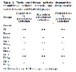
    Table 2: Testis seminiferous epithelial degeneration, seminiferous epithelial necrosis and desquamation scores in control and treatment groups

    Boldine is a natural substance with an alkaloid structure and has been reported to have a cell-protective, antitumor, anti-inflammatory and antipyretic effect 6. Dexpanthenol is an alcohol derivative of pantothenic acid (PA), also known as Provitamin B5, and is an important vitamin for the biosynthesis of coenzyme A 11. Pantothenic acid and its derivatives increase the level of glutathione (GSH), especially mitochondrial coenzyme A (Co A) and ATP, in cells and therefore play an important role in cellular defense, oxidative stress and inflammatory systems 14.

    Fernández et al. 29 reported that Boldo (Peumus boldus molina) has an important antioxidant effect against oxidative damage caused by cisplatin and reduces the MDA increase caused by cisplatin. Previous studies, it was reported that the MDA level increased with hydrochloric acid administration and serum oxidative parameters changed with cisplatin administration approached normal with dexpanthenol administration. It has also been reported that the liver damage caused by cisplatin is reduced by the administration of dexpanthenol 14,22,30.

    Oxidative stress parameters at the end of our study are shown in Figure 1, 2, 3, and 4 is investigated. It was observed in this study MDA level, one of the oxidative stress parameters, increased significantly in the cis group compared to the control group. However MDA levels decreased in other groups. The most significant reduction in MDA level was in the bold 40 and dex 500 groups. In our study, antioxidative enzyme levels (GSH-Px, SOD and CAT) were determined. The treatment boldine 40 mg/kg and dexpanthenol 500 mg/kg altered enzyme activities in cisplatin-treated rat testes tissue, but no significant change in enzyme activities was observed with at a dose boldine 20 mg/kg and dexpanthenol 250 mg/kg. In addition, similar results were obtained in sperm parameters. These results show that the effect of alkaloid on antioxidant enzyme activities becomes significant with increasing high doses. These results suggest that normalization of enzyme activities through ROS scavenging actions may have a significant contribution for the protective efficacy created by 40 mg/kg boldine and 500 mg dexpanthenol treatment. It was understood that these results were in line with the results of previous studies.

    Changes in the amount of enzymes such as MDA, GSH-Px, SOD and CAT in testicular tissue are considered as indicators of oxidative damage. It has been reported that apoptotic and degenerative disorders may occur with an increase in abnormal sperm count, change in sperm density and motility, especially due to increased oxidative stress. 29.

    As a result, we think that boldine and dexpanthenol may represent a promising new protective strategy against cisplatin induced testicular injury. And the use of boldine and dexpanthenol as an adjunct agent, may contribute to cisplatin used cancer chemotherapy. Future in-depth studies that may relate to these results will explore additional mechanisms and improve our understanding of testicular injury provided by boldine and dexpanthenol.

  • Başa Dön
  • Özet
  • Giriş
  • Materyal ve Metot
  • Bulgular
  • Tartışma
  • Kaynaklar
  • Kaynaklar

    1) Jahan S, Munawar A, Razak S, et al. Ameliorative effects of rutin against cisplatin-induced reproductive toxicity in male rats. BMC Urology 2018; 18: 1-11.

    2) Hassan SM, Khalaf MM, Sadek SA, et al. Protective effects of apigenin and myricetin against cisplatin-induced nephrotoxicity in mice. Pharmaceutical Biology 2017; 55: 766-774.

    3) Pınar N, Topaloğlu M, Seçinti İE, Buyuk E, Kaplan M. Protective effect of dexpanthenol on cisplatin induced nephrotoxicity in rats. Biotechnic & Histochemistry 2022; 97: 39-43.

    4) Rehman S, Khan H. Advances in antioxidant potential of natural alkaloids. Current Bioactive Compounds 2017; 13: 101-108.

    5) Subramaniam N, Kannan P, Ashokkumar K, Thiruvengadaml. Hepatoprotective effect of boldine against diethylnitrosamine‐induced hepatocarcinogenesis in wistar rats. J Biochem Mol Toxicol 2019; 33: e22404.

    6) O’Brien P, Carrasco-Pozo C, Speisky H. Boldine and its antioxidant or health-promoting properties. Chemico-Biological Interactions 2006; 159: 1-17.

    7) Türk G, Ateşşahin A, Sönmez M, Yüce A. Çeribaşı AO. Lycopene protects against cyclosporine A-induced testicular toxicity in rats. Theriogenology 2007; 67: 778-785.

    8) Tian M, Liu F, Liu H, et al. Grape seed procyanidins extract attenuates Cisplatin-induced oxidative stress and testosterone synthase inhibition in rat testes. Systems Biology in Reproductive Medicine 2018; 64: 246-259.

    9) Belhan S, Özkaraca M, Kandemir FM, et al. Effectiveness of hesperidin on methotrexate-induced testicular toxicity in rats. Kafkas Univ Vet Fak Derg 2017; 23: 789-796.

    10) Ekinci Akdemir FN, Yildirim S, Kandemir FM, et al. The antiapoptotic and antioxidant effects of eugenol against cisplatin‐induced testicular damage in the experimental model. Andrologia 2019;51: e13353.

    11) Ucar M, Aydogan MS, Vardı N, et al. Protective effect of dexpanthenol on ischemia-reperfusion-induced liver injury. Transplantation Proceedings 2018; 50: 3135-3143.

    12) Karadag A, Ozdemir R, Kurt A, et al. Protective effects of dexpanthenol in an experimental model of necrotizing enterocolitis. J. Pediatric Surgery 2015; 50: 1119-1124.

    13) Sohn SI, Rim HK, Kim YH, et al. The ameliorative effect of 23-hydroxytormentic acid isolated from Rubus coreanus on cisplatin-induced nephrotoxicity in rats. Biological and Pharmaceutical Bulletin 2011; 34: 1508-1513.

    14) Bayrak O, Seckiner I, Solakhan M, et al. Effects of intravesical dexpanthenol use on lipid peroxidation and bladder histology in a chemical cystitis animal model. Urology 2012; 79: 1023-1026.

    15) Paydar M, Kamalidehghan B, Wong YL, et al. Evaluation of cytotoxic and chemotherapeutic properties of boldine in breast cancer using in vitro and in vivo models. Drug design, Development and Therapy 2014;8: 719-733.

    16) Lau YS, Tian XY, Huang Y, et al. Boldine protects endothelial function in hyperglycemia-induced oxidative stress through an antioxidant mechanism. Biochem. Pharm 2013; 85: 367-375.

    17) Linder RE, Strader LF, McElroy WK. Measurement of epididymal sperm motility as a test variable in the rat. Bull. Environ. Cont. and Toxicol 1986; 36: 317-324.

    18) Sönmez M, Türk G, Yüce A. The effect of ascorbic acid supplementation on sperm quality, lipid peroxidation and testosterone levels of male Wistar rats. Theriogenology 2005; 63: 2063-2072.

    19) Ohkawa H, Ohishi N, Yagi K. Assay for lipid peroxides in animal tissues by thiobarbituric acid reaction. Analytical Biochemistry 1979; 95: 351-358.

    20) Kruger NJ. The Bradford method for protein quantitation. The protein protocols handbook 2009; 17-24.

    21) Kandeil MA, Mahmoud MO, Abdel-Razik ARH, et al. Thymoquinone and geraniol alleviate cisplatin-induced neurotoxicity in rats through downregulating the p38 MAPK/STAT-1 pathway and oxidative stress. Life sciences 2019; 228: 145-151.

    22) Bilgic Y, Akbulut S, Aksungur Z, et al. Protective effect of dexpanthenol against cisplatin‐induced hepatotoxicity. Exper Therap Med 2018; 16: 4049-4057.

    23) Agarwal A, Tadros H, Panicker A, et al. Role of oxidants and antioxidants in male reproduction. Oxidative Stress and Antioxidant Protection: The Science of Free Radical Biology and Disease 2016; 221-252.

    24) Yucel C, Arslan FD, Ekmekci S, et al. Protective effect of all-trans retinoic acid in cisplatin-induced testicular damage in rats. World J Mens Health 2019; 37: 249-256.

    25) Silici S, Ekmekcioglu O, Eraslan G, et al. Antioxidative effect of royal jelly in cisplatin-induced testes damage. Urology 2009; 74: 545-551.

    26) Yadav YC. Effect of cisplatin on pancreas and testes in Wistar rats: biochemical parameters and histology. Heliyon 2019; 5: e02247.

    27) Simsek N, Koc A, Karadeniz A, et al. Ameliorative effect of selenium in cisplatin-induced testicular damage in rats. Acta Histochemica 2016; 118: 263-270.

    28) İlbey YO, Ozbek E, Simsek A, et al. Potential chemoprotective effect of melatonin in cyclophosphamide-and cisplatin-induced testicular damage in rats. Fertility and Sterility 2009; 92: 1124-1132.

    29) Fernández J, Lagos P, Rivera P, et al. Effect of boldo (Peumus boldus Molina) infusion on lipoperoxidation induced by cisplatin in mice liver. Phytotherapy Research: An International Journal Devoted to Pharmacological and Toxicological Evaluation of Natural Product Derivatives 2009; 23: 1024-1027.

    30) Toplu Y, Parlakpinar H, Sapmaz E, et al. The protective role of molsidomine on the cisplatin-induced ototoxicity. Indian Journal of Otolaryngology and Head & Neck Surgery 2014; 66: 314-319.

  • Başa Dön
  • Özet
  • Giriş
  • Materyal ve Metot
  • Bulgular
  • Tartışma
  • Kaynaklar
  • [ Başa Dön ] [ Özet ] [ PDF ] [ Benzer Makaleler ] [ Yazara E-Posta ] [ Editöre E-Posta ]
    [ Ana Sayfa | Editörler | Danışma Kurulu | Dergi Hakkında | İçindekiler | Arşiv | Yayın Arama | Yazarlara Bilgi | E-Posta ]