[ Ana Sayfa | Editörler | Danışma Kurulu | Dergi Hakkında | İçindekiler | Arşiv | Yayın Arama | Yazarlara Bilgi | E-Posta ]
Fırat Üniversitesi Sağlık Bilimleri Veteriner Dergisi
2022, Cilt 36, Sayı 3, Sayfa(lar) 183-187
[ Özet ] [ PDF ] [ Benzer Makaleler ] [ Yazara E-Posta ] [ Editöre E-Posta ]
Koyun Korpus Luteumun'da PGF2a ile İndüklenmiş luteoliziste Kemik Morfogenetik Proteinlerinin (BMP) Ekspresyonu
Mustafa HİTİT
Kastamonu University, Faculty of Veterinary Medicine, Department of Animal Genetics Kastamonu, TÜRKİYE
Anahtar Kelimeler: Korpus luteum, gen ifadesi, luteolizis, BMP
Özet
Bu çalışmanın amacı, koyun korpus luteum'unun (KL) indüklenmiş luteolizisinde BMP sisteminin mRNA ekspresyonunu değerlendirmekti. İndüklenmiş luteolizis gruplarından KL örnekleri alındı ve indüklenmiş luteolizis modeli için, koyunlara döngünün 12. gününde PGF2a enjekte edildi ve luteal dokular 0. saatte (PGF2a enjeksiyonu yok, n= 4), 4. saatte (PG4, n=4) ve 16. saate enjeksiyondan sonra toplandı (PG16, n=4). BMP bileşenlerinin mRNA ekspresyon seviyeleri, RT-qPCR kullanılarak değerlendirilmiştir. SDHA, qPCR verilerinin normalleştirilmesi için referans gen kontrol olarak kullanıldı. BMP4 mRNA ekspresyonu PG16'da PG4'ten daha fazla olduğu bulundu (P<0.01). BMP6 ekspresyonunun PG16'da C12'den daha fazla olduğu gösterildi (P<0.01) ancak C12 ve PG4 arasında farklılık bulunmadı (P>0.05). BMP6 ve BMPR1A mRNA ekspresyonu PG16'da PG4'e göre daha fazla olduğu bulundu (P<0.05). BMPR1B mRNA ekspresyonu PG16'da PG4 ve C12'den daha büyük iken (P<0.0001), PG4'te C12'den daha düşük olduğu gösterilmiştir (P<0.05). BMPR2 mRNA'sının PG16'da PG4 ve C12'ye göre daha düşük bulunmasına rağmen, bu istatistiksel olarak anlamlı değildi (P>0.05). Bu çalışma, bazı BMP genlerinin ekspresyonunun koyun KL'sinde luteolizisini düzenleyebileceğini önermektedir.
  • Başa Dön
  • Özet
  • Giriş
  • Materyal ve Metot
  • Bulgular
  • Tartışma
  • Kaynaklar
  • Giriş
    Corpus Luteum (CL), temporary endocrine structure, can be resulted from functional remodeling of follicular structure following ovulation. The key function of CL is to generate and secrete progesterone (P4) for early embryonic development and maintenance of pregnancy (1, 2). When pregnancy is not established, luteolysis initiates immediately to allow another upcoming estrous cycle to occur (3, 4). In ruminants, luteolysis is defined as functional whereby decrease in P4 and structural regression of CL (5), and mediated by the pulsative release of prostaglandin F2α (PGF2α) in elevating concentrations from the non-pregnant uterus (6). On the contrary, P4 released from CL is needed to sustain pregnancy (7-9).

    Bone morphogenetic proteins (BMPs) are among the subfamily members of the transforming growth factor and are regarded as multifunctional growth factors. BMPs have functional roles in embryonic development, including cartilage, neural development, and postnatal bone formation (10). Besides their function in organs, BMPs function in different biological events in the ovary such as folliculogenesis, ovulation, and steroidogenesis (11, 12). BMPs compose the extensive part of the transforming growth factor (TGFβ) superfamily members. Some of the member of BMPs (BMP-2, - 3, -4, -6, -7) which forms heterodimer initiate BMP signaling through receptors of BMP; type I (BMP-IA and BMPR-IB) and type II (BMPR-II). Once ligand interaction implemented, signal transduction is accomplished via phosphorylation of type I receptor by type II receptor.

    Recently, during spontaneous regression, some of BMPs and their own receptors have been shown to be expressed in the human corpus luteum and are adversely modulated by the human chorionic gonadotropin (13). Contrary to the widespread role in folliculogenesis, limited number of studies (13-15) have focused on the modulation and function of BMPs throughout luteinization and luteolysis. Accordingly, we aimed to assess the expression patterns of BMP genes in the induced luteolysis of ovine CL.

  • Başa Dön
  • Özet
  • Giriş
  • Materyal ve Metot
  • Bulgular
  • Tartışma
  • Kaynaklar
  • Materyal ve Metot
    Animal Design of the Study: The experimental design of animal procedures was carried out in the breeding season of 2016. Animal diets for 3- to 5-year-olds ewes were adjusted to satisfy the National Research Council (NRC) Nutrient Requirements of Small Ruminants (16), including grass, dry clover, and concentrated feed. All other supplementals (vitamin and mineral with salt mix, and marble powder) were given ad libitum during the study 16. Bahri Dağdaş Research Center Ethical Committee (Number: 29/01/2016–49–7) approved the study procedures. In this study, we used induced luteolysis design. The ewes were divided into three groups following injection of PGF2α and PGF2α exposure was at 4 (PG4, n= 4) and 16 (PG16, n= 4) hours on day 12 of the cycle. We used cFOS and steroidogenic pathway mRNA expression to validate early challenge of CL to PGF2α treatment. The CL samples obtained on day 12 were allocated as zero h (C12, without PGF2a treatment, n= 4). We kept CL tissues at –80°C. RNA Isolation and cDNA Synthesis: We extracted RNA as defined earlier 17. Shortly, 20 mg of CL tissues in 800 mL Trizol (PureZOL™ RNA Isolation Reagent-Bio-Rad) were smashed and 260 μL chloroform was used to obtain separation phase by centrifugation at 11,000 G for 12 min. We precipitated RNA using 500 μL isopropanol (Sigma-Aldrich) and removed supernatant and subsequently washed the pellet with 70% ethanol. Immediately, we eluted RNA in 40 μL of RNAse-DNAse free water. We confirmed RNA concentration and quality using NanoDrop (Thermo Scientific™ NanoDrop™ 2000) checking the absorbance values. We used one microgram RNA for conversion to cDNA through a kit (I-Script, BioRAD).

    Gene Expression: We evaluated BMPs mRNA expression levels with the aid of qPCR with specific primers of BMP genes (Table 1). We prepared qPCR mix as follows: 10 μl qPCR master mix (Luna® Universal Protocol, #M3003, NEB), 2 μL cDNA, 2 μL primer (10 pMol), and 6 μL water to final volume of 20 μL 18. PCR conditions were arranged as previously defined 19. We ran melting analysis as follows: 95°C for 2 min, the fluorescence signal was captured at 1°C increment starting from 55°C up to 95°C (Bio-Rad CFX96; Bio-Rad, USA). To normalize the gene expression data, SDHA was employed as a housekeeping gene for gene expression analysis 20. Positive and negative control runs were included to eliminate genomic contamination. We performed each gene duplicate.


    Büyütmek İçin Tıklayın
    Table 1: BMP gene primers for qPCR

    Statistical Analysis: qPCR (Ct) data were used to calculate relative expression 18. The method of 2−ΔΔCt was employed to calculate relative expression 21. qPCR normalized data were analyzed by t-test and ANOVA with Tukey’s post hoc test. P<0.05 value was set as significance.

  • Başa Dön
  • Özet
  • Giriş
  • Materyal ve Metot
  • Bulgular
  • Tartışma
  • Kaynaklar
  • Bulgular
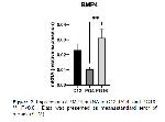
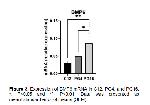
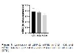
    BMP2 mRNA steady-state levels were demonstrated in C12 and induced luteolysis of PG4, and PG16 in Figure 1. When we evaluated, BMP2 mRNA seemed to be higher in PG4 than in C12 and PG16, but found to be similar among the groups, compared to each other (P>0.05). BMP4 mRNA steady-state levels were demonstrated in C12 and induced luteolysis of PG4, and PG16 in Figure 2. Expression of BMP4 mRNA was found to be greater in PG16 than in PG4 (P<0.05). However, the level of BMP4 mRNA did not change between induced luteolysis groups (PG4 and PG16) and C12 (P>0.05). BMP6 mRNA steady-state levels were demonstrated in C12 and induced luteolysis of PG4, and PG16 in Figure 3. Expression of BMP6 was shown to be greater in PG16 than in C12 (P<0.05) but did not differ between C12 and PG4 (P>0.05). Expression of BMP6 mRNA was found to be higher in PG16 than in PG4 (P<0.05).


    Büyütmek İçin Tıklayın
    Figure 1: Expression of BMP2 mRNA in C12, PG4, and PG16. Data was presented as mean±standard error of means (SEM)


    Büyütmek İçin Tıklayın
    Figure 2: Expression of BMP4 mRNA in C12, PG4, and PG16. **: P<0.01. Data was presented as mean±standard error of means (SEM)


    Büyütmek İçin Tıklayın
    Figure 3: Expression of BMP6 mRNA in C12, PG4, and PG16. *: P<0.05 and **: P<0.01. Data was presented as mean±standard error of means (SEM)

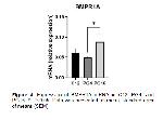
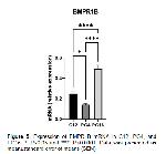
    BMPR1A mRNA steady-state levels were demonstrated in C12 and induced luteolysis of PG4, and PG16 in Figure 4. Expression of BMPR1A mRNA was found to be greater in PG16 than in PG4 (P<0.05). However, the level of BMPR1A mRNA did not change between induced luteolysis groups (PG4 and PG16) and C12 (P>0.05). BMPR1B mRNA steady-state levels were demonstrated in C12 and induced luteolysis of PG4, and PG16 in Figure 5. While expression of BMPR1B mRNA was greater in PG16 than in PG4 and C12 (P<0.05), it was shown to be lower in PG4 than in C12 (P<0.05). BMPR2 mRNA steady-state levels were demonstrated in C12 and induced luteolysis of PG4, and PG16 in Figure 6. Although BMPR2 mRNA was found to be lower in PG16 than in PG4 and C12 (P<0.05), this was not significant (P>0.05). Expression of BMPR2 mRNA did not change between C12 and PG4 (P>0.05).


    Büyütmek İçin Tıklayın
    Figure 4: Expression of BMPR1A mRNA in C12, PG4, and PG16. *: P<0.05. Data was presented as mean±standard error of means (SEM)


    Büyütmek İçin Tıklayın
    Figure 5: Expression of BMPR1B mRNA in C12, PG4, and PG16. *: P<0.05 and ****: P<0.0001. Data was presented as mean±standard error of means (SEM)


    Büyütmek İçin Tıklayın
    Figure 6: Expression of BMPR2 mRNA in C12, PG4, and PG16. Data was presented as mean±standard error of means (SEM)

  • Başa Dön
  • Özet
  • Giriş
  • Materyal ve Metot
  • Bulgular
  • Tartışma
  • Kaynaklar
  • Tartışma
    Our study apparently demonstrated that the mRNA expression of some BMPs genes were detected using qPCR in induced luteolysis (PG4 and PG16). The CL tissues obtained facilitated us to evaluate BMP2, BMP4, BMP6, BMPR1A, BMPR1B, and BMPR2 mRNA expressions in ovine CL.

    Throughout the oestrous cycle, luteal regression in ruminants and other species is induced by the release of PGF2α, which reaches the CL from the uterus. Luteolysis is prevented when the embryo is present and emerges in cyclic animals 21,22. Endogenous or exogenous administration of PGF2α facilitates a signalling of events causing to irreversible death of CL. In the entire process, CL undergoes significant changes in terms of its steroidogenic capacity, vascularization, extracellular matrix regeneration and cell viability 23, 24.

    In recent years, many functional genomic studies have elucidated the underlying mechanism of PGF2α over luteolysis 25, 26. Studies reveal that factors produced by uterine or exogenous PGF2α mediate a variety of processes from decreased steroid production to apoptotic cell death. Factors such as bone morphogenic proteins (BMP), tumor necrosis factor-alpha (TNFα), and activin A may have inhibitory effects on StAR expression 27-30.

    It has been reported that some BMPs cause luteolysis by suppressing StAR expression and P4 generation in women and granulosa cells 13, 31. In our study, this may be consistent with the regulation of BMP genes that we showed BMP6 mRNA were upregulated in PG16 compared to C12. Also, we previously reported that StAR mRNA was shown to be sharply decreased in PG16, thus explaining BMPs’ inhibitory effects on StAR as identified previously in humans CL 13. BMP2 and BMP4 mRNA expression was demonstrated to be increased in induced luteolysis of cattle CL after 12 hours after PGF2α treatment, but BMP6 was after 2 hours 32. BMP2 abundance was reported to link/be linked with diminished P4 in cattle CL 33. However, we have not observed any changes in the expression of BMP2 and BMP4 mRNA levels in our study. BMPRs assessed in the current study were expressed in the ovine CL during luteolysis. We have not detected significant regulation for BMPR2 as in induced luteolysis of bovine CL 32. However, BMPR1A mRNA were not regulated in induced luteolysis against C12, whereas BMPR1B mRNA was upregulated in PG16. In line with our study, in rats, the greater levels of BMPR1B was reported in CL regression which implies its involvement in luteolysis 14.

    In conclusion, considering mRNA expression of BMP2, BMP4, BMP6, BMPR1A, BMPR1B, and BMPR2, BMPs appeared to have functional role in corpus luteum. We may suggest that expression patterns of some BMP genes regulate luteolysis of ovine CL.

  • Başa Dön
  • Özet
  • Giriş
  • Materyal ve Metot
  • Bulgular
  • Tartışma
  • Kaynaklar
  • Kaynaklar

    1) Micks E, Raglan GB, Schulkin J. Bridging progestogens in pregnancy and pregnancy prevention. Endocr Connect 2015; 4: R81.

    2) Stocco C, Telleria C, Gibori G. The molecular control of corpus luteum formation, function, and regression. Endocr Rev 2007; 28: 117-149.

    3) Meidan R, Girsh E, Mamluk R, et al. Luteolysis in ruminants: Past concepts, new insights, and persisting challenges. Life Cycle Corpus Luteum 2017: 159-182.

    4) Spencer TE, Johnson GA, Bazer FW, et al. Implantation mechanisms: Insights from the sheep. Reproduction 2004; 128: 657-668.

    5) Sugino N, Okuda K. Species-related differences in the mechanism of apoptosis during structural luteolysis. J Reprod Dev 2007; 53: 977-986.

    6) Arosh JA, Banu SK, McCracken JA. Novel concepts on the role of prostaglandins on luteal maintenance and maternal recognition and establishment of pregnancy in ruminants1. J Dairy Sci 2016; 99: 5926-5940.

    7) Bazer FW, Spencer TE, Ott TL. Interferon tau: A novel pregnancy recognition signal. Am J Reprod Immunol 1997; 37: 412-420.

    8) Dorniak P, Welsh TH, Bazer FW, et al. Cortisol and interferon tau regulation of endometrial function and conceptus development in female sheep. Endocrinology 2013; 154: 931-941.

    9) Spencer TE, Johnson GA, Bazer FW, et al. Pregnancy recognition and conceptus implantation in domestic ruminants: Roles of progesterone, interferons and endogenous retroviruses. Reprod Fertil Dev 2006; 19: 65-78.

    10) Chen D, Zhao M, Mundy GR. Bone morphogenetic proteins. Growth Factors 2004; 22: 233-241.

    11) Khalaf M, Morera J, Bourret A, et al. BMP system expression in GCs from polycystic ovary syndrome women and the in vitro effects of BMP4, BMP6, and BMP7 on GC steroidogenesis. Eur J Endocrinol 2013; 168: 437-444.

    12) Regan SLP, Knight PG, Yovich JL, et al. Involvement of bone morphogenetic proteins (BMP) in the regulation of ovarian function. Vitam Horm 2018; 107: 227-261.

    13) Nio-Kobayashi J, Narayanan R, Giakoumelou S, et al. Expression and localization of inhibitor of differentiation (ID) proteins during tissue and vascular remodelling in the human corpus luteum. Mol Hum Reprod 2013; 19: 82-92.

    14) Erickson GF, Shimasaki S. The spatiotemporal expression pattern of the bone morphogenetic protein family in rat ovary cell types during the estrous cycle. Reprod Biol Endocrinol 2003; 1: 1-20

    15) Rajesh G, Paul A, Mishra SR, et al. Expression and functional role of bone morphogenetic proteins (BMPs) in cyclical corpus luteum in buffalo (Bubalus bubalis). Gen Comp Endocrinol 2017; 240: 198-213.

    16) NRC, 2007 National Research Council (NRC). Nutrient requirements of small ruminants: Sheep, goats, cervids, and new world camelids. National Academy Press, Washington, DC 2007: 271-280.

    17) Hitit M, Köse M, Bülbül B, et al. The expression of steroidogenic genes in ovine corpus luteum during early pregnancy. Eurasian J Vet Sci; 37: 93-100

    18) Hitit M, Corum O, Ozbek M, et al. Mucus from different fish species alleviates carrageenan-induced inflammatory paw edema in rats. Asian Pac J Trop Biomed 2020; 10: 452.

    19) Hitit M, Kose M, Kocak N, et al. Expression patterns of genes in steroidogenic, cholesterol uptake, and liver x receptor-mediated cholesterol efflux pathway regulating cholesterol homeostasis in natural and PGF2α induced luteolysis as well as early pregnancy in ovine corpus luteum. Anim Reprod Sci 2022; 240: 106988.

    20) Kayis SA, Atli MO, Kurar E, et al. Rating of putative housekeeping genes for quantitative gene expression analysis in cyclic and early pregnant equine endometrium. Anim Reprod Sci 2011;125:124-132.

    21) Livak KJ, Schmittgen TD. Analysis of relative gene expression data using real-time quantitative PCR and the 2-ΔΔCT method. Methods 2001; 25: 402-408

    22) Hansel W, Hickey GJ. Early pregnancy signals in domestic animals. Ann N Y Acad Sci 1988; 541: 472-484.

    23) Spencer TE, Bazer FW. Conceptus signals for establishment and maintenance of pregnancy. Reprod Biol Endocrinol 2004; 2: 1-15.

    24) Meidan R, Milvae RA, Weiss S, et al. Intraovarian regulation of luteolysis. J Reprod Fertil Suppl 1999; 54: 217-228.

    25) Miyamoto A, Shirasuna K, Shimizu T, et al. Regulation of corpus luteum development and maintenance: Specific roles of angiogenesis and action of prostaglandin F2alpha. Soc Reprod Fertil Suppl 2010; 67: 289-304.

    26) Mondal M, Schilling B, Folger J, et al. Deciphering the luteal transcriptome: Potential mechanisms mediating stage-specific luteolytic response of the corpus luteum to prostaglandin F2α. Physiol Genomics 2011; 43: 447-456.

    27) Shah KB, Tripathy S, Suganthi H, et al. Profiling of luteal transcriptome during prostaglandin F2-alpha treatment in buffalo cows: Analysis of signaling pathways associated with luteolysis. PLoS One 2014; 9: e104127.

    28) Chen YJ, Feng Q, Liu YX. Expression of the steroidogenic acute regulatory protein and luteinizing hormone receptor and their regulation by tumor necrosis factor alpha in rat corpora lutea. Biol Reprod 1999; 60: 419-427.

    29) Pierre A, Pisselet C, Dupont J, et al. Molecular basis of bone morphogenetic protein-4 inhibitory action on progesterone secretion by ovine granulosa cells. J Mol Endocrinol 2004; 33: 805-817.

    30) Miyoshi T, Otsuka F, Suzuki J, et al. Mutual regulation of follicle-stimulating hormone signaling and bone morphogenetic protein system in human granulosa cells. Biol Reprod 2006; 74: 1073-1082.

    31) Chang HM, Cheng JC, Klausen C, et al. Effects of recombinant activins on steroidogenesis in human granulosa-lutein cells. J Clin Endocrinol Metab 2014; 99: E1922-1932.

    32) Zhang H, Klausen C, Zhu H, et al. BMP4 and BMP7 Suppress StAR and Progesterone Production via ALK3 and SMAD1/5/8-SMAD4 in Human Granulosa-Lutein Cells. Endocrinology 2015; 156: 4269-4280.

    33) Haas CS, Rovani MT, Ilha GF, et al. Transforming growth factor-beta family members are regulated during induced luteolysis in cattle. Anim Reprod 2019; 16: 1-9.

  • Başa Dön
  • Özet
  • Giriş
  • Materyal ve Metot
  • Bulgular
  • Tartışma
  • Kaynaklar
  • [ Başa Dön ] [ Özet ] [ PDF ] [ Benzer Makaleler ] [ Yazara E-Posta ] [ Editöre E-Posta ]
    [ Ana Sayfa | Editörler | Danışma Kurulu | Dergi Hakkında | İçindekiler | Arşiv | Yayın Arama | Yazarlara Bilgi | E-Posta ]