[ Ana Sayfa | Editörler | Danışma Kurulu | Dergi Hakkında | İçindekiler | Arşiv | Yayın Arama | Yazarlara Bilgi | E-Posta ]
Fırat Üniversitesi Sağlık Bilimleri Tıp Dergisi
2006, Cilt 20, Sayı 4, Sayfa(lar) 297-301
[ Özet ] [ PDF ] [ Benzer Makaleler ] [ Yazara E-Posta ] [ Editöre E-Posta ]
Effects of Egb-761 in Rats with Obstructive Jaundice
Saim BERCIN1, Ziya CETINKAYA2, Yasemin BULUT3, Nusret AKPOLAT4, Erhan AYGEN2
1Harput Devlet Hastanesi, Genel Cerrahi, Elazığ – TÜRKİYE
2Fırat Üniversitesi, Tıp Fakültesi, Genel Cerrahi Anabilim Dalı, Elazığ–TÜRKİYE
3Fırat Üniversitesi, Tıp Fakültesi, Mikrobiyoloji Anabilim Dalı, Elazığ–TÜRKİYE
4Fırat Üniversitesi, Tıp Fakültesi, Patoloji Anabilim Dalı, Elazığ–TÜRKİYE
Keywords: Obstructive jaundice, Liver, İleum, Bacterial translocation, Egb-761
Summary
The aim of this study was to investigate the effects of ginkgo biloba extract (EGb-761) on liver and terminal ileum morphology, biochemical parameters and bacterial translocation in obstructive jaundice. Forty-four Wistar Albino rats weighing in average 200-250 g were divided into four groups: control, sham-operated, normal saline, EGb-761. No surgical procedure and treatment were conducted in the control group (n=10). In sham-operated group (n=10), laparatomy was performed and only common bile duct was mobilized, but no treatment was applied. For the remaining two groups (n=12 in each), after a preliminary treatment including the administration of normal saline and EGb-761 (50 mg/kg) via orogastric intubation for two days, a common bile duct was ligated. Escherichia coli (E. coli) suspension (1010 colony-forming unit bacteria/1 ml) was administered to all groups via orogastric intubation on the day 2 after operation. Twelve hours following the administration of E.coli suspension, blood samples (2 ml) were obtained by cardiac puncture from the rats for polymerase chain reaction (PCR) and biochemical analysis. In addition, the terminal ileum and liver samples were taken for histological examination. There was a significant reduction in histopathological changes and in serum gamma-glutamyl transferase (GGT) and alanin aminotransferase (ALT) level in EGb-761 group compared to the control group (p<0.025). According to the PCR results, although the number of E. coli detected rats was smaller in the groups clinically jaundiced and then treated with EGb-761 compared with the saline group, there was no significant difference between the two groups(p>0.05). Although developmental rate of bacterial translocation in rats treated with EGb-761 and normal saline were not statistically significant, EGb-761 significantly reduced pathological changes in liver and terminal ileum in obstructive jaundice.
  • Top
  • Summary
  • Introduction
  • Methods
  • Results
  • Disscussion
  • References
  • Introduction
    Negative effects of obstructive jaundice (OJ) on the liver and intestine are a major cause of high morbidity and mortality. Retention and accumulation of toxic hydrophobic bile salts in hepatocytes may cause hepatocyte toxicity 1,2. Oxygen free radicals have been implicated as mediators of tissue injury in a variety of diseases. Some authors suggested that to be the role of oxygen free radicals in liver 3-5 and intestine injury 6,7 in rats with obstructive jaundice. Bacterial translocation (BT) has been demonstrated in experimental models of OJ 8-12. Although the pathophysiological mechanisms are not well understood, it is generally thought that removal of bile from the gastrointestinal tract promotes bacterial overgrowth and increased translocation of endotoxin. An important factor which promotes this phenomenon is physical injury of the intestinal mucosa 6,7,13.

    Ginkgo biloba extract (EGb-761) increases peripheral and cerebral blood flow and microcirculation and improves myocardial ischemia reperfusion injury. Some major biochemical/pharmacological activities of EGb-761 are free radical scavenger activity, thereby decreasing tissue levels of ROS and inhibition of membrane lipid peroxidation and anti-PAF activity, contributing to improvements in cerebral insufficiency 14. EGb-761 has protective effect on hepatic endothelial cells and hepatic microcirculation in rats with chronic liver injury induced by CCl4 15,16. These properties of natural antioxidant Egb 761 are thought to provide many benefical effects free radical injuries.

    The goal of treatment in patients with OJ is to relieve the obstruction and obtain the bile flow into gastrointestinal tract. Before and after surgical intervention, reduction of the structural and functional damage in the liver is also an important part of therapy. To the best of our knowledge, there has been no study about the effects of Egb-761 on OJ in the literature. Therefore, we investigated the effects of Egb-761 on the liver and intestine of rats in which OJ was induced.

  • Top
  • Summary
  • Introduction
  • Methods
  • Results
  • Disscussion
  • References
  • Methods
    Forty-four female adult Wistar Albino rats (200-250 g) were randomly assigned to four groups as follows: G1- Control (n=10), G2- Sham-operated (n=10), G3- Normal saline (n=12), G4- EGb-761(n=12).

    All surgical procedures and sampling were performed under general anesthesia. Anesthesia was induced by intramuscular injection of ketamin HCL 12.5mg/100gr (Ketalar flk, Eczacibasi, Turkey) and Xylazine HCl 1mg/100gr (Rompon flk, Bayer). Laparatomy was performed with an average of 2 cm middle line incision under aseptic condition. In control group were applied no surgical procedure. In sham-operated group, only the common bile duct (CBD) was identified and mobilized but not ligated. In other treatment groups, common bile duct was dissected and ligated (CBDL) with 4/0 silk for twice and excised between the two knots in order to avoid recanalisation. Abdominal incision were closed with 3/0 silk sutures.

    The rats in the control and sham groups received no medical treatment. Group 3 received 1 ml normal saline, Group 4 were given 50 mg/kg dose EGb-761 (Tebokan drops 50 ml Abdi Ibrahim, Turkey) via orogastric intubation twice daily for 2 days before and after the operation. Following this treatment, 1 ml of E.coli suspension containing 1 x 10¹⁰ bacteria in phosphate buffer saline was administered to each group through orogastric intubation on the postoperative day 2. Twelve hours after the administration of E.coli suspension, samples were obtained and the rats were sacrificed.

    Following the laparatomy, blood samples were taken by cardiac puncture which were preserved at 20 ºC until assayed. Distal terminal ileum and the caudad (omental) lobes were excised and soaked in 10% formaldehyde for histopathological examination.

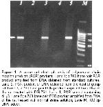
    For PCR analysis, DNA was isolated from the blood samples of the rats using a commercial DNA extraction kit (Wizard Genomic DNA Purification System, Promega Corp., Madison, WI). The PCR protocols were used as described previously (17). Ten microliters of the amplification products were run on a 2% agarose gel and the product was visualized by ethidium bromide staining (Figure- 1).


    Click Here to Zoom
    Figure 1: Agarose gel electrophoresis of polymerase chain reaction products (PCR) products. Lane 1; a 760 base pair PCR product amplified from DNA obtained from standard cultures, Lane 2; PCR product of DNA obtained from the control group rat, Lane 3; a 760 base pair PCR product amplified from DNA of the rat treated with EGb761, Lane 4; PCR control containing dH2O. Lane 5; a 760 base pair PCR product amplified from DNA of the rat treated with normal saline solution, Lane M; 100 bp DNA ladder.

    During analyses, previously confirmed colonies obtained from standard cultures were used as positive control for E. coli. The sensitivity of PCR was determined with 10-fold dilution of the DNA extracted from the isolated colonies E. coli. In order to overcome both false positivity and negativity, we included negative and positive controls in each experiment.

    Liver and terminal ileum tissue samples were fixed in formaldehyde solution and divided into 0.5 cm sections. After the standard laboratory proceedings, 5 µm slices were prepared for subsequent histopathological examination, by fixing the sections in paraffin and staining them with hemotoxiline-eosin. Liver tissue samples were examined by light microscopy and the results were scored in aspects hepatocyte bloating, multinucleation, degeneration, cell necrosis, Kupffer cell proliferation, cholestasis, portal edema, portal region extension, portal PNL infiltration, bile duct proliferation, bile duct dilatation and portal fibrosis: 0 (n/a), 1 (mild), 2 (moderate), 3 (intensive) and 4 (severe).

    Terminal ileum samples were examined by light microscopy and the results were scored in aspects of mucosal damage, PNL infiltration in lamina propria, lenfoid follicular hyperplasia, goblet cell reduction, increase in the number of veins and capillary expansion: 0 (n/a), 1 (mild), 2 (moderate), 3 (intensive) and 4 (severe).

    Serum urea, creatinin, total bilirubin, gamma-glutamyl transferase (GGT), alkaline phosphatase (ALP) and alanin aminotransferase (ALT) were analyzed outomatically using a kit Olympus Diagnostica GmbH'dan (Wendenstrabe, Hamburg-Deutschland).

    The data were given as avarage value ± standart error. For the statistical comparison of biochemical and histopathological results of trial groups, “Bonferroni adjusted Mann Whitney U test” was used and P<0.025 was considered to be significant. “Pronounced chi-square test” was used for the comparison of microbiological results and P<0.05 was considered to be significant.

  • Top
  • Summary
  • Introduction
  • Methods
  • Results
  • Disscussion
  • References
  • Results
    PCR Results: Limit of detection for PCR assay of the E. coli DNA extracted from the isolated colonies was found to be about 5 bacteria per reaction. PCR results of all groups are presented in (Table-1). There was no significant difference between the control and sham-operated groups with respect to blood PCR results (p> 0.05). Results obtained from the groups operated with CBDL were significantly different from that of the control and sham-operated groups (P< 0.05). When CBDL groups were compared with each other, it was found that the difference between them was not significant.


    Click Here to Zoom
    Table 1: Presence of Escherichia coli genetic material in blood samples determined by polymerase chain reaction assay.

    Biochemical results: Urine and creatine values of all groups were very close and did not significantly differ. In CBDL groups, ALT, ALP, GGT, and billirubin levels were found to be significantly increased compared to control and sham-operated groups (P< 0.025). The increase in ALT and GGT levels of EGb-761 group was significantly less than G3 (p< 0.025) (Table-2).


    Click Here to Zoom
    Table 2: Serum biochemistry values

    Histopathological results: No change in cellular or portal level was observed in liver sections of rats in control and sham-operated groups. In jaundiced groups, pathological changes in liver were smaller in groups G4, according to G3. This difference was statistically significant (p< 0.025). Bile duct proliferation and dilatation and hepatocyte degeneration were more rarely seen in G4 (Figure 2).


    Click Here to Zoom
    Figure 2: Histopathologic evaluation of study groups.

    During microscopical evaluation of terminal ileum, no pathology was detected in control and sham-operated groups. Mild lenfoid follicular hyperplasia was observed only in one sample obtained from sham-operated group. In jaundiced groups, mild to severe mucosal damage was observed. There was a statistically significant reduction in mucosal damage in groups treated with EGb-761 compared to normal saline group (p<0.025) (Figure-2).

  • Top
  • Summary
  • Introduction
  • Methods
  • Results
  • Disscussion
  • References
  • Discussion
    A variety of histological grading and scoring systems have been used to evaluate tissue injury 18-20. Classically, histopathological diagnosis is a descriptive process dependent on the observer's interpretation of a specific visual image although morphological changes were not converted into numerical values and were not statistically analyzed. In our study, morphologic changes converted into numerical values were statistically analyzed.

    Traditional methods used for detection of BT in experimental animals include demonstration of live bacteria in blood and in organs/tissues distal from intestines through culturing; or measurement of radioactivity in organs following the administration of radioisotope marked bacteria/endotoxin through enteric route 21. Translocated bacteria cannot be always grown successfully in a suitable culture medium or it may take a long period of time. The radioactive method carries the risk of giving false negative and false positive results. PCR method to amplify nucleic acid of microorganisms has been introduced. PCR method is a quite specific technique and is not affected by conditions of sampling and shipping 22. We therefore believe that PCR method is an effective method for the detection and identification of bacterial translocation.

    Retention and accumulation of toxic hydrophobic bile salts in hepatocytes may cause hepatocyte toxicity by inducing apoptosis 1,2. Without proper treatment in rats with CBDL, hepatocellular injury is an invariant feature of cholestasis causing liver dysfunction, promoting fibrogenesis, and ultimately leading to liver failure. Injury of the liver has been suggested to be mediated by increased xanthine dehydrogenase and oxidase activity with production of oxygen free radicals and reduced inactivation of xanthine oxidase activity3.

    Bile acids reduce the bacterial overgrowth both clinically and experimentally. Bile acids act as detergent in bacteria cell wall and inactivate endotoxins. In the absence of bile flow an increase in gram (-) aerobic bacteria population (by biliary diversion) has been shown. Endotoxins increased in the absence of bile flow damage intestinal mucosa by stimulating ileal xanthine dehydrogenase and oxidase activity 23. Xu et al. 13 have been show that blood flow to the distal ileum and caecum is selectively reduced by 35-50% following endotoxin administration, whereas blood flow to the rest of the intestine, liver, spleen, and pancreas is normal. In addition, small bowel permeability is increased at sites of decreased blood flow (ileum) but not at sites of normal blood flow (jejunum). Other studies have reported significantly reduced blood flow in the mesenteric artery during experimental endotoxaemia 24 or sepsis 25, even when cardiac output is normal or elevated. Changes in gastrointestinal morphology associated with obstructive jaundice are most marked in the distal ileum 8-12. Some authors have described sub epithelial edema, with lifting of the epithelium from the lamina propria 8,9, dilated lymph vessels and edematous sub epithelial regions, with infiltration of inflammatory cells in the villi 10-12.

    The studies have shown that mucosal damage facilitates BT by giving harm to intestinal barrier function. Both non-specific defense mechanisms and immune system depression have a role in the development of BT in obstructive jaundice 19-23. The response of T lymphocytes to pathogens decreases in rats after CBDL operation, while T suppressor cell activity increases and interleukin 2 production decreases 26 and chemotaxis and fagocytosis becomes depressed 27.The present study has demonstrated an increased incidence of BT with obstructive jaundice. The physiopathological mechanism of BT has not been studied.

    EGb-761 prevents extreme free radical formation by inhibiting lipid peroxidation. Preventive effect of EGb-761 on free radical formation in brain ischemia, destruction of cell membrane structure and vascular endothelium damage is well known 14. Additionally flow cytometry and DNA fragmentation studies have shown that EGb-761 decreases apoptosis caused by hydroxyl radicals 28. In a study that chronic liver injury was induced by CCl4, EGb-761 has protective effect on hepatic endothelial cells and hepatic microcirculation in rats. The mechanisms may involve its inhibition on ET-1, PAF and lipid peroxidation 16. In another study on ischemia reperfusion injury of intestine concluded that EGb-761 pre-treatment before ischemia-reperfusion decreased malondialdehyde and myeloperoxidase levels and attenuated the mucosal damage 29. We suggest that EGb-761 does not reduce the development rate of bacterial translocation in rats with CBDL. When focusing on the histopathological changes in terminal ileum observed in our study, there was a significant reduction in mucosal damage in EGb-761 groups compared to the control group.

    According to the histopathological findings in the present study, it was shown that EGB-761 administration decreased the damage in liver and ileum of obstructive jaundice. Level of histopathological changes may be connected to the number of translocated E. coli together with presence of E. coli in the rats. However, we detected presence only of E. coli from the rats by PCR method. Therefore, additional studies are required to investigate the relationship between the number of translocated E. coli and level of histopathological changes.

    Serum liver enzymes (ALT, ALP, GGT) were significantly higher in CBDL Groups than in the sham and control groups (P<0.025). GGT and ALP are better indicators of liver damage due to OJ. Significant decrease was observed in GGT and ALP values in EGb-761 group in comparison to normal saline group (P<0.025). We concluded that EGb-761 treatment decreased the liver damage due to OJ.

    We conclude that in rats which OJ is induced, EGb-761 administration decreases the damage in liver and terminal ileum. However, reduction of bacterial translocation rate in rats was not statistically significant. It is expected that this study will lead to further studies on the effects of EGb-761 on morbidity and mortality following surgery in the presence of OJ.

  • Top
  • Summary
  • Introduction
  • Methods
  • Results
  • Discussion
  • References
  • References

    1) Rodrigues CM, Fan G, Ma X, Kren BT, Steer CJ. A novel role for ursodeoxycholic acid in inhibiting apoptosis by modulating mitochondrial membrane perturbation. J Clin Invest 1998; 101: 2790-99.

    2) Miyoshi H, Rust C, Roberts PJ, Burgart LJ, Gores GJ. Hepatocyte apoptosis after bile duct ligation in the mouse involves Fas. Gastroenterology 1999;117: 669-77.

    3) Singh S, Shackleton G, Ah-Sing E, Chakraborty J, Bailey ME. Antioxidant defenses in the bile duct-ligated rat. Gastroenterology 1992; 103: 1625-29.

    4) Castro V, Muriel P. Comparative study of colchicine and trimethylcolchicinic acid on prolonged bile duct obstruction in the rat. J Appl Toxicol 1996; 16: 269-75.

    5) Tsai LY, Lee KT, Lu FJ. Biochemical events associated with ligation of the common bile duct in Wistar rats. J Formos Med Assoc 1997; 96: 17-22.

    6) Parks RW, Stuart Cameron CH, Gannon CD, Pope C, Diamond T, Rowlands BJ. Changes in gastrointestinal morphology associated with obstructive jaundice. J Pathol 2000; 192: 526-32.

    7) Reynolds JV, Murchan P, Leonard N, Clarke P, Keane FB, Tanner WA. Gut barrier failure in experimental obstructive jaundice. J Surg Res 1996; 62: 11-16.

    8) Deitch EA, Sittig K, Ma L, Berg R, Specian RD. Obstructive jaundice promotes bacterial translocation from the gut. Am J Surg 1990; 159: 79-84.

    9) Slocum MM, Sittig KM, Specian RD, Deitch EA. Absence of intestinal bile promotes bacterial translocation. Am Surg 1992; 58: 305-10.

    10) Ding JW, Andersson R, Soltesz V, Willen R, Bengmark S. The role of bile and bile acids in bacterial translocation in obstructive jaundice in rats. Eur Surg Res 1993; 25: 11-19.

    11) Ding JW, Andersson R, Soltesz V, Willen R, Loft S, Poulsen HE, et al. The effect of biliary decompression on bacterial translocation in jaundiced rats. HPB Surg 1993; 7: 99-110.

    12) Ding JW, Andersson R, Soltesz V, Willen R, Bengmark S. Obstructive jaundice impairs reticuloendothelial function and promotes bacterial translocation in the rat. J Surg Res 1994; 57: 238-45.

    13) Xu D, Qi L, Guillory D, Cruz N, Berg R, Deitch EA. Mechanisms of endotoxin-induced intestinal injury in a hyperdynamic model of sepsis. J Trauma 1993; 34: 676-83.

    14) Smith JV, Luo Y. Studies on molecular mechanisms of Ginkgo biloba extract. Appl Microbiol Biotechnol 2004; 64: 465-72.

    15) Bahçecioğlu H, Ustundağ B, Ozercan I, Erçel E, Baydaş G, Akdere T, et al. Protective effect of Ginkgo biloba extract on CCl4-induced liver damage. Hepatology Research 1999; 15: 215-24.

    16) Zhang C, Zu J, Shi H, Liu J, Qin C. The effect of Ginkgo biloba extract (EGb 761) on hepatic sinusoidal endothelial cells and hepatic microcirculation in CCl4 rats. Am J Chin Med 2004; 32: 21-31.

    17) Cetinkaya Z, Ulger H, Akkus MA, Dogru O, Cifter C, Doymaz MZ, et al. Influence of some substances on bacterial translocation in the rat. World J Surg 2002; 26: 9-12.

    18) Dunnill MS, Whitehead R. A method for quantitation of small intestinal biopsy specimens. J Clin Pathol 1972; 25: 243-246.

    19) Slavin G, Sowter C, Robertson K, McDermott S, Paton K. Measurement in jejunal biopsies by computer-aided microscopy. J Clin Pathol 1980; 33: 254-261.

    20) Illyes G, Hamar J. Sequence of morphological alteration in small intestinal ischaemia-reperfusion model of anaesthetised rat: a light microscopic study. Int J Exp Pathol 1992; 72: 161-172.

    21) Alexander JW, Boyce ST, Babcock GF, Gionotti L, Peck MD, Dunn DL, et al. The process of microbial translocation. Ann Surg 1990; 212: 496-510.

    22) Kane TD, Johnson SR, Alexander JW, Babcock GF, Ogle CK. Detection of intestinal bacterial translocation using PCR. J Surg Res 1996; 63: 59-63.

    23) Clements WD, Parks R, Erwin P, Halliday MI, Barr J, Rowlands BJ. Role of the gut in the pathophysiology of extrahepatic biliary obstruction. Gut 1996; 39: 587-593.

    24) Navaratnam RL, Morris SE, Traber DL, Flynn J, Woodson L, Linares H, et al. Endotoxin (LPS) increases mesenteric vascular resistance (MVR) and bacterial translocation (BT). J Trauma 1990; 30: 1104-1113.

    25) Whitworth PW, Cryer HM, Garrison RN, Baumgarten TE, Harris PD. Hypoperfusion of the intestinal microcirculation without decreased cardiac output during live Escherichia coli sepsis in rats. Circ Shock 1989; 27: 111-122.

    26) Ding XZ, Li H, Xiong ST, Zhang SX, Lu KZ, Shao JF, et al. Effects of cimetidine on IL-2 and T suppressor cell fonction in rats with obstructive jaundice. J Tongji Med Univ 1994; 14: 94-97.

    27) Karan B, Kama NA, Hascelik G, Ercan M. Effects of vitamin A on immunological deficiencies in rats with obstructive jaundice. Eur J Surg 1996; 162: 217-222.

    28) Ni Y, Zhao B, Hou J, Xin W. Preventive effect of gingko biloba extract on apoptosis in rat cerebellar neuronal cells induced by hydroksyl radicals. Neurosci Lett 1996; 214: 115-118.

    29) Pehlivan M, Dalbeler Y, Hazinedaroglu S, Arikan Y, Erkek AB, Gunal O, Turkcapar N, Turkcapar AG. An assessment of the effect of Ginkgo Biloba EGb 761 on ischemia reperfusion injury of intestine. AG. Hepatogastroenterology 2002; 49: 201-204.

  • Top
  • Summary
  • Introduction
  • Methods
  • Results
  • Discussion
  • References
  • [ Başa Dön ] [ Özet ] [ PDF ] [ Benzer Makaleler ] [ Yazara E-Posta ] [ Editöre E-Posta ]
    [ Ana Sayfa | Editörler | Danışma Kurulu | Dergi Hakkında | İçindekiler | Arşiv | Yayın Arama | Yazarlara Bilgi | E-Posta ]