[ Ana Sayfa | Editörler | Danışma Kurulu | Dergi Hakkında | İçindekiler | Arşiv | Yayın Arama | Yazarlara Bilgi | E-Posta ]
Fırat Üniversitesi Sağlık Bilimleri Veteriner Dergisi
2013, Cilt 27, Sayı 2, Sayfa(lar) 075-079
[ Özet ] [ PDF ] [ Benzer Makaleler ] [ Yazara E-Posta ] [ Editöre E-Posta ]
Kör farenin (Spalax leucodon) İntestinal Pleksüslarında Bazı Nörotransmiterlerin İmmunohistokimyasal Lokalizasyonu
Mine YAMAN, Ali BAYRAKDAR, Berrin GENÇER TARAKÇI
Fırat Üniversitesi, Veteriner Fakültesi, Histoloji ve Embriyoloji Anabilim Dalı, Elazığ, TÜRKİYE
Anahtar Kelimeler: Enterik sinir sistemi, immunohistokimya, intestinal pleksüs, kör fare
Özet
Bu çalışmanın amacı kör fare (Spalax leucodon) intestinal pleksüslarında kalsitonin gene-related peptid (CGRP), substance P (SP), nöropeptid Y (NPY), vasoactive intestinal peptid (VIP) ve serotonin immunoreaktivitesinin varlığını araştırmaktır. İmmunohistokimyasal incelemeler ince ve kalın bağırsağın ileum ve kolon bölümlerinde yapıldı. CGRP immunoreaktif nöronal yapılar ileum ve kolonun miyenterik pleksüsları içinde enterik gangliyonlar şeklinde belirlendi. SP immunoreaktivitesi bu bağırsak bölümlerinin her iki kas tabakası içinde uzayan sinir telleri ve miyenterik ganglionlar şeklinde ayıtedildi. Ayrıca NPY sinir hücre cisimcikleri ve iplikleri ileum ve kolonun miyenterik pleksüs ve lamina propriyasında mevcuttu. Buna karşın VIP ve serotonin immunoreaktif yapılar ince ve kalın bağırsağın herhangi bir bölümünde tespit edilemedi. Sonuç olarak, bu çalışma kör farenin ince ve kalın bağırsaklarının mukozal ve gangliyoner pleksüslarında farklı nöronal popülasyonların varlığını ortaya koydu.
  • Başa Dön
  • Özet
  • Giriş
  • Materyal ve Metot
  • Bulgular
  • Tartışma
  • Kaynaklar
  • Giriş
    The enteric nervous system (ENS) is the intrinsic nervous system of the gastrointestinal tract. It contains complete reflex circuits that detect the physiological condition of the gastrointestinal tract, integrate information about the state of the gastrointestinal tract, and provide outputs to control gut movement, fluid exchange between the gut and its lumen, and local blood flow1,2. There are several distinct features of enteric neurons which contribute to their ability to regulate gastrointestinal functions3. The function of the ENS is to coordinate the complex interactions of the enteric networks, which consist of sensory, inter, motor and secremotor neurons4.

    The ENS is usually formed from two major plexus: the myenteric plexus mainly regulating muscle activity and the submucous plexus mainly regulating mucosal functions. The myenteric plexus is an endogenous source for motor innervation to the muscular layers and secremotor innervation to the mucosa. Similar to other nervous systems, the functions of the myenteric plexus are mediated by various neurochemical substances2,5,6.

    The extrinsic and intrinsic components of the enteric nervous system appear to be the primary mechanisms involved in motility regulation. It is well known that besides the noradrenergic and cholinergic regulations, serotoninergic and peptidergic ones are present, too7, some of them having a facilitatory action, others having inhibitory effects upon classical neurotransmitters8-11.

    Calcitonin gene-related peptide (CGRP) which was originally identified as a splicing product of the alternative RNA processing of the calcitonin gene in the rat brain, affects a variety of biological activities in the ENS, such as release of gastrointestinal hormone, co-ordination of gastrointestinal motility, excitation of myenteric neurons and vasodilatation12.

    Substance P (SP) is a member of a family of peptides called the tachykinins13 and a neurotransmitter that plays an important role in regulating gastrointestinal motility14. SP was isolated from the gut and can be detected in a dense nerve fiber network within the myenteric plexus. SP-containing nerve fibers are an intrinsic contractor of the longitudinal muscle layer5,14,15.

    NPY is widely distributed in the central and peripheral nervous system and represents one of the most abundant neuropeptides16. It has been functionally related to regulation of blood pressure, circadian rhythms, feeding behavior, anxiety, memory processing, and cognition in the central nervous system, and to vasoconstriction and gastrointestinal tract motility in the peripheral nervous system17,18.

    Although the immunoreactivity of the abovementioned neurotansmitters has been studied in ENS of several laboratory animals14,19-22, these neurotansmitters have not been examined in small and large intestine of mole-rats (Spalax leucodon). Thus, we have investigated the location of CGRP, SP, NPY, VIP and serotonin immunoreactivity in the extrinsic and intrinsic innervation of the mole-rats (Spalax leucodon) intestines.

  • Başa Dön
  • Özet
  • Giriş
  • Materyal ve Metot
  • Bulgular
  • Tartışma
  • Kaynaklar
  • Materyal ve Metot
    Animal and tissue samples: In the present study, six adult mole-rats, trapped by the farmers, were used. After the mole-rats (Spalax leucodon) were anaesthetized with 2.5% pentathol (6 mL/kg), the left carotid artery was cannulated at the base of the neck and allowed to exsanguinate. Tissue samples were taken from the small and large intestine (ileum and colon) and fixed in 10% neutral-buffered formalin for 24 hr. Then tissues were dehydrated through graded ethanol and embedded in paraffin. Five μm thick sections, mounted on poly-l-lysine coated glass slides, were obtained and processed for immunohistochemical staining.

    Immunohistochemistry: Immunohistochemical staining was performed by using avidin-biotin-peroxidase complex (ABC) methods. According to the ABC methods, tissues were incubated in citrate buffer (10 mM citric acid, pH 6.0) in a microwave oven at 750 W for 20 minutes to retrieve antigenicity. Quenching of the endogenous peroxidase activity was done with 3% hydrogen peroxide (H2O2) in methanol for 10 minutes. In order to block unspecific binding incubation with normal goat serum (1.10) in 0.1 M phosphate buffered saline (PBS), pH 7.2 was performed. Sections were incubated with primary antibodies for 16–20 hours at 4°C. The working dilutions and the sources of the primer antibodies were listed in Table 1. The primary antibodies were diluted in PBS containing 0.25% sodium azide and 2.5% bovine serum albumin (BSA). Negative control sections were performed by replacing the primary antibodies with PBS. The sections were then incubated with biotinylated goat anti-rabbit immunoglobulin G (Sigma) followed by avidin-biotin-peroxidase complex (Dako) both at dilution of 1.50 in PBS for 1 hour at room temperature. Sections were washed in PBS for 30 minutes after each incubation step and finally immersed in glucose oxidase–DAB (diaminobenzidine)–nickel ammonium sulphate substrate23 for 10 minutes. After washing in distilled water and counterstaining with hematoxylin, sections were dehydrated and coverslips mounted with mounting medium. Primary antibodies were omitted in negative controls; no immunoreactivity was detected in these preparations.

    The specificity of each immunohistochemical reaction was determined as recommended by Sternberger24 including the replacement of specific antiserum by the same antiserum which had been preincubated with its corresponding antigen. Sections were examined with microscope (Olympus BX–51, New York) and the photographs were taken.

  • Başa Dön
  • Özet
  • Giriş
  • Materyal ve Metot
  • Bulgular
  • Tartışma
  • Kaynaklar
  • Bulgular
    In the present study CGRP, SP and NPY immunoreactivities were found in the nerve cell bodies and fibers of the intestinal myenteric plexus and submucous plexus of the mole-rats (Spalax leucodon). However, VIP and serotonin immunoreactivity were not observed in any intestinal region.

    CGRP immunoreactivity was very numerous in the small and large intestine. These immunoreactive neuronal structures were detected within the myenteric plexus. The myenteric plexus was located between the inner and outer musculature. In addition, structures belonging to the myenteric plexus were often embedded in the circular musculature of the ileum. This plexus was arranged in an almost continuous layer which contained both nerve cell bodies and fibers in the ileum (Figure 1A). Also, the neuronal cell bodies exhibiting strong CGRP immunoreactivity was in the form of enteric ganglia in the colon myenteric plexus (Figure 1B).


    Büyütmek İçin Tıklayın
    Figure 1: CGRP-immunoreactive neuronal cell bodies (arrows) are observed in the myenteric plexus of ileum (A) and colon (B)

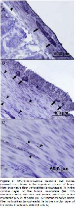
    SP immunoreactivity was detected in the tunica muscularis of the small and large intestine. The immunoreactivity was discernible with particular evidence within nerve fibers running in both the musculature and myenteric ganglia of ileum (Figure 2A). SPimmunoreactive cell bodies of myenteric neurons were localized in the small groups within longitudinal musculature of colon (Figure 2B). In addition, these immunoreactive nerve fibers were found in the circular musculature of this intestinal region (Figure 2C).


    Büyütmek İçin Tıklayın
    Figure 2: SP-immunoreactive neuronal cell bodies (arrows) are shown in the myenteric plexus of ileum. Note that nerve fiber varicosities (arrowheads) lie in the circular layer of the tunica muscularis (A). SPimmunoreactive neuronal cell bodies are seen in the myenteric plexus of colon (B). SP immunoreactive nerve fiber varicosities (arrowheads) lie in the circular layer of the tunica muscularis colon (B and C)

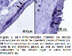
    NPY immunoreactive nerve cell bodies and fibers were present in the lamina propria, myenteric and subserosal plexus of small and large intestine. The immunoreactive nerve cell bodies were seen between the inner and outer musculature layers of ileum, sometimes opposed to negative nerve cell bodies. Also, it was observed in the sub-serosal plexuses of this intestinal region (Figure 3A). In the colon, NPY immunoreactivity was detected in subtle nerve fibers of the lamina propria. Additionally, IR nerve cell bodies were mostly grouped in small numbers, forming tiny ganglia in the myenteric plexus of the colon (Figure 3B).


    Büyütmek İçin Tıklayın
    Figure 3: NPY-immunoreactive neuronal cell bodies (arrows) are shown in the myenteric plexus of ileum (A). NPY-immunoreactive neuronal cell bodies (arrows) are seen in the myenteric plexus as well as nerve fiber varicosities in the circular layer of colon tunica muscularis (B).

    VIP and serotonin immunoreactive neuronal structures were not detected any other region of the small and large intestine.

  • Başa Dön
  • Özet
  • Giriş
  • Materyal ve Metot
  • Bulgular
  • Tartışma
  • Kaynaklar
  • Tartışma
    The basic organisation and function of the ENS in all species is much the same25. However, there are some differences between large and small mammals26. CGRP27,28 and various other peptides such as SP, NPY and VIP are expressed in the myenteric neurons of the variety mammals19,29-33. No information concerning the occurrence of the neuronal structures in the gastrointestinal tract in subspecies of mole-rats (Spalax leucodon) is available apart from the regional distribution and relative frequency of the endocrine cells in the gastrointestinal tract34. The present study was identified the existence and distribution of neuronal structures in the small and large intestine of the molerats. This is the first report showing that the CGRP, SP and NPY innervation in the mole-rat (Spalax leucodon) intestines.

    Neurons expressing the CGRP were present in the myenteric ganglia of the intestine in many mammalian species22,35,36. CGRP immunoreactivity was reported in the myenteric plexus and submucosal plexus in the human small intestine37. CGRP-IR was also localized to neurons of the myenteric plexus and to the fibers in the mucosal folds of the mice small intestine32. Chiocchetti et al.33 reported that the existence of CGRP-IR in fibers and nerve cell bodies located in the myenteric and submucosal plexuses of lamb ileum. İn addition, CGRP-containing neurons were observed in the myenteric plexus of the rat colon19,22,30. CGRPcontaining neurons in many species, including the rat, exhibit morphological similarities. However, in this study, CGRP-IR was observed in wide ganglioner form within myenteric plexus in both the small and large intestine. In the submucosal plexus, the neuronal elements were not present. The neuronal densitiy was too high particulary in the circular musculature. The results of this study were in agreement with the immunohistochemical studies reported by some authors19,22,30,32.

    SP immunoreactive neurons that innervate the longitudinal muscle of the guinea pig ileum were identified in the longitudinal muscle plexus31. A study in the canine ileum demonstrated SP-IR varicose nerve fibers in the myenteric plexus and both external smooth muscle layers29. Sang et al.38 reported that SP immunoreactive nerve fibers were located in the circuler musculature of the mouse large intestine. In addition, some studies reported that the presence of SP-IR nerve fibers in the myenteric plexus and longitudinal musculature of the guinea pig colon39. Similar to the findings reported for canine, SP-IR nerve fibers showed the same localisation in ileum in the present study29. Furthermore, as in the guinea pig colon39, SP –IR was found in the myenteric plexus and the longitudinal musculature of the mole rats.

    Autoradiographic studies had shown the presence of NPY receptor in the submucosal and myenteric ganglia in the intestine40,41. In the rat intestinal tract, Y1 receptor immunoreactivity was detected in the submucosal and myenteric nerve cell bodies20. In addition, NPY-IR nevre cell bodies were only observed in the myenteric plexus of the rat small intestine and canine ileum21,29. The other study was reported that NPY-IR were present immunoreactive fibers forming a dense network and immunoreactive nevre cell bodies in the lamina propria and submucosal plexus of the rat small intestine; they were few in this location in the colon42. Also, another study was demonstrated the presence of NPY receptor immunoreactivity in the myenteric and submucosal nerve plexuses of the human colon43. As in the some studies20,21,42,43, NPY immunoreactivity was detected in the lamina propria, myenteric and sub-serosal plexuses of small and large intestine in this study. Our findings showed the presence of dense immunoreactive localization in the colon myenteric plexus by contrast with Sundler42.

    In conclusion, this study demonstrated the presence of different neuronal populations in the mucosal, submucosal and ganglionated plexuses in the small and large intestine of the mole rats. This is the first report of neuronal localization of CGRP, SP and NPY in the mole rat intestines.

  • Başa Dön
  • Özet
  • Giriş
  • Materyal ve Metot
  • Bulgular
  • Tartışma
  • Kaynaklar
  • Kaynaklar

    1) Gershon MD. Nerves, reflexes, and the enteric nervous system. J Clin Gastroenterol 2005; 38: 184-193.

    2) Furness JB. The Enteric Nervous System. Oxford: Blackwell, 2006.

    3) Brookes SJ. Retrograde tracing of enteric neuronal pathways. Neurogastroenterol Motil 2001; 13: 1-18.

    4) Hansen MB. The enteric nervous system I: Organization and classification. Pharmacol Toxicol 2003; 92: 105-113.

    5) Furness JB, Lloyd KCK, Sternini C, Walsh JH. Projections of substance P, vasoactive intestinal peptide and tyrosine hydroxylase immunoreactive nerve fibers in the canine intestine, with special reference to the innervation of the circular muscle. Arch Histol Cytol 1990; 53: 129-140.

    6) Lees GM, MacKenzie GM, Pearson GT. Complex correlations between the morphology, electrophysiology and peptide immunohistochemistry of guinea-pig enteric neurones. Eur J Morphol 1992; 30: 123-36.

    7) Furness JB, Young HM, Pompolo S, et al. Plurichemical transmission and chemical coding of neurones in the digestive tract. Gastroenterology 1995; 108: 554-563.

    8) Lundberg JM, Hökfelt T. Coexistence of peptides and classical neurotransmitters. Trends Neurosci 1983; 6: 325-333.

    9) Surprenant A. Control of the gastrointestinal tract by enteric neurons. Annu Rev Physiol 1994; 56: 117-140.

    10) Lundberg JM. Pharmacology of cotransmission in the autonomic nervous system. Integrative aspects on amines, neuropeptides, adenosine triphosphate, amino acids and nitric oxide. Pharmacol Rev 1996; 48: 113-178.

    11) Maggi CA, Giuliani S, Zogorodnyuk V. Calcitonin generelated Peptide (CGRP) in the circular muscle of guinea-pig colon: role as inhibitory transmitter and mechanisms of relaxation. Regul Pept 1996; 6: 27-36.

    12) Scheuermann DW, Stach W, De Groodt-Lasseel MH, Timmermans JP. Calcitonin gene-related peptide in morphologically well-defined type II neurons of the enteric nervous system in the porcine small intestine. Acta Anat (Basel) 1987; 129: 325-328.

    13) Maggioj E, Mantyh PW. The Gastrointestinal System, Gut Tachykinins. In: Schultz SG, Makhlouf GM, Rauner BB, (Editors). Handbook of Physiology. Bethesda: American Physiological Society1989: 661-690.

    14) El-Salhy M, Spangeus A. Substance P in the gastrointestinal tract of non-obese diabetic mice. Scand J Gastroenterol 1998; 33: 394-400.

    15) Wathuta EM. The distribution of vasoactive intestinal polypeptide-like, substance P-like and bombesin-like immunoreactivity in the digestive system of the sheep. Q J Exp Physiol 1986; 71: 615-631.

    16) Mannon P, Taylor IL. The pancreatic polypeptide family. In: Walsh JH, Dockey GJ, (Editors). Gut Peptides: Biochemistry and Physiology. New York: Raven Press 1994: 341-370.

    17) Mutt V, Fuxe K, Hokfelt T, Lundberg JM. Karolinska Institute Nobel Conference Series. Neuropeptide Y. New York: Raven Press, 1989.

    18) Allen JJ, Koenig JI. Central and peripheral significance of neuropeptide Y and its related peptides. Ann NY Acad Sci 1992; 611.

    19) Sundler F, Ekblad E, Hakanson R. Projections of enteric peptide containing neurons in the rat. Arch Histol Cytol 1989; 52: 181-189.

    20) Jackerott M, Larsson LI. Immunocytochemical Localization of the NPY/PYY Y1 Receptor in Enteric Neurons, Endothelial Cells, and Endocrine-like Cells of the Rat Intestinal Tract. J Histochem Cytochem 1997; 45: 1643- 1650.

    21) Matsuda H, Brumovsky PR, Kopp J, Pedrazzini T, Hokfelt T. Distribution of Neuropeptide Y Y1 Receptors in Rodent Peripheral Tissues. J Comp Neurol 2002; 449: 390-404.

    22) Mitsui R. Characterisation of calcitonin gene-related peptide-immunoreactive neurons in the myenteric plexus of rat colon. Cell Tissue Res 2009; 337: 37-43.

    23) Shu S, Ju G, Fan L. The glucose oxidase -DAB-nickel method in peroxidase histochemistry of the nervous system. Neurosci Lett 1988; 85: 169-171.

    24) Sternberger LA. The unlabelled antibody peroxidaseantiperoxidase (PAP) method, In: Sternberger LA, (Editors). Immunocytochemistry. New York: J Wiley, Sons 1979: 104-169.

    25) Hens J, Schrödl F, Brehmer A, et al. Mucosal projections of enteric neurons in the porcine small intestine. J Comp Neurol 2000; 421: 429-436.

    26) Smith VC, Dhatt N, Buchan AM. The innervation of the human antro-pyloric region: organization and composition. Can J Physiol Pharmacol 2001; 79: 905-918.

    27) Clague JR, Sternini C, Brecha NC. Localization of calcitonin gene-related peptide-like immunoreactivity in neurons of the rat gastrointestinal tract. Neurosci Lett 1985; 56: 63-68.

    28) Sternini C, Reeve JR Jr, Brecha N. Distribution and characterization of calcitonin gene-related peptide immunoreactivity in the digestive system of normal and capsaicin-treated rats. Gastroenterology 1987; 93: 852- 862.

    29) Daniel EE, Furness JB, Costa, Belbeck L. The projections of chemically identified nerve fibers in canine ileum. Cell Tissue Res 1987; 247: 377-384.

    30) Ekblad E, Ekman R, Hakanson R, Sundler F. Projections of peptide-containing neurons in rat colon. Neuroscience 1988; 27: 655-674.

    31) Brookes SJH, Song ZM, Steele PA, Costa M. Identification of motor neurons to the longitudinal muscle of the guinea pig ileum. Gastroenterology 1992; 103: 961-973.

    32) Schütz B, Mauer D, Salmon AM, Changeux JP, Zimmer A. Analysis of the Cellular Expression Pattern of α-CGRP in β-CGRP-Deficient Mice. J Comp Neurol 2004; 476: 32-43.

    33) Chiocchetti R, Grandis A, Bombardi C, et al. Extrinsic and intrinsic sources of calcitonin gene-related peptide immunoreactivity in the lamb ileum: A morphometric and neurochemical investigation. Cell Tissue Res 2006; 323: 183-196.

    34) Yaman M, Bayrakdar A, Tarakçı BG. Existence of serotonin and neuropeptides-immunoreactive endocrine cells in the small and large intestines of the mole-rats (Spalax leucodon). Tissue Cell 2012; 44: 257-263.

    35) Furness JB, Costa M, Gibbins IL, Llewellyn-Smith IJ, Oliver JR. Neurochemically similar myenteric and submucous neurons directly traced to the mucosa of the small intestine. Cell Tissue Res 1985; 241: 155-163.

    36) Lee Y, Shiotani Y, Hayashi N, et al. Distribution and origin of calcitonin generelated peptide in the rat stomach and duodenum: an immunocytochemical analysis. J Neural Transm 1987; 68: 1-14.

    37) Timmermans JP, Scheuermann DW, Barbiers M, et al. Calcitonin gene-related peptide-like immunoreactivity in the human small intestine. Acta Anat 1992; 143: 48-53.

    38) Sang Q, Williamson S, Young HM. Projections of chemically identified myenteric neurons of the small and large intestine of the mouse. J Anat 1997; 190: 209-222.

    39) Messenger JP, Furness JB. Projections of chemicallyspecified neurons in the guinea-pig colon. Arch Histol Cytol 1990; 53: 467-495.

    40) Walsh DA, Wharton J, Blake DR, Polak JM. Localization and characterization of neuropeptide Y binding sites in porcine and human colon. Br J Pharmacol 1993; 108: 304-

    41) Mao YK, Wang YF, Ward G, et al. Peptide YY receptor in submucosal and myenteric plexus synaptosomes of canine small intestine. Am J Physiol 1996; 271: 36-41.

    42) Sundler F, Moghimzadeh E, Hakanson R, Ekelund M, Emson P. Nerve fibers in the gut and pancreas of the rat displaying neuropeptide-Y immunoreactivity. Intrinsic and extrinsic origin. Cell Tissue Res 1983; 230: 487-493.

    43) Peaire AE, Krantis A, Staines WA. Distribution of the NPY receptor subtype Y1 within human colon: Evidence for NPY targeting a subpopulation of nitergic neurons. J Auton Nerv Syst 1997; 67: 168-175.

  • Başa Dön
  • Özet
  • Giriş
  • Materyal ve Metot
  • Bulgular
  • Tartışma
  • Kaynaklar
  • [ Başa Dön ] [ Özet ] [ PDF ] [ Benzer Makaleler ] [ Yazara E-Posta ] [ Editöre E-Posta ]
    [ Ana Sayfa | Editörler | Danışma Kurulu | Dergi Hakkında | İçindekiler | Arşiv | Yayın Arama | Yazarlara Bilgi | E-Posta ]