
Köpeklerde Ağız Boşluğu Muayenesi ve Diş Problemlerinin İnsidansının Belirlenmesi
Cilt 38, Sayı 2
Temmuz 2024 · Sayfalar 147-156
Köpeklerde Ağız Boşluğu Muayenesi ve Diş Problemlerinin İnsidansının Belirlenmesi
Mouth Cavity Examination and Determination of the Incidence of Dental Problems in Dogs
Esma ERER1, Gültekin ATALAN1
Çalışmada, sahipsiz köpeklerin ağız boşluğu ve diş problemi hastalıklarının Erciyes Üniversitesi Veteriner Fakültesi Cerrahi Anabilim Dalı Kliniklerinde muayene edilerek diş hastalıklarının insidansının belirlenmesi amaçlandı. Sunulan çalışma kapsamına, çeşitli ırk, yaş ve cinsiyette olan 90’ı sahipsiz ve 10’u sahipli olmak üzere toplam 100 adet köpek dahil edildi. Her bir hayvan için rutin anestezi protokolü uygulandı. Baş ve boyun bölgesi incelendi. Ağız boşluğu makroskopik olarak ayrıntılı bir şekilde incelendi. Her diş detaylı muayene edilerek olası diş kırıkları, diş sayı eksiklikleri, çürük, tartar oluşumları gibi dental ve periodontal hastalıklar yönünden değerlendirildi. Her bulgu diş muayene kartlarına işlendi. Mevcut tartar oluşumları manuel aletler ve ultrasonik scaler yardımıyla temizlendi. Tespit edilen kırıklar diş çekim tekniğine uygun olarak çekildi. Çalışmada gingivitis %79, dental plak %76, dental kalkulus %48, dental atrisyon %46, dental abrazyon %42, kırık diş %39, eksik diş %31, açık kron kırığı %29, kapalı kron kırığı %25, çürük diş %18, periodontitis %17, mine hipokalsifikasyonu %17, hareketli diş %14, gingival resesyon %13, çapraşık diş %13, gömülü diş %9, oklüzyon %9, furkasyon açılması %6, fazla diş %4, sıkışık diş %3, epulis %3 oranında istatistiksel olarak belirlendi. Çalışmada 4 köpekte oral mukoza ve dilde yaralanma ve 1 köpekte yabancı cisim görüldü. Ancak sayılarının az olması nedeniyle değerlendirilmeye alınmadı. Sonuç olarak, sahipsiz köpeklerin yüksek oranda diş sorunlarına sahip olduğu ve tedavi edilmesi gerektiği sonucuna varıldı.
In this study, it was aimed to determine the incidence of dental diseases by examining the oral cavity and dental problems of stray dogs in Erciyes University Faculty of Veterinary Medicine, Department of Surgery Clinics. A total of 100 dogs, 90 stray and 10 owned, of various breeds, ages and genders were included in the study. Routine anesthesia protocol was applied for each animal. The head and neck region was examined. The oral cavity was examined macroscopically in detail. Each tooth was examined in detail and evaluated for dental and periodontal diseases such as possible tooth fractures, missing teeth, caries and tartar formations. Each finding was recorded on dental examination cards. Tooth tartars formations were removed with the help of manual instruments and ultrasonic scalers. The detected fractures were extracted in accordance with the tooth extraction technique. At the end of the study, gingivitis 79%, dental plaque 76%, dental calculus 48%, dental attrition 46%, dental abrasion 42%, broken tooth 39%, missing tooth 31%, open crown fracture 29%, closed crown fracture 25%, decayed tooth 18%, periodontitis 17%, enamel hypocalcification 17%, mobile teeth 14%, gingival recession 13%, crowded teeth 13%, impacted teeth 9%, occlusion 9%, furcation opening 6%, excess teeth 4%, compressed teeth 3% were determined statistically. Oral mucosa and tongue injuries were recorded in 4 dogs and foreign substance in 1 dog. As a result, it was concluded that stray dogs have high rate of dental problems and needs to be treated.
Giriş
There have been significant developments in cat and dog care in our country, especially in recent years. With these developments, progress has been made in the field of oral and dental health in cats and dogs. Pet medicine has become a field that develops and grows very rapidly at the international level day by day and provides great advantages for both patients and veterinarians. Pet owners have become more conscious about care and their perspectives on the dental health of their pets have changed. Thus, the value they attach to the health of animals has increased and they have begun to be more interested in oral and dental health[1].The most common disorders in dogs are oral cavity (such as tongue, gingivitis, palate) and dental diseases. Untreated oral and dental problems can lead to local or systemic diseases[1,2]. Failure to maintain dental health can affect the animal's general health, well-being, and interaction with its owner, without showing obvious clinical signs[3]. There are many factors in the formation of oral cavity and dental diseases, and various diseases occur accordingly[1,2].
The literatures presented shows that the prevalence of dental diseases in dogs is also increasing in many other countries[4-6]. For instance, in a study carried out in the Czech urban area revealed dental changes in 348 out of 408 dogs[6]. Similarly, in the United States, dental diseases were detected in 1350 out of 1436 dogs, with 769 having periodontal disease, 407 undergoing tooth treatment (non-periodontal disease), and the remaining 175 dogs receiving treatment for non-oral diseases[7]. Therefore, dental surgery holds a significant place in the work of veterinarians dealing with pet animals[8,9]. Studies on oral cavity and dental diseases in dogs have been supported by numerous articles and have progressively increased over time[9]. For example, studies in the literature have focused on topics such as dental caries[5,10], periodontal diseases[7,
The aim of this project was to determine the incidence of oral cavity and dental diseases in dogs by detailed examination at Erciyes Faculty of Veterinary Medicine, Department of Surgery Clinics. With this study, it can be determined which teeth are more affected and the percentage dental disease can be revealed.
Animal Material: In the study, 100 dogs (90 stray and 10 owned) of various breeds, ages and genders brought by municipal teams from Kayseri and surrounding provinces were included in to study. The dogs were fasted for 10 hours before anesthesia. Body temperature, heart rate, and respiratory rate were measured and noted, with any abnormal findings noted on the chart.
Anesthesia Protocol: Body temperature, heart rate, and respiratory rate were measured and noted, with any abnormal findings recorded on the chart. Each dogs was sedeated by Xylazine-HCl (1 mg/kg IV, Xylazine Bio 2%, 20 mg/mL, Bioveta, Czech Republic). Ketamine hydrochloride (Ketasol 10%, 100 mg/mL, Ricter Pharma, Austria) was injected IV at a dose of 10 mg/kg after 15 minutes of premedication.
Oral Cavity and Dental Examination: A specific dental record chart was prepared for each anesthetized dog. Before proceeding to the examination of the oral cavity and teeth, the oral cavity was inspected by visually.
Macroscopically, the structure, symmetry, swelling, and occlusion (alignment between maxillary and mandibular teeth) of the animal's head were examined. The presence of occlusion determined the class of occlusion in the following way:
Class 1 malocclusion: Lower and upper jaw lengths are normal, but the teeth are not in normal positions.
Class 2: The lower jaw is shorter than the upper jaw.
Class 3: The lower jaw length is longer than the upper jaw.
Class 4 malocclusion: There is a discrepancy between jaw lengths, with one jaw sliding in the midline as Class 2 and the other as Class3.
After the general examination of the animal, the mouth was opened with a mouth speculum. The oral cavity was examined first generally and then in detail. Lips, mucous membranes, salivary glands, frenulums, and the palate were individually evaluated for color, shape, swelling, tumor, moisture, fistula, mass, ulcer, foreign body, bad odor, salivary flow amount, papillomas, edema, hyperemia, and bleeding. Abnormal findings were noted.
After the detailed examination of the oral cavity, dental examination was started. The teeth were examined in terms of number. Decayed, broken, mobile, crowded, missing and extra teeth were identified. Teeth with enamel hypoplasia, dental abrasion, and attrition (tooth wear) were noted.
Following the dental examination of the teeth, periodontal tissues were examined with the help of a probe and their indices were determined. The periodontal tissues, plaque, and calculus index were determined according to Loe and Silness's technique, while mobility and furcation index were evaluated according to Lobprise and Dodd's technique. According to these indices, a grading of healthy (0), mild (1), moderate (2), and severe (3) was assigned. Gingival tissues were evaluated for gingivitis, gingival recession (gum withdrawal), gingival hyperplasia, and gingival sulcus depth during the periodontal examination. Periodontal tissues were checked for the presence of periodontal pocket formation, periodontal abscess, endodontic problems, degree of bone loss, mobility, and furcation opening (destruction between tooth roots). Radiographic images of the suspected teeth were taken in anterio-posterior and latero-lateral positions. The presence of periapical lesions, resorption, pulpitis, and epulis involvement were evaluated. The identified abnormal findings were recorded in the dental chart.
Dental plaque and calculus were removed using hand instruments (hoe-shaped hand instrument, sickle-shaped scaler, subgingival and supragingival curettes) and a device (ultrasonic universal-tipped scaler). Mobile and broken teeth were extracted (using a 1-5 degree wing elevator and hammer). After the treatment, 10% glycerin iodine was applied into the mouth.
Statistical Analysis: All statistical analyses for dental problems were performed using the IBM SPSS Statistics 2020 software package. Arithmetic mean, standard deviation and median values were determined for continuous data. Percentage values for categorical data were summarized using basic descriptive statistics. Pearson correlation analyses were conducted to assess the relationships between dental diseases and age, gender, and body weights. Additionally, Pearson Correlation analysis was used to explore the relationship between dental problems (gingivitis, periodontitis, dental plaque and calculus, furcation opening, gingival recession). Significance values was P<0.05.
Oral Cavity Examination Findings: The statistical significance of periodontal and dental diseases concerning age, gender, and body weight is shown in the Table 1.
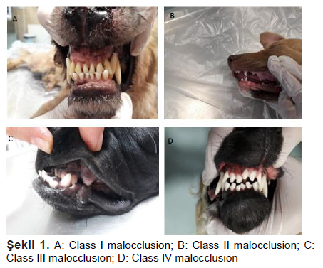
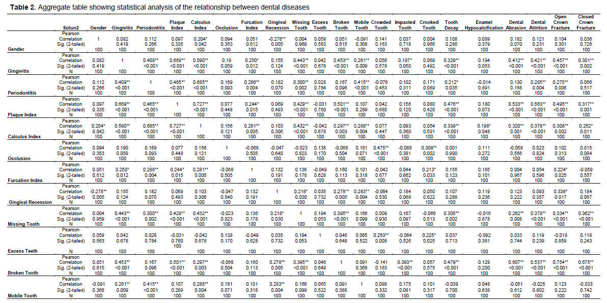
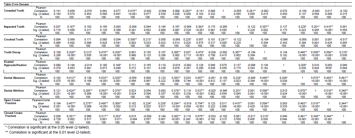
Occlusion: In the evaluation of malocclusion regarding the misalignment of upper and lower jaw teeth in 9 dogs, malocclusion was identified. Among them, 3 dogs had normal upper and lower jaw lengths, but their teeth were not in the correct position (Class I). One dog had a significantly shorter lower jaw compared to the upper jaw (Class II). In 3 dogs, the lower jaw was considerably longer than the upper jaw (Class III). Two dogs exhibited discrepancies in jaw length, with one jaw shifting to the midline as Class II and the other as Class III (Class IV), (Figure 1). In Class I and Class IV malocclusion, the teeth were misaligned and crowded, with no evidence of abnormal wear. Dogs with Class II and Class III malocclusion showed normal findings, but they experienced difficulty in feeding. The teeth did not cause damage to palatal and lingual tissues. The relationship between age, gender, and body weight with occlusion was statistically examined (Table 1). According to the data, there was a significant negative correlation between occlusion and body weight (r = - .24; P<0.01). According to Table 2, a positive and significant relationship between occlusion and crowded and misaligned teeth was observed, suggesting that a dog with malocclusion may also have crowded and misaligned teeth.
Epulis: In the study, epulis (oral tumor) was detected in 3 dogs. The tumor, approximately the size of a walnut, was located at the incisor level of the maxillary and mandibular regions in 2 dogs. Additionally, radiographs revealed erosions in teeth and bone tissue. In one dog, it was found that the epulis originated from the maxillary premolar tooth border, covering the entire hard palate surface, leading to respiratory difficulties. The identified mass exhibited a tendency to bleed during examination.
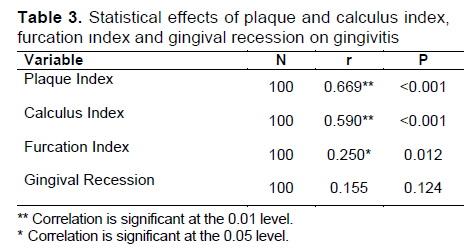
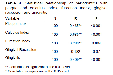
Plaque Index: Plaque accumulation was recorded as 76%, making it the most common dental disorder after gingivitis. Among the dogs, 46% had mild plaque accumulation (n = 46), 23% had moderate accumulation (n = 23), and 7% had advanced plaque accumulation (n = 7). The accumulation was most prominent on maxillary Premolar 3 (P3), Premolar (P4), Molar 1 (M1) and mandibular P4, M1, Molar 2 (M2) teeth. Subsequently, this was followed by all canines, premolars, molars, and to a lesser extent, incisor teeth. Soft, easily removable accumulations on the teeth varied in color from yellow to brown, sometimes exhibiting gray-green discolorations. An analysis of the impact of age and body weight on plaque accumulation revealed a positive correlation (Table 1). Additionally, statistically significant and positively correlated relationships were observed between the plaque index and gingivitis as well as periodontitis, as presented in Tables 3 and 4. The overall relationship between the plaque index and dental disorders was statistically presented in Table 2.
Calculus Index: Macroscopic examination and probe inspection revealed a calculus accumulation rate of 48%. Among these, 31% (n = 31) were classified as mild, 16% (n = 16) as moderate, and 1% (n = 1) as advanced. Lesions ranging in color from yellow to brown/green, hard, and not easily removable without the aid of an instrument were observed. Bad breath was evident, and changes (red, swollen, and round) were noted at the gum margins. Some exhibited a tendency to bleed, and teeth showed mobility. According to the examinations, it was determined that intense dental calculus led to tooth mobility, gingival recession, and furcation. Statistical analyses revealed a significant positive correlation between age, gender, and body weight with the calculus index (P<0.01), as shown in Table 1. The impact of the calculus index on gingivitis and periodontitis showed significant differences according to the data in Table 2 (P<0.01). Statistical analysis data illustrating the impact of the calculus index on dental diseases were collectively presented in Table 2.
Furcation Index: In 6 dogs in the study (6%), opening of the tooth roots was observed. The opening was particularly localized on multi-rooted teeth such as premolars and molars. It was prominent in animals with heavy dental calculus and periodontitis. Among the dogs, 5 (5%) had mild, and 1 (1%) had moderate furcation involvement. Exposed tooth roots were easily visible macroscopically. Teeth exhibited mobility, and the structure of periodontal tissues was compromised. Statistical analysis revealed a significant positive correlation between the furcation index and levels of dental plaque, calculus, gingivitis, and periodontitis (Table 3 and 4).
Gingival Recession: Gingival recession (17%) was noticeable on maxillary incisors. Of these, 9 (9%) were mild, and 4 (4%) were of moderate severity. In affected animals, tooth roots were visible. The relationship between gingival recession and dental diseases was collectively presented in the Table 6. According to the data, there was a significant positive correlation between gingival recession and missing, broken, and mobile teeth (Table 2).
Gingivitis: Overall, gingivitis was present in 79% of the dogs. Lesions were equally distributed and bilateral in the maxilla and mandible. Dogs with gingivitis showed varying degrees of plaque and calculus accumulation. Mild symptoms were observed in 49% of the dogs (n = 49), with red and slightly swollen gum margins. However, there was no bleeding during probe examination. Moderately affected dogs constituted 29% (n = 29) of the study, with rounder and more adherent gums, noticeable redness, and a tendency to bleed. Severe gingivitis was observed in only 1% of the dogs (n = 1), with difficulty in eating and hyperplastic, swollen gums. Bleeding occurred during probe examination.
The effectiveness of dental plaque, calculus, furcation index, and gingival recession on the severity of gingivitis was statistically examined, determining possible significance values. The relationship between them was evaluated using the Pearson correlation coefficient. For this purpose, Table 3 displays the analysis results.
According to Table 3, there was a statistically significant and positive difference (P<0.001) in the impact of plaque, calculus index, and furcation opening on the severity of gingivitis. However, no significant relationship was found between gingivitis and gingival recession (r = .155; P>0.001). It was determined that a dog with gingivitis might also have plaque/calculus accumulation and furcation opening. The relationship between gingivitis and dental diseases was collectively presented in Table 2.
Periodontitis: Periodontitis was evident in 17% of the dogs in the study, with most cases (15%) being mild. Moderate-level affliction was present in 2% of the animals. Periodontitis was particularly observed on maxillary P3, P4, M1, and mandibular P4, M1, Molar 2 (M2) teeth, where intense supra/subgingival plaque and calculus accumulation occurred. Lesions were also bilateral. Supragingival plaques were visible macroscopically, and subgingival plaques were detected during probe examination. Miniature breeds with excessive dental calculus exhibited severe periodontitis.
Table 4 statistically illustrates the effectiveness of dental plaque, calculus, furcation index, gingival recession, and gingivitis on the severity of periodontitis. According to the data, no significant relationship was found between gingival recession and periodontitis. However, a statistically significant positive relationship was observed between periodontitis and the plaque, calculus indices, furcation opening, as seen in the table, especially with gingivitis. Periodontitis may progress in conjunction with one or more of these variables. Statistical analysis data between periodontitis and dental diseases were collectively presented in Table 2.
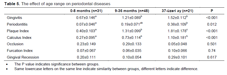
The Effect of Age Range on Periodontal Diseases: The impact of age on periodontal diseases was shown in Table 5 based on the examined data analyses.
Gingivitis and age were statistically assessed, revealing differences between animals aged 0-8 months, 9-36 months, and 37 months and older. In the evaluation between age and periodontal disease, it was observed that there were progressively increasing differences among age groups (P<0.05). The plaque index showed a statistically strong relationship with each age group (P<0.001). It was determined that index averages increased progressively from 0-8 months to 37 months and older. The calculus index had a significant difference between animals aged 0-8 months and those aged 9-36 and 37 months and older (P<0.001). According to the data, gingivitis, periodontitis, plaque, and calculus accumulation increased proportionally with age. No significant relationship was found between age groups and occlusion, gingival recession, and furcation index. It was concluded that age groups did not have an impact on these diseases (P<0.05).
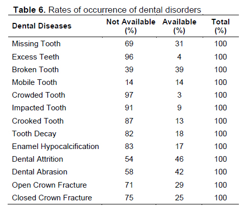
Rates of Occurrence of Dental Disorders: The dental disease rate of the dogs participating in the study was shown in the Table 6. According to this;
In the study, 31 dogs (31%) had missing teeth, considering teeth that had not yet erupted in animals younger than 9 months as missing. Additionally, it was observed that tooth loss decreased proportionally with age, particularly in the 2.5-10 age range. Excess teeth, resulting from retained deciduous teeth, were found in 4% of the dogs, especially in Yorkshire, Terrier, and Pekingese breeds. Broken teeth were present in 39 dogs (39%), with fractures predominantly occurring in incisors and canines. Moreover, tooth fractures were more common in male dogs over two years old. Mobile teeth were observed in 8% of the dogs, developing with advancing age and periodontal disease. However, the cause remained undetermined in the remaining 6%. Crowded teeth were observed in only 3 dogs, particularly in miniature breeds (Yorkshire, Terrier). Crooked teeth were present in 13 dogs and were common in miniature breeds. However, they were also observed in animals with brachycephalic skull structures and malocclusion.
Tooth decay was detected in 18 animals. Especially on the occlusal surfaces of maxillary 1st molars and canine teeth, dental caries were encountered, with 8% of them identified in dogs aged 5-10. Dogs with enamel hypoplasia exhibited pigmented, soft, hollow, and pitted areas on their enamel. The dentin layer was easily distinguishable. Dental attrition was observed in 46 dogs aged over 12 months. Wear was particularly prominent on the palatal surface of upper incisors and the labial surface of lower incisors, followed by canines, premolars, and molars. Abrasion was noted in 42 dogs, especially on the lingual and palatal surfaces of canines and incisors. The affected teeth showed varying degrees of enamel wear on their crowns, with most cases having a normal pulp chamber. Open crown fractures were present in 29 dogs, with necrotic and infected pulp chambers and canals. The central part appeared dark brown/black. Closed crown fractures were identified in 25 dogs, with a normal-looking pulp chamber. In one dog, an awn embedded in the gum tissue at the level of the upper left canine tooth was observed. Additionally, injuries to the oral mucosa and tongue were detected in 2 dogs, but due to their low numbers, they were not included in the assessment.
It is well-known that a significant portion of periodontal diseases progresses through the stages of gingivitis and periodontitis[7,20,21].
In our study, the incidence of periodontitis was determined to be 17%, which is comparatively lower when compared to other studies. For instance, a study conducted in the United States reported periodontitis in over 80% of dogs aged three and older [22]. Another study in the Czech urban area with 408 dogs showed a periodontitis rate of 60%[6]. Other studies reported varying prevalences, such as 68.9% in India (103 dogs);[23], 56.8% in Tirana/Albania (776 dogs);[24], 41% in Africa (37 dogs);[25], and cases of periodontitis in 15 out of 74 dogs in Brazil[14]. The onset of periodontal disease is typically around two years of age[26]. Our study's lower incidence might be attributed to the inclusion of dogs aged 0-8 months (n = 31), potentially reducing the overall incidence. There was a positive correlation between increasing age and the prevalence of periodontal disease in our study, aligning with literature showing a rise in disease severity with age. The presence of inadequate oral hygiene, plaque, and dental calculus is closely associated with periodontal disease, which could influence the incidence level.
In our study, it was found that 79% of the examined dogs had gingivitis, which was identified as the disease with the highest incidence. In previous studies, this rate was observed to be 35.5% in Brazil[13] and 17.1% in Bulgaria[5], where researchers emphasized it as the most commonly encountered disease. According to the findings, the most affected areas of the gums in dogs are, in order, the premolar, molar, and incisor regions. The labial/buccal surface of the gums was more affected than the lingual/palatal surface, and inflammation was observed to shape equally in the maxilla and mandible. These results are consistent with similar studies[6]. The high incidence of gingivitis in stray dogs is believed to be due to poor oral hygiene, inadequate veterinary care, lack of dental examinations, and the inability to remove accumulated plaque and calculus on the teeth.
In our study, despite being less common in small breeds (Yorkshire, Pekingese, Terrier), periodontal disease was more frequently encountered. Penman, Isogai, and their colleagues also reported a higher prevalence of periodontal disease in small dog breeds compared to larger breeds in their studies, aligning with our findings. Additionally, it was observed that periodontal disease develops at an earlier age in small breeds compared to large breeds, which is consistent with existing literature.
Similar to humans, dogs also experience cases of dental decay, with an increase in dental decay with age[6]. Dental decay was found in 18% of the dogs included in the study which is higher compared to other studies. In a study, it was mentioned that 5.25% of adult dog patients had one or more decay lesions, and in most cases, the lesions were bilaterally symmetrical[22]. An examination of 776 dogs in Tirana/Albania veterinary hospital[24] and 408 dogs in the Czech urban area [6] only reported one decay case. Similar studies suggest a decay incidence of around 8-10%[27,28]. The high incidence of dental decay in our study is likely attributed to the majority of dogs being stray, poor nutrition, lack of oral hygiene, and age differences among the dogs.
It has been reported that a diet consisting of soft and processed foods increases plaque accumulation, while mechanical cleaning can be achieved by providing chewing materials or using brushing to reduce plaque buildup[29,30]. In our study, plaque accumulation in dogs was found to be 76%. Similar studies reported rates of 61.3% in the Czech urban area[6] and 60.3% in Tirana/Albania city hospital[24]. Our data aligns with these literature findings. In owned animals included in the study, plaque accumulation was observed to be high due to a diet based on soft and processed foods. In the case of street dogs, constituting 90% of the study, poor and irregular nutrition, inability to maintain oral hygiene, and the absence of brushing or mechanical cleaning contribute to a significant presence of plaque formation.
In our study, 48% of dogs were observed to have dental calculus, a rate consistent with similar studies. Dental calculus, like periodontal disease, tends to increase with the age of the animal. The majority of dogs fed with homemade or commercial diets are reported to develop dental calculus and gum inflammation from around 26 months of age[6]. The lack of oral hygiene leads to the accumulation of plaque containing bacteria, eventually causing gum inflammation and calculus formation[6,31,32]. There was a significant increase in calculus accumulation in the dogs in our study, likely attributed to the inability of stray animals to maintain oral hygiene.
In our study, dental fractures were detected in 39% of dogs, with these fractures being particularly localized in the incisor and canine teeth of male dogs over the age of two. Thus, our findings exhibited clinical symptoms consistent with similar literatüre[7,13]. Dental fracture frequencies vary across studies, such as 2.4% in Belem/Brazil[13], 8.5% in Bulgaria[5], 48% in Africa [25], and frequencies ranging from 35.5% to 57.1% reported in other studies[14]. Butkovič reported a 19.3% incidence of dental fractures, with the majority resulting from traffic accidents, playing, wrestling with other dogs, catching hard objects, and hunting, leading to incisor fractures in both upper and lower jaws[33]. The fractures encountered in our study were similar to the studies in terms of causes.
Tooth wear is often observed in working dogs and dogs fed with a hard diet who enjoy playing with stones[6]. Estimates of age based on tooth wear should be made carefully, as wear varies depending on the dog's care conditions and diet[6]. In our study, dental attrition was identified in 46% of dogs, and it was found that these animals were over 12 months old. Wear was particularly evident on the palatal surface of upper incisor teeth and the labial surface of lower incisor teeth, followed by canine, premolar, and molar teeth. Compared to other studies, tooth wear was reported to be 5.9% in the Czech urban area[6], 6.4% in an examination of 776 dogs in Tirana/Albania city veterinary hospital[24], 83% in Africa[25], and only in 25 dogs in Brazil[14]. The significance of age in tooth wear and the inclusion of dogs under 12 months old in the study were believed to have influenced the incidence rate.
Periodontal disease, agenesis, trauma, and age-related factors can lead to tooth loss[6,7]. The number of missing teeth is believed to increase with age, showing a significant relationship [32]. In our study, dogs with missing teeth constituted 31% of the total population. A study in Brazil with 74 dogs reported missing teeth in 63.5% (47 dogs);[14]. An assessment of 408 dogs in the Czech urban area found missing teeth in 33.8% of dogs[6], while an examination of 776 dogs in Tirana/Albania city veterinary hospital revealed a 31% incidence of missing teeth [24]. Unlike other studies, our findings indicate that missing teeth were most common in incisor and canine teeth, with fewer occurrences in premolar and molar teeth. This pattern was thought to be due to weaker attachment of maxillary and mandibular incisor teeth to the alveolar bone compared to other canine, premolar, and molar teeth. In our study, tooth loss was not observed for reasons other than age, fighting with other dogs, and trauma.
Tooth enamel is highly mineralized and cannot be reshaped or repaired. Hereditary imperfections arise when there is insufficient enamel matrix during tooth development. They can occur due to systemic infections like distemper virus, poor nutrition, or damage to the enamel during tooth eruption[34]. In our study, enamel hypoplasia was observed, particularly in canine teeth, at a rate of 17%. It was determined to have a higher incidence compared to similar studies. This condition was believed to be shaped by factors such as dogs being stray and exposed to the streets, vulnerability to distemper disease, imbalanced and poor nutrition, and trauma during tooth development. In a study conducted on 540 dogs at Iran Razi University, enamel hypoplasia was observed in 11 dogs [34], while other studies reported it in 3 dogs[14], 2 dogs [24], and 1 dog[6].
The conclusion of our study demonstrates a significant incidence of dental health issues in dogs. Particularly, the high prevalence of gingivitis indicates a substantial portion of periodontal diseases progresses through this stage. The concentration of missing teeth, especially in incisors and canines, the prominence of enamel hypoplasia, and the exploration of other dental health problems provide crucial data for veterinary practice. By offering important insights into the overall oral health of dogs, our study provided an opportunity to develop more effective interventions and preventive strategies in veterinary practice. Furthermore, the agreements of our study with existing literature on similar topics enhanced the reliability of the obtained results, making a substantial contribution.
Gereç ve Yöntem
Research and Publication Ethics: The study was carried out at the Surgery Department of the Faculty of Veterinary Medicine at Erciyes University. Approval for the study was obtained from the Erciyes University Local Ethics Committee.Bulgular
The gender distribution of randomly selected dogs was determined as 39 females and 61 males. The dogs consisted of 79 mixed breeds, 3 German Shepherds, 3 Chihuahuas, 1 Shepherd, 1 Pekingese, 1 Yorkshire Terrier, 1 Pharaoh Hound, 2 Golden Retrievers, 2 Pointers, 3 Terriers, and 5 Kangals. The age of the dogs ranged from one month to 120 months. For ease of the study, they were divided into three categories: 0-8 months with 31 dogs (n = 31), 9-36 months with 48 dogs (n = 48), and over 36 months with 21 dogs (n = 21). The dogs' b 3 kg to 65 kg, with an mean of 30 kg.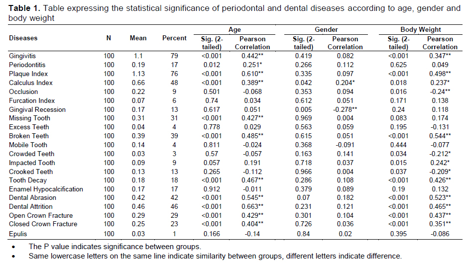
Tartışma
The study examined dental diseases in dogs, noting frequent occurrences of tooth fractures, missing teeth, broken teeth, dental attrition, dental abrasion, gingivitis, dental plaque, and dental calculus. Similar findings have been reported in previous studies [6,18-20]. Our study found that 87% of the dogs had multiple anomalies, including periodontal and dental diseases. References
- The Effects of Antioxidants Vitamins on Clinical and Some Haemotological Parameters, Antioxidant Enzyme and Lipid Peroxidation Levels of Beef Cattle Vaccinated with Foot and Mouth Disease Vaccine
- ELAZIĞ’DA KÖPEKLERDE TESPİT EDİLEN TRAVMATİK MYİASİSLER
- STUMP PYOMETRALI BİR KÖPEKTE OVARİAN REMNANT SENDROMU
- Incidence of Foot Diseases in Cattle in and around Elazığ
- ELAZIĞ VE ÇEVRESİNDE SIĞIRLARDA GÖRÜLEN AYAK HASTALIKLARININ İNSİDANSI ÜZERİNE GÖZLEMLER