
Tavşanlarda Bazı Sedatif ve Hipnotiklerin Gözyaşı Miktarı Üzerine Etkilerinin Araştırılması
Cilt 39, Sayı 3
Ekim 2025 · Sayfalar 195-199
Tavşanlarda Bazı Sedatif ve Hipnotiklerin Gözyaşı Miktarı Üzerine Etkilerinin Araştırılması
Investigation of the Effects of Some Sedatives and Hypnotics on Tear Volume in Rabbits
Nimet KILIÇ1, Cavit KUM2, Kerem UZUN3
2Aydın Adnan Menderes University, Faculty of Veterinary Medicine, Department of Pharmacology and Toxicology, Aydın, TÜRKİYE
3Ministry of Agriculture and Forestry, Çaldıran Directorate of Agriculture and Forestry, Van, TÜRKİYE
Bu çalışmanın amacı, Asepromazin, Haloperidol, Detomidin, Medetomidin ve Ksilazin gibi bazı sedatif ve hipnotik ilaçların tavşanlarda gözyaşı miktarı üzerine etkilerini araştırmaktır. Çalışma materyali dört erkek ve dört dişi Yeni Zelanda beyaz tavşanından oluşmaktadır. Enjeksiyondan önce, her tavşanda gözyaşı miktarı Schirmer 1 gözyaşı şeritleri (T-0) kullanılarak ölçülmüştür. Daha sonra her grupta quadriceps femoris kasına intramusküler olarak Asepromazin 3.3 mg/kg, Haloperidol 5 mg/kg, Detomidin 0.30 mg/kg, Medetomidin 0,25 mg/kg ve Ksilazin 5 mg/kg uygulandı. Enjeksiyonu takiben, sedasyondan 15. (T-1), 30. (T-2), 45. (T-3), 60. (T-4) ve 24. (T-5) saatlerde gözyaşı hacmi ölçüldü. Enjeksiyondan 15 dakika sonra Xylazine (p=0.024) ve Detomidin (p=0.001) gruplarında gözyaşı hacminde istatistiksel olarak anlamlı azalmalar gözlenirken, Haloperidol (p=0.002) ve Asepromazin (p<0.0001) enjeksiyonundan 15 dakika sonra yapılan ölçümlerde gözyaşı hacminde artışlar gözlendi. Xylazine, Detomidin ve Medetomidin gruplarında enjeksiyondan 30, 45 ve 60 dakika sonra hafif bir artış gözlenirken, 24 saat sonra yapılan ölçümlerde başlangıç değerine yakın değerler elde edildi. Bu çalışma sonucunda tavşanlarda gözyaşı hacmindeki azalmanın, veteriner hekimlikte bu çalışmada kullanılan sedatif ve hipnotiklerle yapılacak ilerideki kısa süreli cerrahi işlemler sırasında yol gösterici olacağı, kornea kserozisi ve buna bağlı keratit, keratokonjunktivit, kornea ülseri ve gözyaşı üretiminin azalmasına bağlı göz kayıpları gibi hastalıkların önlenebileceği sonucuna varılmıştır.
This study aimed to investigate the effects of some sedatives and hypnotics such as Acepromazine, Haloperidol, Detomidine, Medetomidine and Xylazine, on tear quantity in rabbits. The study material comprised four male and four female New Zealand white rabbits. Before injection, tear quantity was measured in each rabbit using Schirmer 1 tear strips (T-0). Then, Acepromazine 3.3 mg/kg, Haloperidol 5 mg/kg, Detomidine 0.30 mg/kg, Medetomidine 0.25 mg/kg and Xylazine 5 mg/kg were administered intramuscularly to the quadriceps femoris muscle in each group. Following the injection, tear volume was measured at 15 (T-1), 30 (T-2), 45 (T-3), and 60 minutes (T-4), and 24 hours (T-5) after sedation. Statistically significant decreases in tear volume were found in the Xylazine (p=0.024) and Detomidine (p=0.001) groups at 15 minutes following the injection compared to the pre-sedation times. On the other hand, increases were observed in tear volumes in measurements taken 15 minutes after the Haloperidol (p=0.002) and Acepromazine (p<0.0001) injection. In the Xylazine, Detomidine, and Medetomidine groups, a slight increase was observed 30, 45, and 60 minutes after injection, while measurements taken 24 hours later were found to be close to the initial value. As a result of this study, it was concluded that the reductions in tear volume in rabbits may serve as a reference during future short-term surgical procedures performed with the sedatives and hypnotics used in this study in veterinary medicine, and that corneal xerosis and related diseases such as keratitis, keratoconjunctivitis, corneal ulcers, and eye loss due to decreased tear production can be prevented.
Giriş
Experimental studies conducted on laboratory animals, as in the past, are of great importance to humanity in many areas, including vaccine studies, the discovery of new drugs and treatment methods, and cancer studies in both human and veterinary medicine. One of the laboratory animals important for scientific studies is the rabbit. The precorneal tear film is a complex, dynamic structure composed of lipids, proteins, and mucins that lie on the hydrophobic surface of the epithelium[1]. The precorneal tear film is vital for conjunctival and corneal health, maintenance of normal functions[2,3], and integrity of the ocular surface[4]. It is approximately 7 microns thick. It completely covers the cornea and conjunctiva[5].The Schirmer tear test (STT) is commonly used in ophthalmic examination to assess and diagnose keratoconjunctivitis sicca or excessive tear volume. The general clinical use of the test involves placing a standard notched filter paper in the lower conjunctival fornix of the eye and reading the amount of wetness produced after 1 minute[6]. Specific diagnoses are made by comparing the recorded value with the average normal value for the species. STT-1, performed without topical anesthesia, measures basal and reflex tear volume, while STT-2 measures only basal tear volume after topical anesthesia with one drop of tetracaine. Basal tears are those produced without stimulation and are lacrimal secretions that protect the ocular surface. Reflex tears are produced after stimulation, usually due to a substance that irritates the ocular surface, such as a foreign body or inflammatory response[7].
In this study, the hypothesis that investigate the effects of some sedative and hypnotic drugs such as Acepromazine, Haloperidol, Detomidine, Medetomidine, and Xylazine, on tear volume in rabbits during surgical procedures was established, and the data obtained after the study were aimed at forming a guide for future studies. Thus, it is aimed to prevent corneal xerosis and the subsequent process of keratitis, keratoconjunctivitis, corneal ulcus, and loss of the eye due to the decrease in tear volume in the short-term surgical interventions where sedatives and hypnotics are used, as mentioned in academic studies conducted with rabbits. In this way, it is aimed that the study will also contribute to animal health and welfare.
Gereç ve Yöntem
Research and Publication Ethics: This study was conducted at the Aydın Adnan Menderes University Experimental Animals Unit with the approval of ADU-HADYEK dated 18.05.2023 and numbered 64583101/2023/65.Animal Material: The study material consisted of 8 New Zealand white rabbits (Oryctolagus cuniculus), four males and four females, between the ages of 8 months and 3 years, and weighing between 2.5 and 3.5 kg. After being brought from the rabbit farm, all rabbits were placed in numbered individual cages by a simple random assignment method. The cages were cleaned daily, and wheat straw and wood shavings were used as bedding material. The environment where the animals were housed was 18-22 °C, with 30%-70% humidity, centrally ventilated, and rabbits were housed in the light for 12 hours and automatically in the dark for 12 hours. The animals were fed ad libitum, and feed and water were always available in the cage. Standard rabbit feed and dried alfalfa were used as feed material. The animals were allowed to acclimate for 7 days before the study and were examined clinically to ensure that they were healthy.
Method: Eight rabbits, four males and four females, were used for each sedative or hypnotic substance in the study. A two-week washout period was implemented between experiments with various sedative or hypnotic substances. This study determined an initial sample size of five groups and forty animal subjects by conducting a power test using the G Power program (Version 3.1.9.7, F test, ANOVA). Before the study, all animals underwent a clinical examination, and individual standard forms were created and filled out for each animal. The body weight of each animal was regularly measured and documented before the study. The inclusion criteria for this study were healthy males aged 1 to 3 years, weighing between 2.5 and 3.5 kg. The exclusion criteria included individuals with eye problems, general health issues, or those who are elderly. These criteria were determined before the study. Before the study began, criteria were established. A 20-24 gauge intravenous catheter (AYSET, Adana, Turkey) was then sterilely fixed in the marginal ear vein, depending on the animal's size.
The study was repeated after the rabbits had rested for two weeks, using five different sedative or hypnotic substances. Before the injection, the tear volume was measured in each rabbit using Schirmer 1 tear strips (ERC®, Ankara, Turkey) (T-0). The Schirmer tear strip was removed from its sterile packaging, folded from the marked part, and placed in the lower conjunctival fornix in the middle of the distance between the medial and lateral canthus in the right and left eyes. The mean value obtained in both eyes was evaluated statistically. Then, Acepromazine (Sedan®, Bioveta) at a dose of 3.3 mg/kg[8,9] was injected to the rabbits in the acepromazine group, Haloperidol (Norodol®, Aris) at a dose of 5 mg/kg[10] was injected to rabbits in the Haloperidol group, Detomidine (Domosedan®, Pfizer) at a dose of 0.30 mg/kg[11] was injected to the rabbits in Detomidine group, Medetomidine (Domitor®, Pfizer) at a dose of 0.25 mg/kg[12] was injected to the rabbits in Medetomidine group, and Xylazine (Rompun®, Bayer) at a dose of 5 mg/kg[13,14] was injected to the rabbits in the Xylazine group into the quadriceps femoris muscle intramuscularly. All male and female animals received all five drugs by simple randomization. Following the injection, the amount of tears was measured and recorded at the 15th minute (T-1), 30th minute (T-2), 45th minute (T-3), 60th minute (T-4), and 24th hour (T-5) after sedation[15]. The health status of the rabbits was regularly checked during and after sedation, and interventions were made when deemed necessary.
Statistical Analysis: The study evaluated statistical data using the SPSS 22.0 statistical package program (Inc., Chicago, IL, USA). The conformity of variables to normal distribution was examined using visual (histogram) and analytical methods (Shapiro-Wilk). Descriptive analyses were expressed as mean ± standard error (SE) for normally distributed variables. Although both male and female rabbits were included in the study, sex was not incorporated as a factor in the statistical models. This decision was made because the primary objective was to evaluate the effects of treatments and time, and the limited sample size (n=8 per group) did not provide sufficient statistical power for reliable subgroup analyses based on sex. Since the experimental design included repeated measurements across multiple independent groups, the data were analyzed using a mixed-model Repeated Measures Analysis of Variance (RM-ANOVA) with time as a within-subjects factor and treatment group as a between-subjects factor, thereby accounting for both temporal changes and intergroup differences. Sphericity assumptions were checked with Mauchly’s test, and when necessary, Greenhouse–Geisser corrections were applied. Homogeneity of variances was assessed using the Levene’s test. Differences within and between groups were determined, and p<0.05 was considered statistically significant. The sample size consisting of a total of 5 groups and 40 animals subjects, was determined by performing a power test using G*Power software (Version 3.1.9.7, F test, ANOVA).
Bulgular
In this study, the effects of some sedatives and hypnotics on tear volume were comparatively examined; no complications or deaths related to sedatives and hypnotics were encountered, and all rabbits continued their normal lives after the study.Statistically significant decreases in tear volume were found in the Xylazine (p=0.024) and Detomidine (p=0.001) groups and statistically insignificant decreases Medetomidine (p=0.136) groups at 15 minutes following the injection compared to the pre-sedation times. On the other hand, increases were observed in tear volumes in measurements taken 15 minutes after Haloperidol (p=0.002) and Acepromazine (p<0.0001) injection (Table 1).
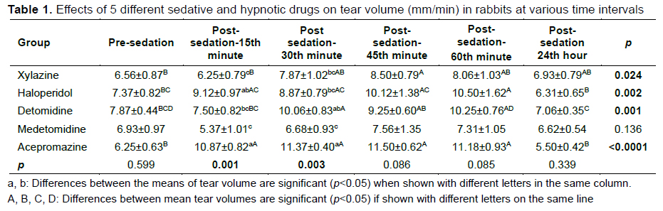
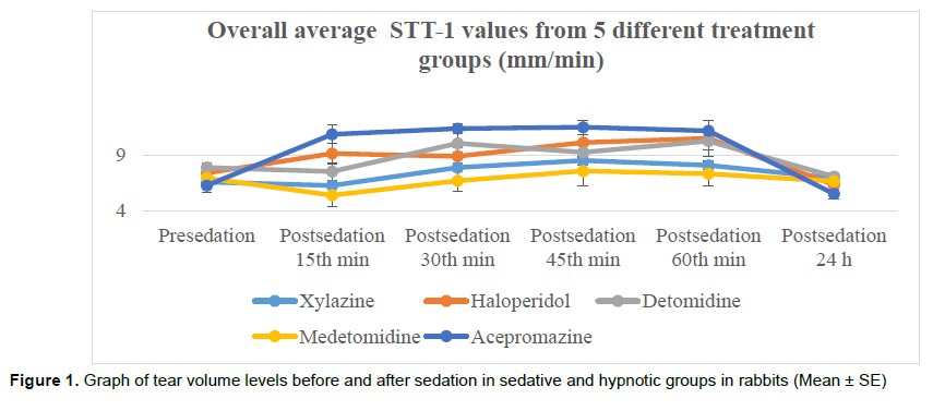
In the Xylazine, Detomidine, and Medetomidine groups, a slight increase was observed 30, 45, and 60 minutes after injection, while measurements taken 24 hours later were found to be close to the initial value. Mild increases continued after 30, 45, and 60 minutes of sedation with Haloperidol and Acepromazine, and this increase was found to be statistically significant in all sedation groups at the 30th minute of sedation (Figure 1). When comparing the groups tear amount measurement times, there is no statistical difference between the groups before sedation (p=0.599). At 15 minutes after sedation, there was a statistically significant difference between the Xylazine group and the Haloperidol and Acepromazine group, between the Haloperidol group and the Xylazine and Medetomidine group, between the Detomidine group and the Acepromazine group, between the Medetomidine group and the Haloperidol and Acepromazine group, and finally between the Acepromazine group and all groups except Haloperidol (p=0.001). At 30 minutes after sedation, there was a statistically significant difference between the Xylazine group and the Acepromazine group, between the Haloperidol group and the Acepromazine group, between the Detomidine group and the Medetomidine group, between the Medetomidine group and the Detomidine and Acepromazine groups, and finally between the Acepromazine group and all groups except the Detomidine group (p=0.003). There is no statistical difference between the groups at 45 minutes after sedation (p=0.086). There is no statistical difference between the groups at 60 minutes after sedation (p=0.085). There is no statistical difference between the groups 24 hours after sedation (p=0.339).
Tartışma
Xylazine, Medetomidine, and Detomidine are pharmacologically classified as alpha-2 adrenoreceptor agonists. This group of drugs has a wide range of uses in animals as sedatives and analgesics. They are frequently used in laboratory animals, pet patients, farm animals, and exotic patients. Xylazine and Medetomidine are commonly used in rabbits, either in combination with Ketamine or alone. In this study, decreases in tear volume were observed in the Xylazine, Detomidine, and Medetomidine groups 15 minutes after injection. These decreases were minimal in the Xylazine and Detomidine groups, while significant reductions in tear volume were observed after the Medetomidine injection. This expected effect has also been reported in other animals.One study administered acepromazine-butorphanol, diazepam-butorphanol, xylazine-butorphanol combinations, and butorphanol and xylazine alone to dogs and investigated their effects on tear volume[16]. While all three combinations caused statistically significant decreases in tear volume, the depressive impact on the xylazine group was minimal, parallel to our study, and it was concluded that xylazine can be used in healthy dogs. Another study observed that xylazine alone did not affect the tear volume in horses[17]. In our research, reductions were noted in the medetomidine group, which has also been reported in other animal species. Another study investigated the effects of medetomidine and medetomidine-butorphanol combinations on tear volume in dogs. Similar to our research, they found a depressant impact on tear volume and attributed this to vasoconstriction in the lacrimal gland ducts[18].
Another study examined the effects of propofol-sevoflurane, midazolam-sevoflurane, and medetomidine-ketamine-sevoflurane combinations on tear volume in rabbits and reported that the medetomidine-ketamine-sevoflurane combination reduced tear volume. This study observed a decrease in tear volume in the detomidine group at 15 minutes, which was reversed after 30 minutes[19].
In another study, detomidine was used alone and with butorphanol in horses. In parallel with our research, an initial decrease was observed, and after this temporary decrease, the depressive effect on tear volume disappeared[20]. This study observed increases in tear volume after acepromazine injection. One study investigated the effects of intramuscular administration of diazepam and acepromazine in rabbits and demonstrated a reduction in tear volume[21].
Another study found that intramuscular injection of acepromazine and xylazine in cats resulted in a statistically significant decrease in tear discharge[22]. Furthermore, atropine administration after acepromazine-ketamine administration in cats resulted in statistically significant decreases in tear discharge[23]. The effect of L-methadone and acepromazine administration before general anesthesia on tear discharge was also investigated in dogs, and it was observed that these preanesthetic agents caused a decrease in tear discharge in dogs[24].
Another study in dogs examined the effect of chlorpromazine alone and in combination with morphine on tear discharge and observed that it caused statistically significant reductions in tear discharge[25]. Haloperidol, together with droperidol, is among the major tranquilizers and it finds usage in veterinary medicine in dogs. It finds a rare use in rabbits, and its effect on tear volume has been reported for the first time, based on our literature review. In dogs, haloperidol was used with ketamine at a 1:1 ratio[26]. It was also used in dogs combined with thiopental[27]. In this study, increases in tear discharge in rabbits were observed after the haloperidol injection.
In this study, the effects of the alpha-2 adrenoceptor agonists Xylazine, Detomidine, and Medetomidine, which are used as sedatives and hypnotics, and the tranquilizer Acepromazine from the phenothiazine group, and the butyrophenone derivative haloperidol on tear production were investigated after intramuscular administration in rabbits. While Medetomidine injection caused statistically significant decreases in tear production, increases in tear discharge were observed after Acepromazine and haloperidol injection. In light of the data obtained from this study, which investigated the effects of some sedatives and hypnotics on tear production in 4 male and 4 female New Zealand White rabbits, it is thought that the use of intraoperative and postoperative artificial tear preparations may be beneficial to prevent dry eyes in all groups, especially in the Medetomidine group. More comprehensive results can be obtained by conducting future studies with different preanesthetic and anesthetic agents.
Acknowledgments
The authors would like to thank Mehmet KAYA for assistance with the statistical analyses.
References
- Foulks GN. The correlation between the tear film lipid layer and dry eye disease. Survey of Ophthalmology 2007; 52(4): 369-374.
- Alkan F, Izci C, Tepeli C, Koc Y. Evaluation of the Schirmer tear test in two Turkish breeds of shepherd dogs. Revue Méd Vét 2004; 155(2): 67-70.
- Peiffer RL, Petersen-Jones SM. Small Animal Ophthalmology: A Problem-Oriented Approach. 4th Edition, Saunders, 2008.
- Sullivan DA, Krenzer KL, Sullivan BD, et al. Does androgen insufficieny cause lacrimal gland inflammation and aqueous tear deficiency. Invest Ophthalmol Vis Science 1999; 40: 1261-1265.
- Akın F, Samsar E. Göz Hastalıkları. Malatya: Medipres, 2005.
- Occhipinti JR, Mosier MA, Lamotte J, Monji GT. Fluorophotometric measurement of human tear turnover rate. Current Eye Research 1988; 7(10): 995-1000.
- Petznick A, Evans MD, Madigan MC, et al. A comparison of basal and eye‐flush tears for the analysis of cat tear proteins. Acta Ophthalmologica 2011; 89(1): 75-81.
- Sarwar MS, Khan NU, Ali H, Gohar A. Pharmacodynamics of xylazine, acepromazine and diazepam on various physiological parameter in rabbits. Pak-Euro Journal of Medical and Life Sciences 2022; 5(1): 147-154.
- Flecknell RA. Laboratory Animal Anaesthesia: An Introduction for Research Workers and Technicians. London: Academic Press, 1987.
- Iglauer F, Beig C, Dimigen J, et al. Hereditary compulsive self-mutilating behaviour in laboratory rabbits. Laboratory Animals 1995; 29(4): 385-393.
- Hurley RJ, Marini RP, Avison DL, et al. Evaluation of detomidine anesthetic combinations in the rabbit. Lab Anim Sci 1994; 44(5): 472-478.
- Ko JC, Thurmon JC, Tranquilli WJ, Benson GJ, Olson WA. A comparison of medetomidine-propofol and medetomidine-midazolam-propofol anesthesia in rabbits. Lab Anim Sci 1992; 42(5): 503-507.
- Sanford TD, Colby ED. Effect of xylazine and ketamine on blood pressure, heart rate and respiratory rate in rabbits. Lab Anim Sci 1980; 30(3): 519-523.
- Green CJ. Neuroleptanalgesic drug combinations in the anaesthetic management of small laboratory animals. Lab Anim 1975; 9(3): 161-178.
- Giannetto C, Macrì F, Falcone A, et al. Evaluation of tear production as measured by Schirmer test I in dogs after acepromazine and acepromazine–methadone premedication. Animals 2021; 11(11): 3015.
- Dodam JR, Branson KR, Martin DD. Effects of intramuscular sedative and opioid combinations on tear production in dogs. Vet Ophthalmol 1998; 1(1): 57-59.
- Brightman AH, Manning JP, Benson GJ, Musselman EE. Decreased tear production associated with general anesthesia in the horse. J Am Vet Med Assoc 1983; 182(3): 243-244.
- Sanchez RF, Mellor D, Mould J. Effects of medetomidine and medetomidine-butorphanol combination on Schirmer tear test 1 readings in dogs. Vet Ophthalmol 2006; 9(1): 33-37. 19. Erol H, Erol M, Atalan G, Ceylan C, Yönez MK. The effects of propofol-sevoflurane, midazolam-sevoflurane, and medetomidineketamine-sevoflurane anesthetic combinations on tear production measured by the schirmer tear test I (STT I) in healthy rabbits. Turkish Journal of Veterinary & Animal Sciences 2021; 45(2): 330-335.
- Leonardi F, Costa GL, Dubau M, et al. Effects of intravenous romifidine, detomidine, detomidine combined with butorphanol, and xylazine on tear production in horses. Equine Veterinary Education 2018, 32: 53-57.
- Selk Ghaffari M, Moghaddassi AP, Bokaie S. Effects of intramuscular acepromazine and diazepam on tear production in rabbits. Vet Record 2009; 164(5): 147-148.
- Ghaffari MS, Malmasi A, Bokaie S. Effect of acepromazine or xylazine on tear production as measured by Schirmer tear test in normal cats. Vet Ophthal 2010; 13(1): 1-3.
- Arnett BD, Brightman AH, Musselman EE. Effect of atropine sulfate on tear production in the cat when used with ketamine hydrochloride and acetylpromazine maleate. J Am Vet Med Assoc 1984; 185(2): 214-215.
- Volk HA, West E, Linn-Pearl RN, et al. Effect of methadone and acepromazine premedication on tear production in dogs. Veterinary Record Open 2018; 5 (1): e000298.
- Ghaffari MS, Madani S, Trbolova A, Ghaffari MS, Akbarein H. Effects of intramuscular chlorpromazine alone and chlorpromazine–morphine combination on Schirmer tear test results in clinically normal dogs. Comparative Clinical Pathology 2011; 20(4): 413-415.
- Kumar A, Sobti VK, Singh KI. Evaluation of haloperidol–ketamine mixture (1:1) anaesthesia in dogs. J Vet Med A 2001; 48: 65-73.
- Sobti VK, Singh K, Bansal PS, et al. Haloperidol as a pre-medicant for thiopental anaesthesia in the dog. J Vet Med A 1990; 37: 170-173.
- YENİ ZELANDA TAVŞANLARINDA ÜÇÜNCÜ GÖZ KAPAĞI (PALPEBRAE TERTİA) ÜZERİNDE HİSTOLOJİK ARAŞTIRMALAR
- İÇME SUYU İLE BELİRLİ DOZLARDA FLOR VERİLEN TAVŞANLARIN KARACİĞER DOKUSUNDAKİ YAPISAL VE BİYOKİMYASAL DEĞİŞİKLİKLER
- PRENATAL VE POSTNATAL DÖNEMDE TAVŞAN AKCİĞERLERİNİN HİSTOLOJİK VE ELEKTRON MİKROSKOBİK İNCELENMESİ*
- TAVŞANLARDA SİNİR DEFEKTLERİNİN OTOJEN SİNİR GREFİ VE SİLİKON TUBULİZASYON İLE ONARILMASI ÜZERİNE KARŞILAŞTIRMALI ÇALIŞMALAR
- TAVŞANLARDA (Oryctolagus cuniculus) ERKEK EKLENTİ BEZLERİNİN POSTNATAL DÖNEMDE MORFOLOJİK GELİŞİMİ*