
Zingeron, TRPM2/PARP-1 Yolak Modülasyonu Yoluyla PTZ Kaynaklı Oksidatif Stresi ve İnflamasyonu Hafifletir
Cilt 40, Sayı 1
Mart 2026 · Sayfalar 077-083
Zingeron, TRPM2/PARP-1 Yolak Modülasyonu Yoluyla PTZ Kaynaklı Oksidatif Stresi ve İnflamasyonu Hafifletir
Zingerone Alleviates PTZ-Induced Oxidative Stress and Inflammation Through TRPM2/PARP-1 Pathway Modulation
Ramazan ÇINAR1, Mahmut YARDIMCI2, Kenan YILDIZHAN3
2Muş Alparslan University, Bulanık Vocational High School, Muş, TÜRKİYE
3Van Yüzüncü Yıl University, Faculty of Medicine, Department of Biophysics Van, TÜRKİYE
Bu çalışma, zingeron’nun (ZGN) SH-SY5Y nöronal hücrelerinde pentylenetetrazol (PTZ) kaynaklı oksidatif stres, inflamasyon ve apoptoz üzerindeki nöroprotektif etkilerini; PARP-1/TRPM2 sinyal yolunun rolüne odaklanarak araştırmayı amaçlamıştır. SH-SY5Y hücreleri, ZGN ön uygulaması (5–25 µM) ile ya da ZGN’siz şekilde PTZ’ye (30 µM, 24 saat) maruz bırakılmıştır. Hücre canlılığı CCK-8 testiyle değerlendirilmiş; oksidatif (MDA, GSH) ve inflamatuar (IL-1β, TNF-α) belirteçler ELISA yöntemiyle ölçülmüştür. Apoptoz yanıtları kaspaz-3/9 aktivite analizleriyle incelenmiş, TRPM2 ve PARP-1 protein ekspresyon düzeyleri Western blot yöntemiyle belirlenmiştir. PTZ maruziyeti hücre canlılığını anlamlı derecede azaltırken MDA, IL-1β, TNF-α, kaspaz-3/9 aktiviteleri ile TRPM2 ve PARP-1 ekspresyonlarını artırmıştır (p<0.05). ZGN tedavisi bu değişiklikleri doz bağımlı olarak tersine çevirmiş; oksidatif dengeyi yeniden sağlamış, inflamasyon ve apoptozu azaltmıştır. PARP-1/TRPM2 sinyal yolunu inhibe ederek, PTZ’nin neden olduğu nöronal hasara karşı güçlü bir nöroprotektif etki göstermektedir. Bu etkiler, oksidatif stresi, sitokin salınımını ve kaspaz aktivasyonunu azaltarak gerçekleşmektedir. Bulgular, ZGN’nin TRPM2 aracılı oksidatif nörotoksisiteyi hedefleyen nörodejeneratif hastalık tedavilerinde umut verici bir ajan olabileceğini göstermektedir.
This study aimed to investigate the neuroprotective effects of zingerone (ZGN) against pentylenetetrazol (PTZ) -induced oxidative stress, inflammation, and apoptosis in SH-SY5Y neuronal cells, focusing on the involvement of the PARP-1/TRPM2 signalling pathway. SH-SY5Y cells were exposed to PTZ (30 μM, 24 h) with or without ZGN pretreatment (5–25 μM). Cell viability was assessed by CCK-8 assay, while oxidative stress (MDA, GSH) and inflammatory (IL-1β, TNF-α) markers were quantified by ELISA. Apoptotic responses were assessed through caspase-3/9 activity assays. TRPM2 and PARP-1 protein expression were determined by Western blot analysis. PTZ exposure significantly decreased cell viability and increased MDA, IL-1β, TNF-α, and caspase-3/9 activities, as well as TRPM2 and PARP-1 expression (p<0.05). ZGN treatment significantly reversed these changes in a dose-dependent manner, reducing oxidative stress, apoptosis, and inflammation. ZGN provides potent neuroprotection against PTZ-induced cell damage by inhibiting the PARP-1/TRPM2 signalling axis, thereby reducing oxidative stress, cytokine release, and caspase activation. These findings suggest that ZGN may be a promising agent for targeting TRPM2-mediated oxidative neurotoxicity in neurodegenerative disorders.
Giriş
Oxidative stress (OS) is a central mechanism in the pathogenesis of many neurological disorders, as overproduction of reactive oxygen species (ROS) disrupts redox homeostasis, damages macromolecules, and triggers cell death cascades[1]. Neuronal cells under excitotoxic or proconvulsant stimuli are particularly vulnerable to such effects[2]. One widely used in vitro paradigm is pentylenetetrazole (PTZ)-induced neurotoxicity, which reliably induces ROS overproduction, mitochondrial dysfunction, Ca²? dysregulation, and apoptotic signals in neuronal cell lines such as SH-SY5Y[3,4].Studies indicate that Transient Receptor Potential Melastatin-2 (TRPM2), a nonselective Ca²? permeable ion channel, contributes to OS, leading to intracellular Ca²? overload and cell death in neurons[5]. TRPM2 is subsequently activated by ROS, leading to ADP-ribose production (often via PARP-1 activity) and intracellular Ca²?, creating a feedback loop that increases calcium influx and oxidative damage[6,7]. Indeed, TRPM2-mediated Ca²? influx has been shown to exacerbate ROS accumulation, mitochondrial membrane permeability, and caspase cascade activation in various neuronal and non-neuronal systems[8,9]. Recent studies have further highlighted the dual regulatory role of TRPM2, which depends on the cellular redox status; in this context, modulation can either alleviate or exacerbate oxidative damage[10].
Currently, there is increasing interest in natural phytochemicals with antioxidant, anti-inflammatory, and cytoprotective properties. Among these, zingerone (ZGN) (4-(4-hydroxy-3-methoxyphenyl)-2-butanone), a pungent compound derived from ginger (Zingiber officinale), is emerging as a promising candidate. Recent preclinical studies and systematic reviews have documented the ability of ZGN to prevent neuroinflammation, OS, and behavioral deficits in models of cognitive impairment, heavy metal toxicity, and mood disorders[11,12]. For example, it has been found to attenuate cadmium-induced oxidative damage and cognitive decline and also to attenuate inflammation in pain models through modulation of Ca²? signaling and neuronal excitability (13, 14). However, its direct effects on TRPM2-mediated pathways in neuronal cells under proconvulsant stress have not yet been addressed in the literature.
The present study investigated the neuroprotective effects of ZGN in SH-SY5Y cells exposed to PTZ through inhibition of the PARP-1/TRPM2 signaling cascade. Cell viability, OS markers (MDA and GSH), proinflammatory cytokine expression (IL-1? and TNF-?), apoptotic mediators (caspase-3 and -9), and TRPM2 and PARP-1 protein expression were examined. The study aimed to elucidate the mechanistic link between the antioxidant/anti-inflammatory effects of ZGN and the modulation of ionic signalling in a neuronal toxicity model.
Gereç ve Yöntem
Research and Publication Ethics: This study utilized commercially available cell culture lines; therefore, ethics committee approval was not required.Cell Culture: The SH-SY5Y cells were sourced from the ATCC. The cells were grown in DMEM medium supplemented with 10% fetal bovine serum (FBS), penicillin (100 U/mL), and streptomycin (100 ?g/mL) (Sigma?Aldrich). They were incubated at 37°C in a humidified atmosphere containing 5% CO?. To maintain optimal bioavailability, the culture medium was renewed every other day, and the cells were seeded at suitable densities according to each experimental design.
Cell Viability Assay and Study Groups: Cell viability was assessed using the Cell Counting Kit-8 (CCK-8, Abbkine, Cat_KTA1020), a sensitive and reproducible colorimetric assay. SH-SY5Y cells were seeded in 96-well plates at a density of 1 × 10⁶ /mL per well in a total volume of 100 ?L. Cells were treated with ZGN (5, 10, 15, 20, 25 ?M) at 85% confluency for 24 h. According to the results obtained, the 25 ?M ZGN dose was determined as the highest safe dose that did not cause a significant decrease in cell viability; cytotoxic effects were observed at higher concentrations. Absorbance values indicating the number of viable cells were measured at 450 nm using a BioTek EL808? microplate reader according to the manufacturer's instructions. The assay was performed independently in triplicate for each experimental group, and results were expressed as a percentage relative to the untreated control group. The study groups were formed as follows: the control group consisted of cells incubated under standard culture conditions without the application of any substance. ZGN group: Cells were treated with only zingerone (25 ?M) for 24 hours. PTZ group: Cells were exposed to PTZ (30 ?M) for 24 hours[4]. PTZ+ ZGN group: Cells were treated with PTZ (30 ?M) and ZGN (25 ?M) together for 24 hours. The experiment was performed independently three times for each experimental group.
Biochemical Analysis: Commercial ELISA commercial kits were used to measure the amounts of MDA, GSH, IL-1?, TNF-?, Caspase 3, and Caspase 9 in the cell supernatants that were collected. Analyses were performed according to the manufacturer?s instructions (Sun. Red Biotech China). The absorbance values were measured using an ELISA spectrophotometer (BioTek EL808?).
Western Blotting Analysis: The total protein concentration in the cells was measured spectrophotometrically using the BCA kit (Thermo Fisher Scientific, 23227). Each well was loaded with 50 µg of protein. Electrophoresis was performed using the Bio-Rad Mini-Protean Tetra Cell Gel Electrophoresis System. Afterwards, the gels were prepared for transfer. A sandwich model was created by layering a sponge, filter paper, gel, nitrocellulose membrane, filter paper, and sponge in the transfer cassette, arranged from cathode to anode. Proteins in the gel were then transferred to a nitrocellulose membrane using the Western blotting technique with the Bio-Rad semi-wet transfer system. The membranes were incubated overnight with primary antibodies diluted in 5% milk powder: TRPM2 (1:1000), PARP-1 (1:1000) and Beta-actin (1:1000). Following this, the membranes were incubated for one hour with secondary antibodies, which were also diluted in 5% milk powder (Anti-Mouse Secondary Antibody: 1:5000, Anti-Rabbit Secondary Antibody: 1:10000). The membranes were treated with a chemiluminescent conjugate (ECL), and band images were captured using the SYNGENE G: Box Chemi XRQ imaging device. The intensities of the bands in the images were analyzed using ImageJ software. Protein expression levels were normalized to beta-actin, which served as an internal control, and compared to the control group.
Statistical Analysis: All quantitative data obtained from cell viability assays, ELISA measurements, and Western blot analyses were expressed as mean ± standard deviation (SD). Prior to experimental procedures, an a priori power analysis was conducted using G*Power software to estimate the minimum sample size required to detect biologically meaningful differences. Assuming a medium effect size, an alpha value of 0.05, a statistical power of 0.80, and three experimental groups, the analysis indicated that at least eight independent samples per group were necessary. Therefore, all experiments were performed with n= 8 independent biological replicates. Data distribution was evaluated using the Shapiro?Wilk test for normality, and homogeneity of variances was assessed using Levene?s test. When parametric assumptions were met, one-way ANOVA followed by Tukey?s post-hoc test was applied. If these assumptions were not satisfied, the Kruskal?Wallis test with appropriate post-hoc comparisons was used as a non-parametric alternative. All statistical analyses were performed using SPSS software (version 17.0, USA). A p-value <0.05 was considered statistically significant.
Bulgular
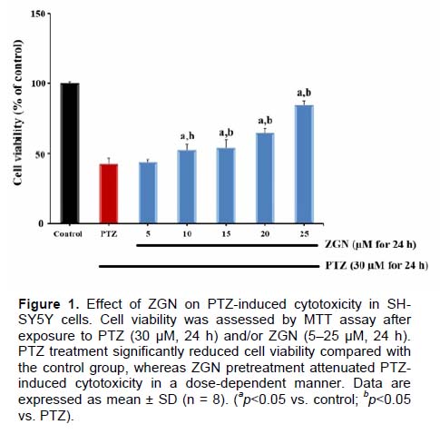
Effect of ZGN on PTZ-Induced Cytotoxicity: Exposure of SH-SY5Y neuronal cells to PTZ (30 µM, 24 h) resulted in a pronounced reduction in cell viability compared to the control group (p<0.05), confirming the cytotoxicity induced by PTZ. Pretreatment with ZGN at concentrations ranging from 5 to 25 µM significantly improved cell viability in a dose-dependent manner (Figure 1). The maximal protective effect was observed at 25 µM. These findings suggest that ZGN exhibits a significant cytoprotective effect against PTZ-induced neuronal damage.
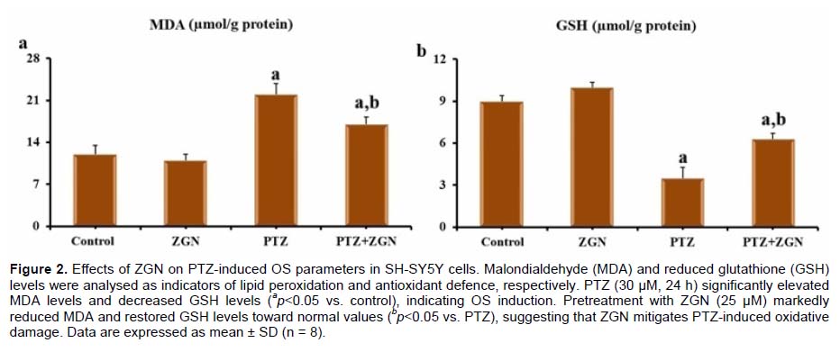
ZGN Attenuates PTZ-Induced OS: To assess the oxidative status of SH-SY5Y cells, intracellular MDA (Figure 2a) and GSH (Figure 2b) levels were quantified. PTZ treatment significantly increased MDA levels and reduced GSH concentrations relative to control (p<0.05), suggesting enhanced lipid peroxidation and impaired antioxidant defence. ZGN pretreatment (25 µM) effectively counteracted these changes by lowering MDA and restoring GSH levels (p<0.05 vs. PTZ). These results demonstrate that ZGN mitigates PTZ-induced oxidative damage by reinforcing the antioxidant defence system of neuronal cells.
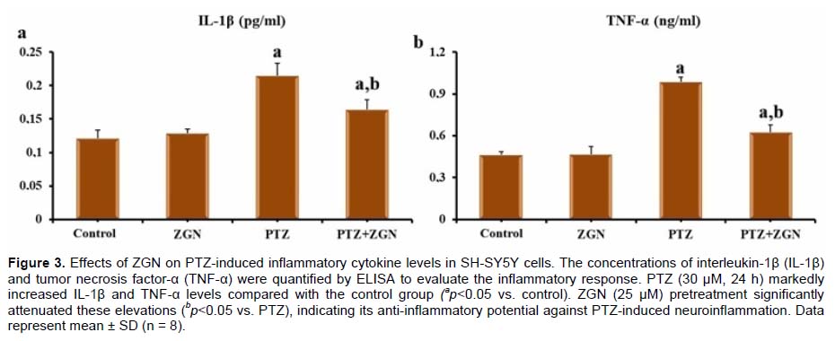
ZGN Suppresses PTZ-Induced Proinflammatory Responses: The inflammatory response triggered by PTZ was evaluated by measuring the cytokine levels of IL-1β (Figure 3a) and TNF-α (Figure 3b). PTZ exposure markedly increased both IL-1β and TNF-α levels compared with the control group (p<0.05), reflecting an acute neuroinflammatory process. Pretreatment with ZGN (25 µM) significantly reduced the elevated cytokine concentrations (p<0.05 vs. PTZ), demonstrating the anti-inflammatory potential of ZGN in preventing PTZ-induced inflammation in SH-SY5Y cells.
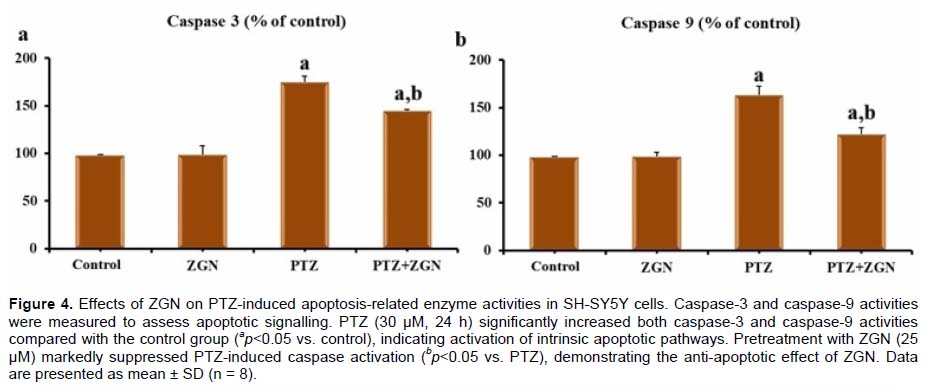
ZGN Inhibits PTZ-Induced Apoptotic Pathways: To explore whether ZGN modulates PTZ-induced apoptotic signalling, the activities of caspase-3 (Figure 4a) and caspase-9 (Figure 4b) were measured. Both enzymes exhibited a significant increase in activity following PTZ exposure compared with control (p<0.05), confirming the induction of intrinsic apoptotic pathways. ZGN pretreatment (25 µM) markedly decreased caspase-3 and caspase-9 activities (p<0.05). These data suggest that ZGN protects neuronal cells from PTZ- mediated apoptosis by inhibiting caspase-dependent pathways.
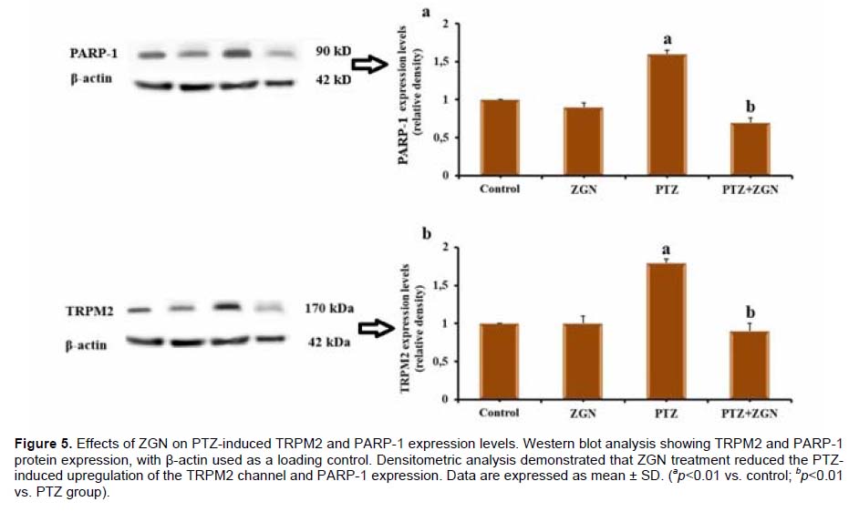
ZGN Regulates TRPM2 and PARP-1 Expression in PTZ-Treated Cells: Western blot analysis was conducted to evaluate the expression of TRPM2 (Figure 5a) and PARP-1 (Figure 5b) proteins, which are known mediators of OS and DNA damage. PTZ exposure resulted in a significant upregulation of both TRPM2 and PARP-1 compared to the control group (p<0.01). ZGN pretreatment markedly downregulated these proteins (p<0.01 vs. PTZ), normalising their expression levels toward control values. These results indicate that the neuroprotective effect of ZGN is mediated, at least in part, through the suppression of TRPM2 channel activation and PARP-1 associated apoptotic mechanisms.
Tartışma
The present study demonstrates, for the first time, that ZGN exerts a robust neuroprotective effect against PTZ-induced oxidative, inflammatory, and apoptotic injury in SH-SY5Y neuronal cells, and that these effects are mediated, at least in part, by the inhibition of PARP-1 and TRPM2 activation.In studies, PTZ exposure significantly reduced SH-SY5Y cell viability, consistent with its ability to induce excitotoxicity, mitochondrial dysfunction, and OS[3,4]. Pretreatment with ZGN significantly restored cell viability in a concentration-dependent manner, demonstrating a potent cytoprotective profile. In a study using a similar model, PTZ was shown to cause neurotoxicity and increase inflammatory cytokine (TNF-?, IL-1?, and IL-6) and MDA levels, while decreasing GSH and GPx levels. Gallic acid treatment has been reported to exert a protective effect against PTZ-induced toxicity in SH-SY5Y cells[15]. Similar findings have been reported in neuronal models of rotenone, cadmium, and amyloid-?-induced cytotoxicity, with ZGN improving survival through modulation of redox signalling and mitochondrial integrity[16,17]. These results confirm the capacity of ZGN to counter neurotoxic insults through both antioxidant and mitochondrial stabilizing mechanisms [18]. In this study, consistent with literature data, PTZ caused cytotoxicity in SH-SY5Y cells by causing a significant decrease in cell viability. However, ZGN treatment significantly increased cell viability in a dose-dependent manner, demonstrating a protective effect (Figure 1). These results demonstrated a potent cytoprotective effect of ZGN against PTZ-induced neuronal damage.
In this study, PTZ caused a significant increase in lipid peroxidation (MDA) and consequent GSH depletion, leading to severe OS. ZGN pretreatment reversed these changes and restored redox balance (Figure 2). This is consistent with recent reports that ZGN enhances endogenous antioxidant systems (SOD, CAT, GPx) and suppresses ROS accumulation in neuronal and hepatic tissues[13,19,20]. Mechanistically, the phenolic hydroxyl group acts as a free radical scavenger, while the methoxy moiety provides membrane permeability, allowing direct ROS neutralization[21,22]. These biochemical properties may explain the observed reduction in PTZ-induced oxidative load.
In the current study, the significant increase in IL-1? and TNF-? following PTZ treatment suggests activation of neuroinflammatory cascades (Figure
As seen in Figure 4, PTZ-induced cytotoxicity was accompanied by increased caspase-3 and caspase-9 activities, hallmarks of mitochondrial (intrinsic) apoptosis. ZGN pretreatment significantly suppressed both enzymes, confirming its anti-apoptotic potential. This is consistent with evidence that ZGN protects hippocampal and cortical neurons from toxin-induced apoptosis by preserving mitochondrial membrane potential and reducing cytochrome c release[14,18,27]. The concomitant decrease in caspase-9 and caspase-3 suggests that ZGN likely acts upstream by stabilising mitochondrial integrity and preventing ROS-induced activation of the intrinsic death pathway.
Upregulation of TRPM2 and PARP-1 expression levels in PTZ-exposed cells (Figure 5) supports the concept that oxidative DNA damage and ADP-ribose overproduction contribute to Ca²-dependent cell death[28,29]. ZGN significantly reduced the levels of both proteins, indicating a downregulatory effect on the PARP-1/ADPR-TRPM2 axis. PARP-1 is known to increase TRPM2 activation via ADP-ribose synthesis, and overactivation of this loop promotes Ca²? overload and apoptosis[28,29]. ZGN can prevent ADPR accumulation by inhibiting PARP-1, thereby blocking TRPM2-mediated Ca²? influx and subsequent apoptotic signalling. These findings are consistent with recent evidence indicating that TRPM2 channel activation serves as a molecular hub sensitive to oxidative stress in neuronal damage[30-33].
This study provides compelling evidence that ZGN mitigates PTZ-induced OS, neuroinflammation, and apoptosis in SH-SY5Y cells through the downregulation of the PARP-1/TRPM2 signalling pathway. By restoring antioxidant capacity, suppressing pro-inflammatory cytokines, and inhibiting caspase-dependent apoptosis, ZGN confers broad neuroprotective effects against chemical-induced cytotoxicity. These findings highlight TRPM2 channel modulation as a mechanistic cornerstone of ZGN?s action, suggesting that natural vanilloid derivatives may represent viable therapeutic adjuvants for OS-related neurological disorders, such as epilepsy, ischemic injury, and neurodegeneration. Future studies employing in vivo PTZ seizure models and specific TRPM2 antagonists will be crucial to confirm causality and translational potential.
References
- Investigation of the Effects of Zingeron on Acetic Acid – Induced Experimental Ulcerative Colitis Model in Rats
- Sıçanlarda Asetik Asit ile Oluşturulmuş Deneysel Ülseratif Kolitis Modelinde Zingeron’un Etkilerinin Araştırılması
- Investigation of the Effects of Zingeron in the Experimental Hepatorenal Toxicity Model Induced with Bisphenol A
- Bisfenol A ile Oluşturulmuş Deneysel Hepatorenal Toksisite Modelinde Zingeron’un Etkilerinin Araştırılması