A 10-years-old male Golden Retriever dog was brought to Firat University Animal Hospital, surgery clinic with a skin swelling on the upper left side of the pelvis. After the clinical and radiographic examinations of the patient, certificate of approval was taken from animal owner and mass was removed surgically and biopsy samples were sent to the pathology department for histopathological and immunohistochemical evaluation. Chemotherapy was initiated 1 week after the operation by using vincristine sulphate 3 times per week. Tumor recurrence started again in the same region about two weeks after the last chemotherapy. Approximately 4 weeks later, the second surgical operation was performed when mass grow up again. Patient died 8 hours after the last operation.
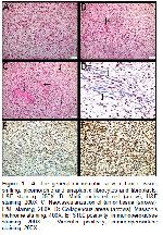
Histopathological and Immunohistochemical Findings: The biopsy material macroscopically was white in color, elastic in structure, pieced, changing sizes and dimensions. Histopathological examination revealed, spindle shaped (fusiform), shifting, pleomorphic and anaplastic fibrocytes and fibroblasts in biopsy tissue (Figure 1-A). Histological features of tumor included spindle shaped cells, scant cytoplasm, elongated to oval shaped nucleoli, marked cellular pleomorphism and increase in cellular density. These cells also showed anisocytosis and anisonucleosis. Mitotic figures and multinucleated cells were also observed rarely (Figure 1-B). Cellular arrangement was herringbone pattern or interwoven arrangement to form eddies. The surrounding stromal tissue was, keloid-like or loose myxoid character. In addition,several small capillar veins were seen in the tumor stroma (Figure 1-C). Massons trichrome staining also showed intense collagen tissue between the anaplastic fibrocytes and fibroblasts forming the tumor paranchyme (Figure 1-D). Since the surgical margins of the tumour are quite large (about 20 cm), each region could not examine in detail, so there was no clear information about whether the surgical borders were clean or not. Avidin-biotin complex (abc) method was used for immunohistochemical examinations with anti-S100 and anti-vimentin primer antibodies. Lymph node was used as positive control for S100 and vimentin antibodies. As negative control, these biopsy samples, without primary antibody application, were used. S100 positivity was found limited to the nuclei of fibrocytes and fibroblasts (Figure 1-E). It was seen that vimentin positivity was common and severe and that both the cytoplasm and the nuclei of the fibrocytes and fibroblasts were intensely stained (Figure 1-F). In the light of these findings, biopsy specimen was diagnosed as fibrosarcoma.
Büyütmek İçin Tıklayın |
Figure 1: A: The general microscobic view of tumor tissue; shifting, pleomorphic and anaplastic fibrocytes and fibroblasts, H&E staining, 200X. B: Multi nucleated cell (arrow), H&E staining, 200X. C: Neovascularization of tumor tissue (arrows), H&E staining, 200X. D: Collagenous areas (arrows), Massons trichrome staining, 400X. E: S100 positivity, immunoperoxidase staining, 200X. F: Vimentin positivity, immunoperoxidase staining, 200X |
Surgical Procedure: The patient owner was informed about the dogs old age and the risk of recurrence of this malignant tumor. Preparations were made for the operation with the approval of the patient owner (Figure 2).
After general anesthesia of the patient, the skin was excised to reach the mass. Tumor mass was gelatinous and semi-fluid. All of the mass was removed from 2 separate incision lines and the skin was closed with sutures. The mass of the tumor was approximately 1570 g. and the size was 20x15x7 cm. One week after the operation, the chemotherapy application was started with using vincristine sulphate. Intravenous administration of vincristine sulphate (Vincristine, DBL®, Mayne Pharma PtyLtd, Melbourne, Australia) 0.025 mg/kg (0.5-0.7 mg/m2, maximum 1 mg) in 500 mL isotonic serum with slow infusion at 3 times per week, but recurrence started in the same area and bilateral region about two weeks after the last chemotherapy. Approximately 4 weeks after the last chemotherapy, tumoral mass size was measured 31x21x9 cm (Figure 3 and 4).
When it was decided for the second operation, low dosage anesthesia was used because of dogs age. In this operation wide and deep surgery was made. The extracted mass was weighed 3020 gr. During the operation, it was seen that there were milimetric protrusions covering the under the skin in one area. This skin part was removed and the wound line was closed with sutures. Parenteral antibiotic and nonsteroidal anti-inflamatory treatment was recommended for 5 days after the operation and the patient was discharged. The patient owner reported that the patient died 8 hours after the second operation. Because the animal owner didnt accept the necropsy, the reason of the death and metastasis could not be understood exactly.


)
)
)
)
