[ Ana Sayfa | Editörler | Danışma Kurulu | Dergi Hakkında | İçindekiler | Arşiv | Yayın Arama | Yazarlara Bilgi | E-Posta ]
Fırat Üniversitesi Sağlık Bilimleri Veteriner Dergisi
2014, Cilt 28, Sayı 3, Sayfa(lar) 151-154
[ Özet ] [ PDF ] [ Benzer Makaleler ] [ Yazara E-Posta ] [ Editöre E-Posta ]
Doğal Şartlarda Kuduz Bir İnekten Doğan Buzağıda Prenatal Enfeksiyonun Araştırılması
Metin GÜRÇAY1, Ahmet SAİT2, Turhan TURAN1
1Elazığ Veteriner Kontrol Enstitüsü, Elazığ, TÜRKİYE
2Pendik Veteriner Kontrol Enstitüsü, İstanbul, TÜRKİYE
Anahtar Kelimeler: Kuduz virusu, doğum öncesi infeksiyon, buzağı
Özet
Kuduz virusu ensefalomiyelitise neden olan son derece nöyrotropik bir virusdur. Gebe on yaşındaki bir hoştayn inek durgunluk, böğürme, inkoordinasyon, dilin dışarı sarkması, kuyruk hareketlerinde azalma, salivasyon, kamburlaşma, aşırı duyarlılık, saldırganlık ve takiben felç gibi klinik belirtiler gösterdi. İnek bir ay öncesinden bir köpek tarafından ısırılmıştı. O ölü bir erkek dana doğdu. İnek doğumdan bir gün sonra öldü. İnek ve buzağının kafatası kemikleri açılarak beyinleri çıkartıldı. Klinik ve otopsi gözlemleri sadece kuduzdan şüphelendirdi. Beyinler kuduz yönünden teşhis laboratuvarında test edildi. İnekte kuduz hastalığı teşhisi yapıldı. Kuduz virusu, fare inokulasyonu ile izole edildi. Rabies virus antijenleri ve spesifik virus genleri flöresan antikor test (FAT) ve reverze transkription polimeraz zincir reaksiyonu (RT-PCR) ile belirlendi. Buzağının cerebellum ve cornu ammonislerinden yapılan FAT testinde negri cisimciklerine rastlanmadı. Buzağı tükrük bezi, lenf düğümleri, böbrek, kalp kası, akciğer, karaciğer, barsak, dalak, spinal cord ve beynin cornu ammonis ve cerebellum dokularından FAT, RT-PCR ve fare inokulasyonu yapıldı. Dokulardan kuduz virusu izole edilemedi. Kuduz virusu antijenleri ve spesifik virus genlerine rastlanmadı. İnekten elde edilen pozitif RT-PCR amplifikasyon ürününden kısmi dizin analizi yapıldı ve GenBank'da TR/EL/753/2009/cattle KM058759 kodu ile yayınlandı. RT- PCR amplifikasyon ürününün virus nükleoproteini N geni kısmi 0-510 bp dizin analizinin NCBI programında değerlendirilmesinde izolatın Rabies Virus (RABV) olduğu doğrulandı. RABV' nin filogenetik analizi belirlendi. Dizinin filogenetik analizi sonucuna göre, Türkiye ve Türkiye'nin Ortadoğuda bulunan komşu ülke izolat dizinleri ile identik olduğu belirlendi. Bu araştırma, kuduz inekten doğan bir buzağıda rabies virusunun, bulunmaması spesifik bir vakada, transplesantal bulaşmanın gözlenmediğini göstermektedir.
  • Başa Dön
  • Özet
  • Giriş
  • Olgu Sunusu
  • Kaynaklar
  • Giriş
    Rabies, a zoonosis that affects the central nervous system (CNS), provokes acute and fatal encephalitis in its mammal hosts. The etiologic agent of the disease is the Rabies Virus (RABV), a neurotropic RNA virus that belongs to the order Mononegavirales, family Rhabdoviridae and genus Lyssavirus1. The most rabies infections of animals and human beings occur following a bite by a rabid animal although infections through aerosols have been reported2-4. Oral and nasal infections are rare but mice and other animals have been experimentally infected via these routes. An epizootic that killed thousands of antelopes in Namibia was thought to have spread horizontally by the oral route5. Vertical transmission is not well defined. In most of the few previous reports, newborns did not contract the infection by vertical transmission from mothers with clinical rabies6-9. However, vertical transmission in mammalian models of experimental or natural rabies infection has also rarely been reported10,11.

    The neurotropic of the virus has been recognised following early work in the nineteenth century. It has been shown that the virus may replicate in muscle fibres before invading the nervous system, which would be a necessary amplification stage to yield sufficient virus to invade peripheral nerves. The virus does not start to replicate in the surrounding muscle fibres immediately and there is a delay, with the virus apparently in a latent form and not stimulating an effective immune response. Subsequent studies have shown that the virus attaches to nerve cells through acetylcholine receptors at the neuromuscular junction. These receptors are the same as those used by some neurotoxins. Once the virus enters the peripheral nerves it travels towards the Central Nervous System (CNS) via the motor and sensory axons. Experimental studies have shown that the perineural structures are not necessary for viral spread to the CNS. Within the CNS, the virus infects neurons and dendrites, with virus budding occurring from neuronal cell surfaces and synapses. Virus dissemination occurs through retrograde axoplasmic flow, cell-to-cell transmission via synaptic junctions and free passage of virus within intercellular spaces. The virus appears to spread progressively by infecting areas adjacent to the entry site until most areas of the brain and spinal cord are affected. There is some evidence to suggest that neurons with long axons may enhance the spread of virus to distant areas. Successive cycles of axoplasmic transmission and replication in the perikarya and dendrites and trans neural spread result in widespread dissemination. Eventually there is also a centrifugal spread of the virus in the axons of peripheral nerves and infection and possible replication in the salivary glands, the skin, heart, and other tissues5.

    The aim of this study is expose that lack of prenatal infection in a calve delivered by a rabid cow in natural conditions.

  • Başa Dön
  • Özet
  • Giriş
  • Olgu Sunusu
  • Kaynaklar
  • Olgu Sunusu
    On 20 May 2009, clinical signs of yawning, bellowing, incoordination, decreased feeling in hind limbs, loss of tail movement, drooling saliva, "cud-dropping", inability to stand, hypersensitivity, and aggression followed by paralysis were observed in a ten year old pregnant holstein cow. She had been bitten by a dog one month earlier. Four days later, she calved a dead male calf. The cow died a day later after parturition. The skull of the cow and her dead male calf were admitted to Elazığ Veterinary Research Control Institute for rabies diagnosis.

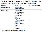
    Reference methods were used for post-mortem laboratory confirmation of rabies infection of the holstein cow4. The fluorescent-antibody test (FAT) was positive on the cow's brain specimen (Table 1), and rabies was isolated in cow's Cerebellum and Ammon's corn by inoculation of suckling mice. Viral RNA extraction, reverse transcription (RT) and PCR were performed on brain sample specimens, hereinafter2 described. The dead male calf was autopsied. There was no autolysis and putrefaction. There was congestion in the lungs and petechial haemorrhages in the endocardium.


    Büyütmek İçin Tıklayın
    Table 1: Laboratory diagnostic and biological testing with samples from the pregnant cow and her dead calf

    Cerebellum and Ammon's corn samples were collected following the opening of the skull belong the cow. Ammon's corn, cerebellum, spinal cord, umbilical cord, salivary gland, lymph nodes, kidney, myocardia, lung, liver, intestine and spleen were collected belong the dead calf in a necropsy room (Table 1).

    Clinical and post mortem observation may only lead to a suspicion of rabies because signs of the disease are not characteristic and may vary greatly from one animal to another12. The only way to perform a reliable diagnosis of rabies is to identify the virus or some of its specific components using laboratory tests. The methods vary in their efficiency, specificity and reliability. They are classically applied to brain tissue, but they can also be applied, though less effectively, to other organs3,13,14.

    Rabies laboratory diagnosis using pregnant cow and in fetal calf samples was according to (WHO, OIE) recommendations using reference methods2-4,14. The most widely used test for rabies diagnosis is the FAT, which is recommended by both WHO and OIE4,14. This test may be used directly on a smear, and can also be used to confirm the presence of rabies virus in cow and in fetal calf samples of mice that have been inoculated for diagnosis.

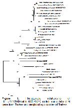
    The RT-PCR assay was performed as described by Heaton et al.2 with modifications. The RT-PCR amplicons were resolved by electrophoresis on 1.5% agarose gels, stained with ethidium bromide (0.5 μg/mL), visualized and photographed by using a Thermo Hybaid Gel Grab imaging system. After electrophoresis, a specific 606 bp amplicon was observed in two cow samples whereas, specific 606 bp amplicon and 582 bp amplicon was not observed in any of the calf samples analyzed. PCR products were purified by a QIA quick gel extraction kit (Qiagen) and sequenced by ABI 3130xl genetic analyzer (Applied Biosystem, USA). The obtained sequence was compared and aligned with previously published Rabies virus sequences15 by using MEGA 516. MEGA 5 was also used to draw the amino acid based phylogenetic trees using the neighbor-joining method, 1000 bootstrap replicates were performed for analysis to assess the likelihood of the tree construction (Figure 1).


    Büyütmek İçin Tıklayın
    Figure 1: Phylogenetic relationship of R/EL/753/2009/cattle KM058759 isolate nucleotids of N gene from Turkey and neighboring countries of Turkey.

    Rabies diseases diagnosed in the cow. Rabies Virus isolated in mouse inoculation test (Table 1). Rabies virus antigens and specific virus gens observed FAT and RT-PCR. The dead male calf was made post mortem examination. There was congestion in the lungs and petechial haemorrhages in the endocardium. Ammon's corn, cerebellum, spinal cord, umbilical cord, salivary gland, lymph nodes, kidney, myocardia, lung, liver, intestine and spleen were collected in a necropsy room. Fluorescent antibody test, Hemi nested RT- PCR and the mouse inoculation test were made for rabies virus diagnosis for this tissues. Rabies virus not isolated in the mouse inoculation test. Rabies virus antigens and specific virus gens not observed FAT and RT-PCR (Table 1). Partial nucleotid sequence of the positive primary PCR amplification was identified from the cow brain sample and published in GenBank with TR/EL/753/2009/cattle KM058759 code. Sequencing results of the positive primary PCR amplifications according to the partial nucleoprotein N gene 0-510 sequences confirmed that the isolate is rabies virus (RABV) by NCBI Programme. Phylogenetic analysis of RABV identified. As a result, nucleotid sequence of the isolate were found as identical with the isolates obtained from province isolates, as well as related with isolates sequences from neighboring countries of Turkey in the Middle East (Figure 1).

    Transmission of infection to the fetus during pregnancy, although theoretically it might lead to immune tolerance and persistent virus carriage in the affected offspring, is nearly always an unimportant matter from the epidemiological point of view. With most viruses and in most species, primary infections during pregnancy are uncommon under natural circumstances. Persistent viruses are sometimes reactivated during pregnancy (e.g. malignant catarrhal fever virus in the wildebeest), but for most viruses this does not appear to be an important source of fetal infection. The virus must be present in the blood of the pregnant animal at the right stage of gestation, and it must reach the placenta and be either carried across, leak across, or grow across this barrier to reach the fetus. The placenta is the obvious route for passage of an infectious agent from mother to offspring, but infection directly from oviduct or uterine wall would also be possible. In any case, the fetus must not be unduly damaged, and throughout this process the virus must contend with both maternal and fetal immune responses17.

    There is little information available about the potential vertical transmission of rabies and the outcome for neonates born to rabid mothers. This hampers informed choice concerning route and conditions of delivery by physicians for such cases, which mostly occur in places where medical facilities are limited. Indeed, these cases of clinical rabies are difficult to manage due to potential risks during nursing, and stress affecting the familial and medical environment18. We report a rare and instructive case: We find non infected newborn calf with rabies virus, borned from rabid cow. It indicates that rabies viral infection of the mother seems not to affect normal in utero (Table 1). Dissemination of rabies virus through peripheral nerves associated with internal organs or by biological fluid is demonstrable in patients with neurological clinical signs of rabies12. Also human rabies infection via the transplacental route, and detection of rabies virus in genital or placental organs, has not been reported6,7,9,18. However, vertical transmission in mammalian models of experimental or natural rabies infection has also rarely been reported10,11. There has been no systematic analysis of virus dissemination6. This research demonstrated the absence of rabies virus in the newborn calf, thus, in this specific case, the transplacentary transmission was not observed.

  • Başa Dön
  • Özet
  • Giriş
  • Olgu Sunusu
  • Kaynaklar
  • Kaynaklar

    1) Fauquet EM, Mayo MA, Maniloff J, Desselgerger U, Ball LA. Virus taxonomy: Classification and nomenclature of viruses. Eighth report of the International Committee on the Taxonomy of Viruses. Virus Taxonomy. 8th Edition, San Diego: Academic Press, 2005.

    2) Heaton PR, Johnstone P, Mc Elhinney LM, et al. Heminested PCR assay for detection of six genotypes of rabies and rabies related viruses. J Clin Microbiol 1997; 35: 2762-2766.

    3) Meslin FX, Kaplan MM, Koprowski H. An Overview of Laboratory techniques in diagnosis and prevention of rabies and in rabies resarch. In: Meslin FX, Kaplan MM, Koprowski H. (Editor). Laboratory Techniques in Rabies, 4th Edition, Geneva: WHO, 1996: 9-27.

    4) Office International des Epizooties (OIE). Manual of Diagnostic Tests and Vaccines for Terrestrial Animals. Paris: OIE, 2008.

    5) Woldehiwet Z. Rabies: Recent developments. Res Vet Sci 2002; 73: 17-25.

    6) Iehl'e C, Dacheux L, Ralandison S, et al. Delivery and follow-up of a healthy newborn from a mother with clinical rabies. J Clin Virol 2008; 42: 82-85.

    7) Müller-Holve W. Leitritz H, Knorr E. Early development of a child following rabies of the mother during pregnancy. Infection 1977; 5: 49-50.

    8) Spence MR, Davidson DE, Dill Jr GS, Boonthai P, Sagartz JW. Rabies exposure during pregnancy. Am J Obstet Gynecol 1975; 23; 655-656.

    9) Swende TZ, Achinge GI. Clinical rabies in pregnancy with delivery of a live fetus: A case report. Niger J Med 2009; 18; 1: 114-115.

    10) Howard DR. Transplacental transmission of rabies virus from a naturally infected skunk. Am J Vet Res 1981; 42: 691-692.

    11) Martell MA, Montes FC, Alcocer R. Transplacental transmission of bovine rabies after natural infection. J Infect Dis 1973; 127: 291-293.

    12) Hemachudha T, Laothamatas J, Rupprecht CE. Human rabies: A disease of complex neuropathogenetic mechanisms and diagnostic challenges. Lancet Neurol 2002; 1: 101-109.

    13) Hemachudha T, Kamolvarin N, Tirawatnpong T, et al. Ante- and postmortem diagnosis of rabies in man and animal by PCR using nested primers. Neurol 1992; 42: 370.

    14) World Health Organization Expert Committee on Rabies Eight Report, WHO Technical Report Series 1992.

    15) Un H, Johnson N, Vos A, et al. Genetic analysis of four human rabies cases reported in Turkey between 2002 and 2006. Clin Microbiol Infect 2009; 15: 1185-1189.

    16) Tamura K, Dudley J, Nei M, Kumar S: MEGA4: Molecular Evolutionary Genetics Analysis (MEGA) software version 4.0. Mol Biol Evol 2007; 24: 1596-1599.

    17) Mims CA. Vertical Transmission of Viruses. Microbiol Rev 1981; 45: 2267-286.

    18) Viazhevich VK. Delivery of a normal infant from a mother in incubation period of rabies. Zh Microbiol Epidemiol Immunobiol 1957; 28: 105-106.

  • Başa Dön
  • Özet
  • Giriş
  • Olgu Sunusu
  • Kaynaklar
  • [ Başa Dön ] [ Özet ] [ PDF ] [ Benzer Makaleler ] [ Yazara E-Posta ] [ Editöre E-Posta ]
    [ Ana Sayfa | Editörler | Danışma Kurulu | Dergi Hakkında | İçindekiler | Arşiv | Yayın Arama | Yazarlara Bilgi | E-Posta ]