[ Ana Sayfa | Editörler | Danışma Kurulu | Dergi Hakkında | İçindekiler | Arşiv | Yayın Arama | Yazarlara Bilgi | E-Posta ]
Fırat University Journal of Health Sciences (Veterinary)
2020, Cilt 34, Sayı 2, Sayfa(lar) 103-108
[ Özet ] [ PDF ] [ Benzer Makaleler ] [ Yazara E-Posta ] [ Editöre E-Posta ]
Siirt İli Köpeklerinde Dirofilariasis, Leishmaniasis, Ehrlichiosis ve Anaplasmosis Üzerine Serolojik Bir Çalışma
Burçak ASLAN ÇELİK1, Özgür Yaşar ÇELİK2, Tekin ŞAHİN 2, Kıvanç İRAK3, Memiş BOLACALI 4, Özlem BİÇİCİ2, Vedat BALDAZ2
1University of Siirt, Faculty of Veterinary, Department of Parasitology, Siirt, TURKEY
2University of Siirt, Faculty of Veterinary, Department of Internal Medicine, Siirt, TURKEY
3University of Siirt, Faculty of Veterinary, Department of Biochemistry, Siirt, TURKEY
4University of Siirt, Faculty of Veterinary, Department of Animal Breeding and Husbandry, Siirt, TURKEY
Anahtar Kelimeler: Anaplazmozis, dirofilariyozis, erlişiyozis, layşmanyozis, Siirt
Özet
Köpeklerin vektör kaynaklı hastalıklarına arthropodlar tarafından nakledilen oldukça geniş çeşitlilikte patojenler neden olmaktadır. Bu çalışmanın amacı Siirt ili köpeklerinde Dirofilaria immitis antijeni ile Ehrlichia canis, Leishmania infantum ve Anaplasma phagocytophilum/platys antikorlarının hızlı tanı testi (Anigen Rapid Caniv-4 Leish, Bionote, Güney Kore) kullanılarak belirlenmesiydi. Çalışmanın materyalini farklı ırklardan 26 erkek ve 24 dişi olmak üzere toplam 50 köpek oluşturdu. Çalışma sonucunda örneklerin 5 (%10)’inde Anaplasma phagocytophilum/platys antikorları tespit edilirken D. immitis antijeni, L. infantum ve E.canis antikorları tespit edilemedi. Sonuç olarak; anaplazmozis ile ilgili koruma ve kontrol tedbirlerinin alınması gerektiği kanısına varılmıştır.
  • Başa Dön
  • Özet
  • Giriş
  • Materyal ve Metot
  • Bulgular
  • Tartışma
  • Kaynaklar
  • Giriş
    Canine vector-borne diseases (CVBD) are caused by a wide range of pathogens transmitted by arthropods. While the CVBD is very important in terms of veterinary practice, they also pose a significant risk for human health due to their zoonotic potentials 1-3. Amongst the diseases transmitted to dogs by the vectors, Dirofilariasis, Leishmaniasis, Ehrlichiosis, and Anaplasmosis are particularly of importance. Anaplasmosis and Ehrlichiosis are transmitted by ticks, while Dirofilariasis is transmitted by mosquitos and Leishmaniasis is transmitted by the biting midges. Presence of all of these diseases in Turkey have been reported4.

    Dirofilariasis is a zoonotic disease caused by Dirofilaria spp, a filarial nematode, and is transmitted by mosquitos5-7. The most common species that can cause the disease are Dirofilaria immitis and Dirofilaria repens. The adult form of D. immitis mostly locates in the right ventricle or right atrium of the heart and in the pulmonary arteries, while it can less commonly be found in the vena cava, camera oculi anterior, and peritoneal cavity as well8-10. Clinical symptoms depend on the severity of the disease and the duration of infection. Weight loss, drowsiness, cough, respiratory distress, and acidosis can be seen in sick animals4. Practical antigenic tests based on the detection of parasite-specific antibodies, native examination, and the modified Knott technique can be used in the clinical diagnosis of dirofilariasis9.

    Ehrlichiosis is a disease encountered in dogs living in tropical and sub-tropical regions and is caused by the strict-intracellular pathogen Ehrlichia canis11,12. The disease may have an acute, subclinical, or chronic course. Fever, weight loss, anorexia, nasal and ocular discharges, dyspnea, hemorrhagic disorders, anemia, thrombocytopenia, and leukocytosis or leukopenia can be seen in the acute phase of the disease. With treatment in the acute phase, the dogs may recover, may become subclinically infected, or may progress to the chronic phase13,14. Blood smears, Indirect fluorescence antibody (IFA), Western blot, and Enzyme-Linked Immuno Sorbent Assay (ELISA) techniques can be used in the diagnosis of the disease15.

    Leishmaniasis is a zoonotic protozoan disease commonly encountered all around the world and is caused by the Leishmania species16-18. In Turkey, two clinical forms of the disease known as the cutaneous leishmaniasis and the visceral leishmaniasis can be seen, which are caused by different Leishmania species19-21. Dogs infected with the disease may not show symptoms or may have one or more of the nine major clinical symptoms like skin lesions, weight loss or loss of appetite, local or general lymphadenopathy, ocular lesions, epistaxis, lameness, anemia, renal failure and diarrhea22,23.

    The anaplasma are small (0.2–0.9 μm) gram-negative, non-motile, unencapsulated, asporogenic, coccoid, ring-shaped, strictly intracellular bacteria that have a zoonotic nature24. The species that cause disease in the dogs are the Anaplasma phagocytophilum (A. phagocytophilum) and the Anaplasma platys (A. platys). A. phagocytophilum locates itself especially to neutrophils and eosinophils, while A. platys is settled in the platelets12. Common clinical findings in animals infected with A. phagocytophilum are high fever, anorexia, depression, and lethargy, all of which occur after 1-2 weeks of incubation. Reluctance to move and lameness are widely detected muscle and skeletal system symptoms25,26. Vomiting, diarrhea, coughing, and hemorrhage can be seen in some animals24,27. In the infections caused by A. platys, fever, anorexia, lethargy, mild anemia, and lenfoadenomegaly can be encountered12. IFAT, ELISA, and Polymerase chain reaction (PCR) tests are used to diagnose anaplasmosis.

    Seroprevalence studies are important in the determination of the geographical, regional, national, and universal distribution of diseases, and preparation and execution of control and eradication programs for them4. The aim of this study, therefore, was to determine the seroprevalences of D. immitis antigen, L. infantum, E. canis, and A. phagocytophilum/platys antibodies in the dogs of Siirt province.

  • Başa Dön
  • Özet
  • Giriş
  • Materyal ve Metot
  • Bulgular
  • Tartışma
  • Kaynaklar
  • Materyal ve Metot
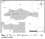
    Study Area: This study was carried out between October 2019 and December 2019 in the Siirt province located in the Southeastern Anatolia Region of Turkey (Figure 1). Siirt province is in the semi-arid climate region. The average, the highest, and the lowest temperatures are 36.9 ⁰C and 18.9 ⁰C in summer, and 8.7 oC and –0.5 ⁰C in winter, respectively. Water shortages and droughts are frequent during the summer28.


    Büyütmek İçin Tıklayın
    Figure 1: The map of Siirt province, in which the study was performed

    Sample Collection and Preparation: The animal material of the study consisted of 26 male and 24 female totals of 50 stray dogs between the ages of 3 months and 6 years.

    Blood samples were collected from the vena cephalica antebrachii of the animals into the non-anticoagulant tubes. The obtained samples were kept in room temperature for 30 minutes and then centrifuged in 3000 rpm for 10 minutes to separate the serum, after which they were kept in –20 ⁰C until they were analyzed.

    Test Procedure: A rapid diagnosis test kit (Anigen Rapid Caniv-4-Leish, Bionote, South Korea) prepared for the simultaneous detection of canine vector-borne diseases (D. immitis antigen, L. infantum antibody, E. canis antibody, and A. phagocytophilum/platys antibody) was used in the study. The specifications of the test report the sensitivity of the test as 94.4%, 97.6%, 95.6% and 88.5% for Dirofilaria, Ehrlichia, Leishmania, and Anaplasma, respectively, while the corresponding specificities are reported as 100%, 99%, 98.0%, and 97.1%, respectively. A micropipette was used to drop 10 μl of serum samples into the sample holes of the rapid test kit. All holes were then added 3 drops of assay diluent solution. Test results were interpreted after 15 minutes (Figure 2).


    Büyütmek İçin Tıklayın
    Figure 2: Test procedure

    Ethical Approval: Ethical approval for this study was obtained from the Siirt University Local Ethics Committee for Animal Experiments (Decision number: 2019/02/05).

  • Başa Dön
  • Özet
  • Giriş
  • Materyal ve Metot
  • Bulgular
  • Tartışma
  • Kaynaklar
  • Bulgular
    Of the 50 dogs examined in the study, 5 (10%) were detected to have the A. phagocytophilum/Platys antibodies, while Dirofilaria antigens, Leishmania and Ehrlichia antibodies were not detected at all. The gender and age distributions of the findings are presented in Table 1.


    Büyütmek İçin Tıklayın
    Table 1: Distribution of seropositive and seronegative animals by gender and age groups

    As it can be seen in Table 1, 3 of the 26 male dogs (11.54%) and 2 of the 24 female dogs (8.33%) were found to have positive indicators. When the dogs are grouped based on their ages, the Anaplasma infection was found to be most common in the over 3 years of age group, followed by the 1-3 years of age group. No infection was found in the under-one-age group.

  • Başa Dön
  • Özet
  • Giriş
  • Materyal ve Metot
  • Bulgular
  • Tartışma
  • Kaynaklar
  • Tartışma
    Besides the environmental factors like temperature, humidity, and the presence of mosquitoes, age and shelter conditions also play significant roles in the development of D. immitis infections9. As a result of the study conducted by Öncel and Vural29 to determine the presence of antigens in the stray dogs of İstanbul, the prevalence of D. immitis was found as 1.52%, while the same researchers reported that no positivity was encountered in the stray dogs in İzmir. While no positivity was found by Civelek et al.8 in their study in Bursa performed by native and modified Knott methods, their study which used the ELISA method reports 2% prevalence for D. immitis. Kozan et al.30 conducted a study using the modified Knott method to determine the prevalence of Dirofilaria sp. in Afyonkarahisar and Eskişehir provinces, for which they determined positivities of 3.6% and 1.4%, respectively. In a study conducted in Diyarbakır with the ELISA method, D. immitis seroprevalence was reported as 2.4%5 while the prevalance was 1.5% in Erzurum31. The Rapid test method was used in a study in Antalya province, in which no D. immitis antigens were encountered32.

    Similar to the findings of researchers8,29,32, all samples in this study were found to be seronegative in terms of D. immitis. The efficient insecticide applications performed in the Siirt province might be the reason why the dogs had no infection with the disease.

    Canine leishmaniasis represents a significant problem for both animal and human health due to its zoonotic nature33. In a study conducted in Manisa, the sera of 490 dogs were investigated with the IFAT method and the seroprevalence of the leishmaniasis was found as 5.3%34. In the study conducted by Voyvoda et al.35 in Antalya, the prevalence of L. infantum was determined as 3.63%, while the same researchers reported the prevalence of the disease for the province of İzmir as 2.5%. In a study conducted in Ankara by Aslantaş et al.36, 116 dog serum samples were analyzed with the IFAT, and the seroprevalence of the disease was reported as 2.58%. In the study conducted by Kilic et al.37 in Sivas, the sera of 50 dogs were analyzed with the IFAT and the seroprevalence of the disease was determined as 2%. Atasoy et al.38 conducted research that included the provinces of Aydın, Manisa, Muğla, and İzmir, and the seroprevalence of the disease were determined as 14.1%, 3.8%, 12%, and 4.6%, respectively.

    A study was conducted by Handemir et al.39 in various locations of İstanbul, and the researchers reported that no seropositivity was encountered. Babür et al.40 conducted a study in Şanlıurfa, in which they reported all of the samples collected from 80 dogs were seronegative. In a study conducted by Ica et al.41 in Kayseri, all of the 300 dog serum samples were found to be seronegative. A study was performed by Tok et al.42 in Çanakkale, and all of the 27 dog serum samples were seronegative. Celik and Sekin17 used IFAT method in their research they conducted in the Dicle and Hani districts of the Diyarbakır province, in which they report no seropositivity was encountered in the 120 samples they inspected.

    Similar to the findings and reports of researchers17,39-42, all samples in this study were seronegative in terms of L. infantum. Within the "cause network" that cause the development of Canine visceral leishmaniasis, the presence of Leishmania species, presence of the biting midges, biting midges stinging the infected host, a susceptible reservoir host being present nearby, the immune system reaction of the host, and environmental factors (humidity, air movements, light) are all present35. It is possible that all samples were found to be negative in this regard due to no leishmania species being present in the environment, or that the insecticide applications against the mosquitoes also affected the phlebotomus and caused a lack of vectors for the disease.

    Canine monocytic ehrlichiosis is amongst the most infectious diseases of dogs worldwide43. It is reported that a Bull terrier breed dog brought to the Veterinary Faculty of the Istanbul University was diagnosed with ehrlichiosis using the IFAT method 15. In the study conducted by Icen et al.5 in Diyarbakır using the ELISA, the prevalence of E. canis was found as 4.8%. In a study conducted by Güneş et al.44 in the Sinop province using the ELISA, the seroprevalence of E. canis was reported as 18.28%. Sari et al.45 conducted a study in the Iğdır province, in which they reported the seroprevalence of 1% for E. canis. Elitok and Ungur46, on the other hand, conducted a study in Uşak and reported a prevalence of 7% for E. canis.

    All the samples analyzed in the present study were found to be seronegative in terms of E. canis. The seropositivity rates of Ehrlichia infection can be dependent on the target population, climate, and the diagnosis method used47. On the other hand, the number of dogs infected with the parasite is reported to be higher in summer and spring months, compared to winter periods48. It is possible that no seropositivity was encountered in this study for the reasons specified by Ansari-Mood et al.47, or the fact that the disease is less frequently encountered in the period the study was conducted, or that the presence of the vector for the disease (Rhipicephalus sanguineas) in the region was not reported in any literature study.

    Anaplasma phagocytophilum and A. platys are the species that cause Anaplasmosis in dogs49. In a study conducted in Erzurum, the rate of Anaplasma spp. antibodies obtained was reported as 0.8%4. The seropositivity of A. phagocytophilum was determined as 30.1% in Sinop50, 7.8% in Kayseri51, and as 7.49% in a study that involved different provinces in the Aegean region52.

    In the current study, seropositivity was detected in five dogs (10%) against Anaplasma spp. It has been reported that the results of studies performed to determine the seroprevalence of Anaplasma spp. in dogs might be dependent on the number of samples used, the analysis method, and the density of the ticks in the region53,54. The seropositivity rate determined in the present study is in line with the studies conducted in Kayseri 51 and the Aegean region52.

    As a result, this is the first study that investigated the seroprevalence of Dirofilariasis, Ehrlichiasis, Leishmaniasis, and Anaplasmosis in the dogs in Siirt province. it was concluded that protection and control measures regarding anaplasmosis should be taken and more detailed studies on vector-borne infections are needed.

    Conflicts of interest
    The authors declare that there is no conflict of interest.

  • Başa Dön
  • Özet
  • Giriş
  • Materyal ve Metot
  • Bulgular
  • Tartışma
  • Kaynaklar
  • Kaynaklar

    1) Bowman D, Little SE, Lorentzen L, et al. Prevalence and geographic distribution of Dirofilaria immitis, Borrelia burgdorferi, Ehrlichia canis, and Anaplasma phagocytophilum in dogs in the United States: Results of a national clinic-based serologic survey. Vet Parasitol 2009; 1-2: 138-148.

    2) Carrade D, Foley J, Sullivan M, Foley CW, Sykes JE. Spatial distribution of seroprevalence for Anaplasma phagocytophilum, Borrelia burgdorferi, Ehrlichia canis, and Dirofilaria immitis in dogs in Washington, Oregon, and California. Vet Clin Pathol 2011; 3: 293-302.

    3) Otranto D, Dantas-Torres F, Breitschwerdt EB. Managing canine vector-borne diseases of zoonotic concern: Part two. Trends Parasitol 2009; 5: 228-235.

    4) Demir A. Erzurum Yöresinde Köpeklerde Dirofilaria İmmitis, Ehrlichia Canis, Borrelia Burgdorferi ve Anaplasma Spp Seroprevalansinin Araştırılması. Master thesis. Erzurum: Ataturk University, 2018.

    5) Icen H, Sekin S, Simsek A, et al. Prevalence of Dirofilaria immitis, Ehrlischia canis, Borrelia burgdorferri infection in Dogs from Diyarbakır in Turkey. Asian J Anim Vet Adv 2011; 4: 371-378.

    6) Vieira AL, Vieira MJ, Oliveira JM, et al. Prevalence of canine heartworm (Dirofilaria immitis) disease in dogs of central Portugal. Parasite 2014; 5: 1-7.

    7) Wang S, Zhang N, Zhang Z, et al. Prevalence of Dirofilaria immitis infection in dogs in Henan province, central China. Parasite 2016; 43-43.

    8) Civelek T, Yıldırım A, Ica A. Prevalence of canine heartworm disease in the Gemlik Area, Bursa. Vet Bil Derg 2006; 1-2: 65-68.

    9) Göz Y, Koltas İS, Altug N, et al. Seroprevalance of Dirofilaria immitis in Dogs in Van District. YYÜ Vet Fak Derg 2007; 2: 5-8.

    10) Montoya-Alonso JA, Morchón R, Falcón-Cordón Y, et al. Prevalence of heartworm in dogs and cats of Madrid, Spain. Parasites Vector 2017; 1: 354.

    11) Rikihisa Y, Ewing S, Fox J, et al. Analyses of Ehrlichia canis and a canine granulocytic Ehrlichia infection. J Clin Microbiol 1992; 1: 143-148.

    12) Shaw SE, Day MJ, Birtles RJ, Breitschwerdt EB. Tick-borne infectious diseases of dogs. Trends Parasitol 2001; 2: 74-80.

    13) Harrus S, Kass PH, Klement E, Waner T. Canine monocytic ehrlichiosis: a retrospective study of 100 cases, and an epidemiological investigation of prognostic indicators for the disease. Vet Rec 1997; 14: 360-363.

    14) Rudoler N, Harrus S, Martinez-Subiela S, et al. Comparison of the acute phase protein and antioxidant responses in dogs vaccinated against canine monocytic ehrlichiosis and naive-challenged dogs. Parasites vector 2015; 1: 175.

    15) Dodurka H, Bakırel U. Case of Ehrlichiosis in a dog. J Fac Vet Med Univ Istanbul 2002; 1: 11-16.

    16) Beyhan Y, Celebi B, Ergene O, Mungan M. Seroprevalance of Leishmaniasis in Dogs from Hatay and Burdur Provinces of Turkey and Northern Cyprus. Turkiye Parazitol Derg 2016; 9-12.

    17) Celik O, Sekin S. Clinical, hemtalogical and biochemical findings of Leishmaniasis in Dicle and Hani distrcircts of Diyarbakır, its serological diagnosis and typology with PCR. Dicle Üniv Vet Fak Derg 2015; 4: 21-29.

    18) Gazyağcı S, Gazyağcı A, Kılıç S, et al. Visceral Leishmaniasis in a Dog in Amasya Province. Kocatepe Vet J 2008; 69-72.

    19) Baneth G, Koutinas AF, Solano-Gallego L, Bourdeau P, Ferrer L. Canine leishmaniosis–new concepts and insights on an expanding zoonosis: part one. Trends Parasitol 2008; 7: 324-330.

    20) Cetin H, Özbel Y. Sand Flies and Their Control Methods. Turkiye Parazitol Derg 2017; 102-113.

    21) Quinnell R, Courtenay O, Garcez L, Dye C. The epidemiology of canine leishmaniasis: Transmission rates estimated from a cohort study in Amazonian Brazil. Parasitology 1997; 2: 143-156.

    22) Desjeux P. Leishmaniasis. Nat Rev Mirobiol 2004; 692-693.

    23) Ferrer LM. Clinical aspects of canine leishmaniasis. International Canine Leishmaniasis Forum. 6-10. Hoechst Roussel Vet 1999.

    24) Carrade D, Foley J, Borjesson D, Sykes J. Canine granulocytic anaplasmosis: A review. J Vet Intern 2009; 6: 1129-1141.

    25) Egenvall A, Hedhammar A, Bjöersdorff A. Clinical features and serology of 14 dogs affected by granulocytic ehrlichiosis in Sweden. Vet Rec 1997; 9: 222-226.

    26) Greig B, Asanovich KM, Armstrong PJ, Dumler JS. Geographic, clinical, serologic, and molecular evidence of granulocytic ehrlichiosis, a likely zoonotic disease, in Minnesota and Wisconsin dogs. J Clin Microbiol 1996; 1: 44-48.

    27) Eberts MD, Vissotto de Paiva Diniz PP, Beall MJ, et al. Typical and atypical manifestations of Anaplasma phagocytophilum infection in dogs. J Am Anim Hosp Assoc 2011; 6: e86-e94.

    28) Celik OY, Aslan-Celik B. Investigation of the Prevalence of Fasciola hepatica in Small Ruminants in the Siirt Region, Turkey. Iran J Parasitol 2018; 4: 627-631.

    29) Öncel T, Vural G. Seroprevalence of Dirofilaria immitis in stray dogs in Istanbul and Izmir. Turk J Vet Anim Sci 2005; 3: 785-789.

    30) Kozan E, Sevimli FK, Birdane FM. Incidence of Dirofilaria sp. in stray dogs in the Afyonkarahisar and Eskisehir provinces. Ankara Univ Vet Fak Derg 2007; 117-119.

    31) Guven E, Avcioglu H, Cengiz S, Hayirli A. Vector-borne pathogens in stray dogs in Northeastern Turkey. Vector Borne Zoonotic Dis 2017; 8: 610-617.

    32) Küçüker S, Şahinduran S. Reasearch in to the seroprevalance of Dirofilariasis, Borreliosis, Ehlichiosis and Anaplasmosis in Antalya. Atatürk Üniversitesi Vet Bil Derg 2018; 2: 191-200.

    33) Hommel M. Visceral leishmaniasis: Biology of the parasite. J Infect 1999; 2: 101-111.

    34) Ozbel Y, Oskam L, Ozensoy S, et al. A survey on canine leishmaniasis in western Turkey by parasite, DNA and antibody detection assays. Acta trop 2000; 1: 1-6.

    35) Voyvoda H, Paşa S, Töz SÖ, Özbel Y, Ertabaklar H. Prevalence of Leishmania infantum and Dirofilaria immitis infection in dogs in Aydın province and the town of Selçuk, İzmir, Turkey. Turk J Vet Anim Sci 2004; 1105-1111.

    36) Aslantaş Ö, Özdemir V, Kiliç S, Babür C. Seroepidemiology of leptospirosis, toxoplasmosis, and leishmaniosis among dogs in Ankara, Turkey. Vet Parasitol 2005; 3-4: 187-191.

    37) Kilic S, Babür C, Özkan AT, Mamak N. Investigation of anti-Toxoplasma gondii and anti-Leishmania infantum antibodies among Sivas Kangal Dogs. Turk J Vet Anim Sci 2008; 4: 299-304.

    38) Atasoy A, Pasa S, Ozensoy Toz S, Ertabaklar H. Seroprevalence of canine visceral leishmaniasis around the Aegean cost of Turkey. Kafkas Univ Vet Fak Derg 2010; 1: 1-6.

    39) Handemir E, Öncel T, Kamburgil K. Seroprevalence of visceral leishmaniasis in stray dogs in Istanbul. Turkiye Parazitol Derg 2004; 3: 123-125.

    40) Babür C, Altaş MG, Çelebi B, et al. Seroprevalance of Toxoplasmosis, Leishmaniosis and Listeriosis in stray dogs in the province of Sanliurfa, Turkey. Turk Hij Den Biyol Derg 2007; 3: 11-16.

    41) Ica A, Inci A, Yıldırım A, Atalay Ö, Düzlü Ö. Investigation of Canine Leishmaniosis by Nested-PCR in Kayseri and Vicinity. Turkiye Parazitol Derg 2008; 3: 187-191.

    42) Tok H, Sevil N, Töz S, et al. The serological and entomological survey of zoonotic visceral leishmaniasis in Ayvacik Region of Canakkale Province, Turkey. Turkiye Parazitol Derg 2009; 2: 109-1113.

    43) Cihan H, Temizel EM, Davoust B, et al. Silent threat: subclinical canine monocytic ehrlichiosis in stray dogs in Turkey. J Res Vet Med 2010; 2: 15-19.

    44) Güneş T, Poyraz Ö, Babacan A. The seroepidemiologic survey of Ehrlichia canis and Rickettsia conorii in clinically healthy dogs from Sinop region. Cumhuriyet Med J 2012; 17-22.

    45) Sari B, Taşçi GT, Kilic Y. Seroprevalence of Dirofilaria immitis, Ehrlichia canis and Borrelia burgdorferi in dogs in Iğdır province, Turkey. Kafkas Univ Vet Fak Derg 2013; 5: 735-739.

    46) Elitok B, Ungur B. Prevalence of Ehrlichia Canis Infection in Uşak and Investigation of Clinical, Hematological and Biochemical Signs in Infected Dogs. Int Biol Biomed J 2016; 4: 134-139.

    47) Ansari-Mood M, Khoshnegah J, Mohri M, Rajaei SM. Seroprevalence and risk factors of Ehrlichia canis infection among companion dogs of Mashhad, North East of Iran, 2009–2010. J Arthropod Borne Dis 2015; 2: 184.

    48) Shoorijeh SJ, Ghasrodashti AR, Tamadon A, Moghaddar N, Behzadi MA. Seasonal frequency of ectoparasite infestation in dogs from Shiraz, Southern Iran. Turk J Vet Anim Sci 2008; 4: 309-313.

    49) Tunç HÖ, Aktaş MS. Tick-borne diseases in dogs in Turkey. Erciyes Üniv Vet Fak Derg 2016; 3: 223-230.

    50) Günes T, Poyraz Ö, Babacan A. The seroprevalence of Borrelia burgdorferi sensu lato and anaplasma phagocytophilum in clinically healthy dogs from Sinop region of Turkey. Cumhuriyet Med J 2011; 396-401.

    51) Düzlü Ö, İnci A, Yıldırım A, Önder Z, Çiloğlu A. Köpeklerde kene kaynaklı bazı protozoon ve rickettsial enfeksiyonların Real Time PCR ile araştırılması ve saptanan izolatların moleküler karakterizasyonları. Ankara Univ Vet Fak Derg 2014; 275-282.

    52) Ural K, Gultekin M, Atasoy A, Ulutas B. Spatial distribution of vector borne disease agents in dogs in Aegean region, Turkey. Revista MVZ Córdoba 2014; 2: 4086-4098.

    53) Farkas R, Gyurkovszky M, Lukács Z, Aladics B, Solymosi N. Seroprevalence of some vector-borne infections of dogs in Hungary. Vector Borne Zoonotic Dis 2014; 4: 256-260.

    54) Kybicová K, Schánilec P, Hulínská D, et al. Detection of Anaplasma phagocytophilum and Borrelia burgdorferi sensu lato in dogs in the Czech Republic. Vector Borne Zoonotic Dis 2009; 6: 655-661.

  • Başa Dön
  • Özet
  • Giriş
  • Materyal ve Metot
  • Bulgular
  • Tartışma
  • Kaynaklar
  • [ Başa Dön ] [ Özet ] [ PDF ] [ Benzer Makaleler ] [ Yazara E-Posta ] [ Editöre E-Posta ]
    [ Ana Sayfa | Editörler | Danışma Kurulu | Dergi Hakkında | İçindekiler | Arşiv | Yayın Arama | Yazarlara Bilgi | E-Posta ]