 |
 |
| [ Ana Sayfa | Editörler | Danışma Kurulu | Dergi Hakkında | İçindekiler | Arşiv | Yayın Arama | Yazarlara Bilgi | E-Posta ] | |
| Fırat University Journal of Health Sciences (Veterinary) | |||||
| 2020, Cilt 34, Sayı 2, Sayfa(lar) 119-120 | |||||
| [ Özet ] [ PDF ] [ Benzer Makaleler ] [ Yazara E-Posta ] [ Editöre E-Posta ] | |||||
| Türkiyede Bir Yaban Domuzunda Askaridiozis Olgusu | |||||
| Yesari ERÖKSÜZ, Canan AKDENİZ İNCİLİ, Burak KARABULUT, Hatice ERÖKSÜZ | |||||
| University of Fırat, Faculty of Veterinary, Department of Pathology, Elazığ, TURKEY | |||||
| Anahtar Kelimeler: Ascaridiosis, Ascaris suum, enteritis, Türkiye, yaban domuzu | |||||
| Özet | |||||
Bu raporda bir yaban domuzunda askaridiozise ilgili karaciğer, akciğer ve bağırsak lezyonları ortaya konuldu. Bağırsaklarda granülomatöz enteritis ve erişkin parazit larvalarına, akciğerde eozinofilik bronşitis ve karaciğerde parazitik göç yollarına bağlı fibrozis ve portal infiltrasyona rastlandı. Sonuç olarak yaban domuzunda askaridiozis ve onunla ilgili lezyonlar Türkiyede ilk kez bu vaka sunumu ile ortaya konmuştur. Yaban domuzu, insan ve evcil hayvanlar için bir enfeksiyon kaynağı olduğundan, yaban domuzlarının sebze üretim birimlerine temasının önlenmesi ve gerekli sıhhi önlemlerden alınması gerekir. |
|||||
| Giriş | |||||
Ascaridea are the largest nematodia which are found in pigs, horses, buffalo dogs, cats, and to a lesser extent in cattle. Adult forms of Ascaris suum are generally present in the small intestine and transitorily in the large intestine during expulsion of the worms. Ascaris suum has a direct life cycle and no need to intermediate host. Ascaris suum is a large parasite; females measured up to 40 cm long. Large numbers of eggs are produced although shed intermittently; they can develop to the infective stage within 34 weeks under optimal conditions1. The eggs are highly resistant to environmental conditions and infective larvae which contained second stage of larvae are present in the resistant egg. After the eggs are taken orally, they hatch in the small intestine and the infective second-stage larvae arrive to the liver through the portal circulation, where the larvae pass to the lungs from the blood. These larvae then migrate to the small intestine via trachea by swallow. After reaching the intestines, the larvae become mature2. Aacaris suum can rarely infect other animals than pigs including sheep and occasionally cattle which are not natural hosts. Bovine infections are characterized by acute, atypical interstitial pneumonia. Information about the A. suum infection in Turkey is quite limited1,3. In Turkey, there is only one report that A. suum eggs were found in 9 of the faecal samples obtained from 208 domestic pigs in the Marmara region4. The current case report describes pathological changes in a wild boar suffered from Ascaris suum in Turkey. |
|||||
| Olgu Sunusu | |||||
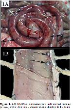
The female wild boar, which was brought to Pathology Department of Veterinary Faculty of Firat University was systematically necropsied. The age of animal is unknown, and certificate of approval couldnt retrieve because of most probably she was hunted by hunters. Macroscopical examination indicated subserosal multifocally distributed nodules in 1-2 mm diameter in jejunum and ileum (Figure 1). In the mucosal surface of small intestinal segments, there were adult ascarids embedded in the intestinal mucosa. The total number of the adult parasites were 17 and measured by 28 to 40 cm in length.
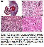
The tissue samples from intestine, liver, lung, brain, and pancreas were fixed in 10% formalin solution. Paraffin blocks prepared by routine methods were cut approximately 5 μm in thickness and stained with hematoxylin-eosin (H-E) (5). Microscopic examination showed that the parasites were embedded in necrotic mass in intestinal mucosa (Figure 2A) and surrounded by an inflammatory infiltrate including eosinophile leukocytes, few epithelioid macrophages and plasma cells (Figure 2B). The infiltrate was covered circumferentially by a thin fibrous capsule (Figure 2C). Mild lymphohistiocytic infiltration was also present in mucosal and muscular surfaces in neigboring intestinal wall. There was a fibrotic parasite tract in the liver (Figure 2D).
Microscopically there was moderate eosinophilic bronchiolitis characterized by the bronchioles surrounded by macrophages and eosinophil leucocytes in the lungs. |
|||||
| Tartışma | |||||
Humans can also be infected by Ascaris suum. Moreover, Ascaris lumbricoides (human roundworm) and Ascaris suum (pig roundworm) are morphologically indistinguishable. A closely related sister species of A. lumbricoides also infects pigs. The swine worm is responsible for significant economic losses by production efficiency, immunosupression and causes organ condemnations at slaughter due to pathologic changes produced by parasite larval stages3. Studies have also indicated that pigs are the main source of ascaris infections in human and it is transmitted by fecal-oral route2,3. As wild boar are source of infection for humans and domestic animals, necessary sanitary measures must be taken and furthermore contact of wild boars to vegetable and animal production units and pasture should be avoided. In conclusion, this report indicates that wild boar with A. suum infection might constitute health hazard for humans and domestic animals unless necessary health precautions are taken. |
|||||
| Kaynaklar | |||||
1) Uzal FA, Plattner BL, Hostetter JM, Maxie GM, Jubb KVF. Jubb, Kennedy and Palmers Pathology of Domestic Animals. 6nd edition, St. Louis: Elsevier, 2016.
2) Crompton DW. Ascaris and ascariasis. Adv Parasitol 2001; 48: 285-375.
3) Miller L, Colby K, Manning SE, et al. Ascariasis in humans and pigs on small-scale farms, maine, USA, 20102013. Emerging Infectious Diseases 2015; 21: 332-334.
|
|||||
| [ Başa Dön ] [ Özet ] [ PDF ] [ Benzer Makaleler ] [ Yazara E-Posta ] [ Editöre E-Posta ] | |||||
 |
| [ Ana Sayfa | Editörler | Danışma Kurulu | Dergi Hakkında | İçindekiler | Arşiv | Yayın Arama | Yazarlara Bilgi | E-Posta ] |

