[ Ana Sayfa | Editörler | Danışma Kurulu | Dergi Hakkında | İçindekiler | Arşiv | Yayın Arama | Yazarlara Bilgi | E-Posta ]
Fırat Üniversitesi Sağlık Bilimleri Veteriner Dergisi
2021, Cilt 35, Sayı 1, Sayfa(lar) 033-036
[ Özet ] [ PDF ] [ Benzer Makaleler ] [ Yazara E-Posta ] [ Editöre E-Posta ]
Hasak Koyununda Arteria Celiaca ve Dallarının Anatomik Özellikleri Üzerine Bir Araştırma
Derviş ÖZDEMİR1, Hülya BALKAYA1, Zekeriya ÖZÜDOĞRU2
1University of Atatürk, Faculty of Veterinary Medicine, Department of Anatomy, Erzurum, TURKEY
2University of Aksaray, Faculty of Veterinary Medicine, Department of Anatomy, Aksaray, TURKEY
Anahtar Kelimeler: Arteria celiaca, makroanatomi, dallanma
Özet
Bu araştırma, hasak koyun ırkının arteria celiaca ve verdiği dalların makro-anatomik olarak incelenmesi amacıyla yapıldı. Çalışmada altı adet hasmer koyunu kullanıldı. Aorta abdominalis’ten renklendirilmiş latex ve polymethyl-methacrylate enjekte edildi. Cavum abdominis’te aorta abdominalis’in verdiği ilk dalın arteria celiaca olduğu gözlendi. Arteria celiaca’nın ilk önce arteria lienalis’i ve daha sonra arteria hepatica ile arteria gastrica sinistra’yı ortak bir kök halinde verdiği belirlendi. Arteria lienalis rami pancreatici, ramus epiploicus, arteria ruminalis sinistra ve arteria ruminalis dextra isimli dallara sahipti. Arteria hepatica’nın, arteria cystica, arteria gastrica dextra ve rami pancreatici ile arteria gastroduodenalis’i verdiği tespit edildi. Arteria gastrica sinistra, arteria celiaca’nın devamı şeklindeydi. Seyri esnasında bu damardan arteria reticularis, arteria gastroepiploica sinistra ile arteria reticularis accessoria ayrılmaktaydı. Arteria reticularis’in, ramus ruminalis ve ramus reticularis isimli dallara sahip olduğu gözlendi. reticulum’un visceral yüzü ile omasum’un parietal yüzüne arteria reticularis accessoria’nın birkaç dal verdiği görüldü. Sonuç olarak; hasak koyun ırkında arteria celiaca’dan orijin alan damarların bazı farklılıklara sahip olduğu tespit edildi. Bu farklılıkların ırkın genetik yapısına bağlı olabileceği kanısına varıldı.
  • Başa Dön
  • Özet
  • Giriş
  • Materyal ve Metot
  • Bulgular
  • Tartışma
  • Kaynaklar
  • Giriş
    In order to increase the production of quality lamb meat, the Hasak genotype was obtained by crossing domestic breeds with low fattening and carcass quality in the Bahri Dagdas International Agricultural Research Institute and meat types with good meat yield and quality 1. In the cross-breeding and selection projects carried out between 1989-2000, F1s, ASB G1 and HD G1s were obtained from the crossbreeding of Akkaraman breed (AKK) with German Black Head (ASB) and Hampshire Down (HD) breeder breeds. A single type was created by applying a balanced seeding program to hybrids from these two paternal lines. Accordingly, this new type includes 31.25% HD, 31.25% ASB and 37.50% Akkaraman genotype. For this type, the name "Hasak" was formed by taking the letters H from HD, AS from ASB and AK from Akkaraman 1-3.

    Arteria celiaca, which gives three main branches: arteria gastrica sinistra, arteria hepatica and arteria lienalis; nourishes the initial part of the liver, spleen, pancreas, stomach and duodenum 4-8. Some sheep species 6,9-12 although there are work-related artery celiaca anatomy, an indigenous race, which Hasak sheep breeds in Turkey where there is not any study about the artery celiac and branches have been identified. With this research, it is aimed to fill the gap on the subject.

  • Başa Dön
  • Özet
  • Giriş
  • Materyal ve Metot
  • Bulgular
  • Tartışma
  • Kaynaklar
  • Materyal ve Metot
    In the research, six Hasak breeders, which were supplied from Konya Bahri Dagdas International Agricultural Research Institute Experimental Animal Unit, were used. After washing with 10% saline, colored latex and polymethyl-methacrylate were injected from the aorta abdominalis. The materials were kept in a 10% formaldehyde solution for 24 hours and dissected. The dissected materials were photographed with Nikon P900 83X Optical Zoom camera. Nomina Anatomica Veterineria (N.A.V.) was based on terminology 13. The necessary permission for the study was obtained from the Atatürk University Veterinary Faculty Unit Ethics Committee (23.10.2015, 8/153).
  • Başa Dön
  • Özet
  • Giriş
  • Materyal ve Metot
  • Bulgular
  • Tartışma
  • Kaynaklar
  • Bulgular
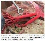
    It was observed that aorta abdominalis passed from cavum thoracis to cavum abdominis through hiatus aorticus. It was determined that arteria celiaca, which was the first branch in cavum abdominis, was progressing towards the ventral at the first lumbar vertebra level. It was observed that the first vessel that left the arteria celiaca. Later, it was determined that arteria lienalis and arteria hepatica and arteria gastrica sinistra took origin as a common root, respectively (Figure 1).


    Büyütmek İçin Tıklayın
    Figure 1: Arteria celiaca’s branches. 1- a. abdominalis, 2- a.celiaca, 3- a.mesenterica cranialis, 4- a.phrenica caudalis, 5- a. lienalis, 6- a. gastrica sinistra, 7- a. hepatica.

    Arteria lienalis: Arteria lienalis first gave the ramie pancreatici to the pancreas. It was later determined that he gave ramus epiploicus and arteria ruminalis sinistra, which fed the omentum majus during the course of the vessel. After giving arteria ruminalis dextra, it continued its course as arteria lienalis (Figure 1, 2).


    Büyütmek İçin Tıklayın
    Figure 2: Arteria lienalis, arteria gastrica sinistra and arteria hepatic's branches. 1- a. celiaca, 2- a.phrenica caudalis, 3- a. lienalis, 4- a. gastrica sinistra, 5- a. hepatica, 6- ramus pancreaticus, 7- ramus epiploicus, 8- a. ruminalis sinistra, 9- a. ruminalis dextra, 10-a.reticularis, 11- a. reticularis accessoria, 12- a. gastroepiploica sinistra, 13- a. cystica, 14- a.gastrica dextra, 15- rami pancreatici, 16- a. gastroduodenalis.

    Arteria ruminalis sinistra: Arteria ruminalis sinistra originated from the left side of arteria lienalis. It was observed that the artery reached the right side of the atrium ruminis by running caudoventrally. Afterwards, it was determined that vascularize the parietal face of saccus cecus craniodorsalis and saccus cecus cranioventralis (Figure 1, 2).

    Arteria ruminalis dextra: It was determined that Arteria ruminalis dextra (Figure 1, 2) originated from the right side of the arteria lienalis and reached the visceral face of the rumen with a caudoventral course. It was observed that the arteria ruminalis dexter, which hovers within the sulcus longitudinalis dexter, was extended to the sulcus caudalis and ended in this groove passing to the parietal face of the rumen. During the course of a. ruminalis dextra, it was observed that this vessel gave coronary arteries vascularizing the saccus cecus on the parietal and visceral face.

    Arteria hepatica: Arteria hepatica was originated from arteria celiaca as a common root with arteria gastrica sinistra. It was determined that the vessel progressed towards porta hepatis in the visceral aspect of liver and continued to progress as arteria gastroduodenalis after giving vessels named arteria cystica, arteria gastrica dextra and rami pancreatici (Figure 2). It was determined that arteria gastroduodenalis was named as arteria gastroepiploica dextra after giving arteria pancreaticoduodenalis cranialis and it anastomosed with arteria gastroepiploica sinistra in the curvatura minor of abomasum.

    Arteria cystica: After leaving arteria hepatica, it was determined that the arteria cystica, which travels on the visceral side of the liver, was distributed to vesica fellea (Figure 2).

    Arteria gastrica dextra: It was determined that the arteria gastrica dextra (Figure 2), which was on the visceral side of the liver, gave the arteria lobi caudati for the caudal lobe of the liver and then anastomosed with arteria gastrica sinistra.

    Arteria gastroduodenalis: Arteria gastroduodenalis, seen as the continuation of arteria hepatica, continued as arteria gastroepiploica dextra on the curvatura major of abomasum, and it gave the arteria pancreaticoduodenalis cranialis (Figure 2). It was determined that Arteria gastroepiploica dextra was ended by anastomosis with arteria gastroepiploica sinistra and vascularized the curvatura major and omentum majus during the course of the vessel.

    Arteria pancreaticoduodenalis cranialis: Arteria pancreaticoduodenalis cranialis (Figure 2), originating from arteria gastroduodenalis, gave branches feeding pancreas and pars cranialis dudodeni.

    Arteria gastrica sinistra, originating from arteria celiaca as a common root with arteria hepatica, was the continuation of the vessel. It was noteworthy as the thickest branch of arteria celiaca. During the course of the vessel, it gave first arteria reticularis, arteria gastroepiploica sinistra and arteria reticularis accessoria in the area between omasum and reticulum. Then, it was determined that continued its course by giving left and right branches on visceral face of omasum (Figure 2). It was ended by anastomosis with arteria gastrica dextra on the curvatura minor of abomasum.

    Arteria reticularis: Arteria reticularis (Figure 2), originating from arteria gastrica sinistra, in the sulcus reticuloruminalis it gave branches named ramus ruminalis and ramus reticularis.

    Arteria gastroepiploica sinistra: Arteria gastroepiploica sinistra (Figure 2), originating from the arteria gastrica sinistra on the atrium ruminis, cranioventrally, passed through the sulcus reticulo-omasi and anastomosed with the arteria gastroepiploica dextra on the curvatura major and the omentum majus. It has been determined that it gave branches to reticulum, omasum and abomasum.

    Arteria reticularis accessoria: Arteria reticularis accessoria originating from arteria gastrica sinistra, immediately after the arteria gastroepiploica sinistra (Figure 2), gave a number of branches on the visceral face of the reticulum and the parietal face of omasum.

  • Başa Dön
  • Özet
  • Giriş
  • Materyal ve Metot
  • Bulgular
  • Tartışma
  • Kaynaklar
  • Tartışma
    It was determined that arteria celiaca was the first branch of aorta abdominalis, which passed from cavum thoracis to cavum abdominis via hiatus aorticus. It was observed that arteria celiaca sailed towards the ventral at the first lumbar vertebra level. Findings that the origin of arteria celiaca and the first branch of aorta abdominalis were also found in some sheep 11 and goat breeds 14,15. Ozudogru et al. 16 in Konya merino; Dursun 5 and Turkmenoglu 12 in ruminant, reported that arteria celiaca originated between the last thoracal and the first lumbar vertebra, while Alsafy 17 and El Gendy 18 reported that the aorta abdominalis originated from the ventral at the 1st and 2nd Lumbal vertebrae levels in the goat.

    Some researchers 6,9,10,17,19 reported that in some of the materials, arteria celiaca originated from aorta abdominalis as a common root with arteria mesenterica cranialis. Ozudogru et al. 16 in Konya merino sheep, Turkmenoglu 12 in three of the sheep used as material, arteria hepatica and arteria gastrica sinistra emerged as a common root and Maala et al. 20 reported that the branching of arteria celiaca in goat has six different forms and in one of them, arteria gastrica sinistra and arteria hepatica emerged as a common root. In this study, it was found that arteria gastrica sinistra and arteria hepatica originated from arteria celiaca as a common root. Some authors 4,6-10 come from arteria celiaca; they stated that arteria lienalis, arteria gastrica sinistra and arteria hepatica emerged as three branches. Some researchers 17,21 from arteria celiaca; they stated that they were divided into four separate branches: arteria lienalis, arteria hepatica, arteria gastrica sinistra and arteria ruminalis sinistra.

    A. lienalis; after giving ramus epiploicus, arteria ruminalis sinistra and arteria ruminalis dextra, it was found to end in the spleen as arteria lienalis. Similar findings were reported to be observed in goat 17 and buffalo 19. The determination that arteria gastrica sinistra is the thickest branch given by arteria celiaca and at the same time is the continuation of the vessel. Kuru 22, Boccaletti and Borelli 23 and Mohamed et al. 14 reported the same findings.

    In the literature 5, it was reported that arteria hepatica is a thicker vein than arteria gastrica sinistra. In this study, it was determined that arteria hepatica was thinner than arteria gastrica sinistra.

    The report of Arteria cystica is similar to the findings of the research that the goat 17, ruminant and sus 24 watched on the visceral face of the liver after extending from arteria hepatica and extended to vesica fellea.

    It was determined that arteria reticularis, which feeds the caudal face of the reticulum and the atrium ruminis, originated from the arteria gastrica sinistra. Nayar et al. 25 in goat, Turkmenoglu 12 in sheep, Smuts and Bezuidenhout 8 in camel, Machado et al. 19 in buffalo, Alsafy 17 in goat, reported that arteria reticularis was originated from arteria gastrica sinistra. Mohamed et al. 11 reported that arteria reticularis was originated arteria gastrica sinistra or arteria ruminalis sinistra.

    As a result in Hasak sheep, which is used as research material, a number of differences were determined from the beginning of arteria celiaca in aorta abdominalis to the branches that vascularize the organs. It was concluded that the detected differences could be caused by race.

  • Başa Dön
  • Özet
  • Giriş
  • Materyal ve Metot
  • Bulgular
  • Tartışma
  • Kaynaklar
  • Kaynaklar

    1) Tekin ME, Gurkan M, Karabulut O, Duzgun H. Performance testing studies and the selection of Hasmer, Hasak, Hasiv and Linmer crossbreed sheep types. III. Fattening Performance. Turk J Vet Anim Sci 2005; 29: 67-73.

    2) Akmaz A, Tekin ME, Kadak R, Akcapınar H. Anadolu (Konya) Merinosu, Hampshire Down x Anadolu Merinosu, Alman Siyahbaş x Anadolu Merinosu F1 ve G1 kuzularında besi ve karkas özellikleri. Turk J Vet Anim Sci 1999; 23: 507-517.

    3) Akmaz A, Tekin ME, Tepeli C, Kadak R. Alman Siyahbaş X Akkaraman ve Hampshire Down X Akkaraman melezi (F1 ve G1) erkek kuzularının besi performansı ve karkas özellikleri. Turk J Vet Anim Sci 2000; 24: 7-17.

    4) Dyce KM, Sack WO, Wensing CJG. Textbook of Veterinary Anatomy. 3rd Edition, Philadelphia: Saunders, 2002.

    5) Dursun N. Veteriner Anatomi II. 11. Baskı, Ankara: Medisan Yayınevi, 2008.

    6) Karmona KH, Kovachev G. Variability of the celiac artery and its branches in sheep. Vet Med Nauki 1985; 22: 31-37.

    7) McCarthy PH. Transruminal palpation of abdominal arteries of the permanently fistulated dairy cow. Am J Vet Res 1984; 45: 1632-1637.

    8) Smuts MMS, Bezuidenhout AJ. Anatomy of the Dromedary. Oxford: Clarendon Press, 1987.

    9) Karadag H. The anatomical comparative study on the celiac artery and its branches in Akkaraman sheep and ordinary goat. J Vet Sci 1988; 12: 196-204.

    10) Langenfeld M, Pastea E. Anatomical variants of the celiac artery in the sheep, with special reference to the celiomesenteric arterial trunk. Anat Anz 1977; 142: 168-174.

    11) Mohamed R, Adogwa A, Driscol M, Rampersad S. Arterial supply of the stomach of the barbados black belly sheep in Trinidad. Int J Vet Sci 2016; 5: 142-147.

    12) Turkmenoglu I. Akkaraman Koyunu ve Ankara Keçisinde Karın Boşluğu Arterleri Üzerinde Karşılaştırmalı Makroanatomik Araştırmalar. Doktora Tezi, Konya: Selçuk Üniversitesi. Sağlık Bilimleri Enstitüsü, 1996.

    13) Internatıonal Commıtteeo Veterınary Gross Anatomıcal NOMENCLATURE – Icvgan. Nomina Anatomia Veterinaria. 5th Edition, Columbia: Editorial Committee Hanover, 2012.

    14) Mohamed R, Adam Z, Gad M. Arterial supply of the stomach of the Egyptian native goat. J Adv Vet Anim Res 2017; 4: 80-87.

    15) Youssef GA. Some Anatomical Studies on the Coeliac, Cranial Mesenteric and Caudal Mesenteric Arteries of Goat. PhD Thesis, Benha, Egypt: Faculty of Veterinary Medicine Moshtohor, Zagazig University, 1991.

    16) Ozudogru Z, Ozdemir D, Balkaya H, Kara H. Konya Merinosunun arteria celiaca’sı ve dalları üzerine macroanatomik bir çalışma. Atatürk Univ Vet Fak Bil Derg 2019; 14: 45-51.

    17) Alsafy M. Celiac trunk and the variability of its branches in goats. J App Biolog Sci 2009; 3: 65-70.

    18) El Gendy SAA. Surgical Anatomical Approach of the Abdomen in the Goat. PhD Thesis, Alexandria: Faculty of Veterinary Medicine, Alexandria University, 2007.

    19) Machado F, Miglino A, Didio A, Oliveira Borges C. The arterial supply of buffalo stomachs (Bubalus Bubalis). Buffalo Journal 2002; 18: 257-265.

    20) Maala C, Reyes EV, Ducusin RJ. Some con-tributions to the anatomy of the arterial supply to the stomach of the goat. Philipp J Vet Med 1988; 25: 47-52.

    21) Perez W, Vazques N, Ungerfeld R. Arterial vasularization of the gastrointestinal tract of the pampas deer. Anat Histol Embryol 2016; 45: 240-245.

    22) Kuru N. Macroanatomic investigations of the course and distribution of the celiac artery in the New Zealand rabbit. Van Vet Journal 2016; 27: 117-122.

    23) Boccaletti D, Borelli V. Ramification of the celiac artery in sheep of Corriedale race. Belo Horizonte, Minas Gerais, Brasil, Socied Brasil de Anatomia, 1981.

    24) Getty R. Sisson and Grossman’s The Anatomy of the Domestic Animals. Volum 2, 5th Edition, Philedephia, London: WB Saunders Company, 1975.

    25) Nayar KN, Singh MG, Singh Y, Singh P, Singh R. Comparative arteriographic anatomy of the abdominal viscera and lumbar region in goats, dogs, pigs and rabbits. Indian j Anim Sci 1983; 53: 1310-1314.

  • Başa Dön
  • Özet
  • Giriş
  • Materyal ve Metot
  • Bulgular
  • Tartışma
  • Kaynaklar
  • [ Başa Dön ] [ Özet ] [ PDF ] [ Benzer Makaleler ] [ Yazara E-Posta ] [ Editöre E-Posta ]
    [ Ana Sayfa | Editörler | Danışma Kurulu | Dergi Hakkında | İçindekiler | Arşiv | Yayın Arama | Yazarlara Bilgi | E-Posta ]