[ Ana Sayfa | Editörler | Danışma Kurulu | Dergi Hakkında | İçindekiler | Arşiv | Yayın Arama | Yazarlara Bilgi | E-Posta ]
Fırat Üniversitesi Sağlık Bilimleri Veteriner Dergisi
2021, Cilt 35, Sayı 1, Sayfa(lar) 053-054
[ Özet ] [ PDF ] [ Benzer Makaleler ] [ Yazara E-Posta ] [ Editöre E-Posta ]
Bir Domuzda Yaygın Pnömatozis Sistoides İntestinalis
Yesari ERÖKSÜZ, Canan AKDENİZ İNCİLİ, Burak KARABULUT, Mehmet AKSOY, Hatice ERÖKSÜZ
University of Fırat, Faculty of Veterinary Medicine, Department of Pathology, Elazığ, TURKEY
Anahtar Kelimeler: Lenf damarları, domuz, pnömatozis sistoides intestinalis, podoplanin
Özet
Yaygın pnömatozis cystoides intestinalis'in morfolojik ve immünohistokimyasal özellikleri yetişkin bir domuzda tanımlandı. Gaz dolu kistler kalın ve ince bağırsak serozasında mevcuttu ve kistlerin duvarı podoplanin antikoruna immünopozitifti, bu da kistlerin aslında gaz dolu lenf damarları olduğunu göstermektedir. Sonuç olarak, mevcut vaka raporu, kistik yapıların genişlemiş lenfatik damarlar olduğunu açıkça göstermiştir.
  • Başa Dön
  • Özet
  • Giriş
  • Olgu Sunusu
  • Tartışma
  • Kaynaklar
  • Giriş
    Pneumatosis cystoides intestinalis (PCI), also known as intestinal emphysema, pneumatosis intestinalis, cystic lymphopneumatosis, and intestinal gas cyst, refers to multiple thin-walled endothelial lining pseudocyst formations which are filled with gas sizing from a few millimeters to several centimeters in diameter in the intestinal wall 1,2. It is reportedly a rare incidental lesion in weanling-pig, dog, rabbit, and human beings 1-3. However, clinically these lesions might lead to an incorrect radiological diagnosis of tumor and signs of intestinal obstruction. It might occur in any portion of small intestine, however also involve in the large intestine, mesenterium, and mesenterial lymph nodes 3-5.

    The condition in human medicine is associated with a large variety of diseases and clinical conditions in which the loss of intestinal mucosal integrity and/or luminal high pressure usually occur. PCI can be classified in terms of pathogenesis as primary (idiopathic without any obvious cause) or secondary to a wide variety of conditions including; intestinal necrosis, intestinal obstruction, mucosal disruption, increased mucosal permeability, and pulmonary disease 1.

    There are mainly 4 sources of gas in the intestinal wall; gas escapes through any disruption of the mucosa, pulmonary gas released from the ruptured alveoli, gas production of some bacteria and excessive fermentation due to maldigestion 6.

    PCI is mostly asymptomatic, however, a spectrum of clinical signs including vomiting, abdominal distention, abdominal pain, diarrhea, constipation, and tenesmus was reported in dogs 4,5.

    This report presents morphological and immunohistochemical findings of a case of PCI in pig.

  • Başa Dön
  • Özet
  • Giriş
  • Olgu Sunusu
  • Tartışma
  • Kaynaklar
  • Olgu Sunusu
    An 18-month-old, male pig was presented to Pathology Department of Veterinary Faculty of Firat University. The pigs were bred for educational purposes and were fed a mixture of barley, wheat and corn. There were 20 pigs in the unit. In history; the present pig quarreled with the other male pig and he was then transferred to hospitalization unit 2 weeks before the death. Although there was no evidence of trauma on the animal's skin surface, the animal was depressed and not drinking water. On the basis of history and clinical examination, prophylactic parenteral antibiotics were administered for 4 days.

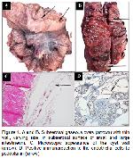
    At necropsy, polyploid thin walled, gas filled cysts in serosal surface of small and large intestineum were seen. Their size of the lesions ranged from 1 mm to a few cm (Figure 1A and B). There was no any other remarkable morphological changes in internal organs or tissues macroscopically.


    Büyütmek İçin Tıklayın
    Figure 1: A and B. Subserosal gaseous cysts (arrows) with thin wall, varying size, in subserosal surface of small and large intestineum. C. Microscopic appearance of the cyst wall (arrow). D. Positive immunoreaction to the endothelial cells to podoplanin (arrow).

    The tissue samples from small and large intestines, liver, lung, brain, and pancreas were fixed in 10% formalin solution. Paraffin blocks prepared by routine methods were cut approximately 5 μm in thickness and stained with hematoxylin-eosin (H&E). Selected sections were stained by podoplanin (Mouse monoclonal, clone D2-40 Dako, Glostrup, Denmark, 1:400) by avidin-biotin peroxidase.

    Histopathological examination showed that the cystic spaces were lined by endothelial cells. There was no inflammatory reacrion in stroma (Figure 1C).

    Immunohistochemical examinations indicated that there was a strong immunopositivity for the podoplanin on the inside of the cystic wall. On higher magnification, the podoplanin-expressing cells were identified to be linearly lined spindle cells (Figure 1D).

  • Başa Dön
  • Özet
  • Giriş
  • Olgu Sunusu
  • Tartışma
  • Kaynaklar
  • Tartışma
    In humans, on the histopathological basis, PCI is divided into 3 main categories including microvesicular, cystic, or diffuse. Microvesicular variant was characterized by the presence of small gas‐filled spaces expanding the lamina propria, and the mucosa was otherwise unremarkable. Cystic variant was characterized by the presence of submucosal or subserosal cysts ranging from a few millimeters to several centimeters, which were lined by macrophages and foreign body type multinucleated giant cells. Lastly, diffuse PI was recognized as a diffusely spongy bowel wall and, these cystic spaces lacked any lining cells, with or without an inflammatory response 7. Microscopically; the presented case was compatible with the diffuse variant of PCI. The absence of inflammatory reaction might be associated with the intact lymphatic cysts in the present report. The rupture of lymphatics would cause inflammatory reaction due to release of lipids and gases.

    The etiology of the present case remains obscure, however based on anamnesis of animal quarrel, the present case of PCI speculatively might be resulted from the translocation of gas as a consequence of the increased mucosal permeability secondary to abdominal trauma. Although there were no skin lesions indicating trauma or ulcer, erosion in intestinal mucosa, blunt trauma might be responsible for the occurrence of the lesions. Similar to present report, a case of posttraumatic pneumatosis intestinalis was reported in human infant 8. However host factors such as carbohydrate intolerance may also have a significant role either alone or in combination with other factors in pigs and humans 9.

    Overall, the present case report clearly demonstrates the cystic structures are dilated lymphatics expressing podoplanin in PCI of a pig.

  • Başa Dön
  • Özet
  • Giriş
  • Olgu Sunusu
  • Tartışma
  • Kaynaklar
  • Kaynaklar

    1) Gui X, Zhou Y, Eidus L, et al. Is pneumatosis cystoides intestinalis gas-distended and ruptured lymphatics? Reappraisal by immunohistochemistry. Arch Pathol Lab Med 2014; 138: 1059-1066.

    2) Uzal FA, Plattner BL, Hostetter JM. Alimentary system. In: Maxie MG, (Editor). Jubb, Kennedy and Palmer’s Pathology of Domestic Animals. 6th Edition, Philadelphia, PA: Elsevier Saunders, 2016: 87.

    3) Peris B, García V, Blas E, et al. Pneumatosis cystoides intestinalis in a rabbit doe (Oryctolagus cuniculus). J Vet Diagn Invest 2005; 17: 256-258.

    4) Morris EL. Pneumatosis coli in a dog. Vet Radiol Ultrasound 1992; 33: 154-157.

    5) Russell NJ, Tyrrell D, Irwin PJ, Beck C. Pneumatosis coli in a dog. J Am Anim Hosp Assoc 2008; 44: 32-35.

    6) Micklefield GH, Kuntz HD, May B. Pneumatosis cystoides intestinalis: Case reports and review of the literature. Mater Med Pol 1990; 22: 70-72.

    7) Hanna P, Kassir R, Tarek D, et al. Pneumatosis cystoidis intestinalis presenting as bowel perforation, a rare entity. Int J Surg Case Rep 2016; 20: 7-9.

    8) Koutouzis T, Lee J. Blunt abdominal trauma resulting in Pneumatosis intestinalis in an infant. Ann Emerg Med 2000; 36: 619-621.

    9) Meyer RC, Simon J. Intestinal emphysema (Pneumatosis cystoids intestinalis) in a gnotobiotic pig. Can J Comp Med 1971; 41: 302-330.

  • Başa Dön
  • Özet
  • Giriş
  • Olgu Sunusu
  • Tartışma
  • Kaynaklar
  • [ Başa Dön ] [ Özet ] [ PDF ] [ Benzer Makaleler ] [ Yazara E-Posta ] [ Editöre E-Posta ]
    [ Ana Sayfa | Editörler | Danışma Kurulu | Dergi Hakkında | İçindekiler | Arşiv | Yayın Arama | Yazarlara Bilgi | E-Posta ]