[ Ana Sayfa | Editörler | Danışma Kurulu | Dergi Hakkında | İçindekiler | Arşiv | Yayın Arama | Yazarlara Bilgi | E-Posta ]
Fırat Üniversitesi Sağlık Bilimleri Veteriner Dergisi
2021, Cilt 35, Sayı 2, Sayfa(lar) 109-113
[ Özet ] [ PDF ] [ Benzer Makaleler ] [ Yazara E-Posta ] [ Editöre E-Posta ]
Beyaz Kas Hastalıklı Kuzularda 4-Hidroksi-2-Nonenal, Ditirozin and 8-Hidroksi-2-Deoksiguanozin Ekspresyonlarının Değerlendirilmesi
Emin KARAKURT1, Özhan KARATAŞ2, Serpil DAĞ1, Enver BEYTUT1, Ali Sefa MENDİL3, Hilmi NUHOĞLU1, Ayfer YILDIZ1
1Kafkas University, Faculty of Veterinary Medicine, Department of Pathology, Kars, TURKEY
2Cumhuriyet University, Faculty of Veterinary Medicine, Department of Pathology, Sivas, TURKEY
3Erciyes University, Health Sciences Institute, Kayseri, TURKEY
Anahtar Kelimeler: 4-HNE, 8-OHdG, ditirozin, kuzu, beyaz kas hastalığı
Özet
Bu çalışmada, beyaz kas hastalıklı kuzularda 4-hidroksi-2-nonenal (4-HNE), ditirozin (DT) ve 8-hidroksi-2-deoksiguanozin (8-OHdG) gibi oksidatif stres belirteçleri ile Reaktif Oksijen Türleri (ROT) kaynaklı lipid peroksidasyonu, protein ve DNA hasarının araştırılması amaçlanmıştır. Bu çalışmanın materyalini 2012-2019 yılları arasında rutin histopatolojik tanı için Patoloji Anabilim Dalı’na getirilen beyaz kas hastalıklı 20 koyundan alınan doku örnekleri ve 6 sağlıklı kontrol grubu oluşturdu. 4-HNE, DT ve 8-OHdG ekspresyonları immünohistokimyasal ve immünofloresan yöntemlerle değerlendirildi. Makroskopik incelemelerde epikardiyal ve özellikle endokardiyal yüzeylerde, sağ ve sol ventrikül duvarlarında tebeşirimsi beyaz büyük nekrotik alanlar gözlemlendi. Mikroskobik incelemelerde kalpteki dejenere olmuş kas liflerinin homojen pembe renkte olduğunu, sitoplazmik sitrifikasyonunu kaybettiğini, şiştiği ve çekirdeklerinin piknotik olduğu gözlemlendi. Nekrotik ve dejenere alanlarda kalsifikasyonlar tespit edildi. Tüm beyaz kas hastalığı vakaları kalp dokularında 4-HNE, DT ve 8-OHdG ekspresyonu yönünden immün pozitifti. İmmünohistokimyasal ve immünofloresan boyama sonuçlarında 4-HNE, DT ve 8-OHdG'ye karşı immünreaktivite açısından gruplar arasında anlamlı farklılık (P<0.05) tespit edildi. Sonuç olarak çalışmamız beyaz kas hastalığının patogenezinde önemli bir faktör olan ROS oluşumunun hem lipid peroksidasyonuna, hem protein modifikasyonuna hem de DNA hasarına neden olduğunu göstermektedir.
  • Başa Dön
  • Özet
  • Giriş
  • Materyal ve Metot
  • Bulgular
  • Tartışma
  • Kaynaklar
  • Giriş
    White muscle disease (WMD), also known as subacute enzootic muscular dystrophy or stiff-lamb disease, is caused by Selenium (Se) and/or vitamin E (Vit E) deficiency. It is an important nutritional disease that is seen in our country as well as all over the world 1-3. The disease, has been reported especially in Central, Eastern and Southeastern Anatolia regions 4,5. It affects not only sheep but also many domestic and wild animals such as goats, cattle, deer, horses, pigs, rabbits, marsupials, monkeys, laboratory and exotic animals and even fish 6. WMD is seen in young sheep grazing (stubble or rank) or being maintained on hay and grain rations 7. Disease is more common in lambs up to 3 months of age, it can also be seen in newborn lambs 2. The main clinical symptoms included weakness, unwillingness to move, difficulty in standing, stiff gait, curvature of the back, short and upright steps 8-10. Both Vit E and Selenium play important roles in protecting cellular membranes to the free radical-induced lipid peroxidation 9,11. Se and Vit E deficiencies cause lipoperoxidation in tissues to muscle degeneration and calcification 3,8,12. Free radicals, which are formed as a result of the decrease in antioxidant defense due to Se and Vit E deficiency, have an important role in the pathogenesis of this disease and causing oxidative damage by causing toxic effects on enzymes, nucleic acids, polysaccharides and unsaturated fatty acids in cell membranes 5,13,14.

    Elevated levels of reactive oxygen species (ROS) act as a marker for oxidative stress and they are associated with lipid peroxidation and imbalance of the redox system 15. Among the many aldehydes produced from lipid peroxidation is the α,β-unsaturated aldehyde, 4-hydroxy-2-nonenal (4-HNE), especially 4-HNE is the most studied lipid peroxidation end product 16,17. 4-HNE, which is a specific marker of oxidative stress, has important electrophilic properties and reacts with many classes of biomolecules such as phospholipids, proteins and nucleotides, forming covalent adducts 18,19. 3,3′-dityrosine, also known as dityrosine (DT), a stable and convenient biomarker for oxidative modifications of proteins, is generated by reactive species, attacking tyrosine residues in proteins and, eventually, generating tyrosyl radicals 20-22. Two tyrosyl radicals can form inter-molecular cross-links producing a dimer called DT 23. The hydroxyl radical attacks DNA strands when it is produced adjacent to cellular and mitochondrial DNA causing the addition of DNA bases radicals, which lead to generation of a variety of oxidation products, one of these oxidative DNA products, 8-hydroxy-2-deoxyguanosine (8-OHdG), is a biomarker that is extensively used to reflect the degree of oxidative DNA damage 24-26.

    The aim of this study was to investigate ROS-induced lipid peroxidation, protein and DNA damage with oxidative stress markers such as 4-HNE, DT and 8-OHdG in lambs with WMD.

  • Başa Dön
  • Özet
  • Giriş
  • Materyal ve Metot
  • Bulgular
  • Tartışma
  • Kaynaklar
  • Materyal ve Metot
    Research and Publication Ethics: The ethics committee report of this study was obtained from Kafkas University Animal Experimentals Local Ethics Committee (Authorization number: KAU-HADYEK-2020/093).

    Animals: The material of this study consisted of tissue samples (heart and gluteal muscle) taken from 20 sheep with WMD and 6 healthy control groups brought to our department for routine histopathological diagnosis between 2012-2019 years.

    Histopathological Investigations: Tissue samples (heart and gluteal muscle) from lambs were fixed in 10% buffered formaldehyde solution, processed routinely, embedded in paraffin and sectioned at 5 μm and stained with Hematoxylin & Eosine (H&E). Sections were examined under a light microscope (Olympus Bx53) and photographed using the Cell^P program (Olympus Soft Imaging Solutions GmbH, 3,4).

    Immunohistochemical and Immunfluorescence Investigations: Paraffine sections of on Poly-L-lysine slides passed through xylol and alcohol series. After washing with PBS, sections were incubated for 10 minutes in 3% H2O2 for inactivation of endogenous peroxidase activity. In order to reveal the antigen in the tissues, the antigen was treated with retrieval solution at 500 watts for 2x5 minutes. The tissues were then washed with PBS and left for incubation for 0 minutes at room temperature with primary antibodies, 4-HNE (Abcam, Catalog no: ab 46545, anti rabbit, polyclonal), 8-OHdG (Santa Cruz, Catalog no: sc 66036, anti mouse, monoclonal) and Anti 3,3'Dityrosine (Biomol, Catalog no: MDT-020P). Secondarily; Mouse and Rabbit Specific HRP/DAB IHC Detection Kit - Micro-polymer kit (Abcam, Catalog No. ab236466, anti mouse, monoclonal) was used as recommended by the manufacturer. DAB (3,3′-Diaminobenzidine) was used as chromogen. After contrast painting with Mayer's Hematoxylin, it was covered with entellan and examined under a light microscope. In the immunohistochemical examination, findings ere classified as no immunoreactivity (-), mild (+), moderate (++) and severe (+++). In the immunofluorescence method, 45 min. Goat Anti-Mouse IgG H & L-FITC (cat. no. 6785, dilution 1/50, Abcam, UK) and Mouse Anti-Rabbit IgG H & L-FITC (cat. no. sc-2359, dilution 1/50, Santa cruz) secondary antibodies were applied. At the end of the incubation period, the washed sections were covered with 4’,6-diamidino-2-phenylindole (DAPI) fluorescence medium. Fluorescence was evaluated as absent (-), mild (+), moderate (++) and severe (+++) under the microscope.

    Statistical Analysis: The data obtained were analyzed with the SPSS 20.00 program. The difference between the groups was determined by Mann Whitney U test, one of the nonparametric tests. A value of P<0.05 was considered statistically significant 27.

  • Başa Dön
  • Özet
  • Giriş
  • Materyal ve Metot
  • Bulgular
  • Tartışma
  • Kaynaklar
  • Bulgular
    Clinical Results: 18 of the 20 animals had an average age of 45 days. The remaining 2 animals were 10 days old. According to the anamnesis obtained from the animal owners, the animals had symptoms including anorexia, inability to get up from the ground, reluctance to move, hunched back, stiff gait, curvature of the back, short and upright steps, shortness of breath, inability to lift the head and limpness.

    Macroscopic Results: In macroscopic examinations, pale, chalky-white large necrotic areas on the epicardial and especially on the endocardial surfaces, right (18/20, %90) and left (2/20, %10) ventricular walls were detected (Figure 1a). Necrotic changes were generally present in the heart muscle, however both the heart and gluteal muscles were affected in some cases. The gluteal muscles were typically looked like chicken meat (Figure 1b).


    Büyütmek İçin Tıklayın
    Figure 1: a: Chalky-white-looking Zenker’s necrosis (arrow) on the endocardial surface of the heart, b: Chicken meat appearance in the gluteal muscles (arrow heads)

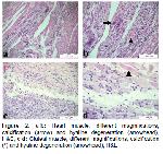
    Microscopic Results: Microscopic examinations showed that the degenerated muscle fibers in heart were homogeneously pink in color, lost their cytoplasmic citriation, were swollen, and their nuclei were pyknotic. We detected calcifications in necrotic and degenerated areas (Figure 2a-b). Fibrosis was prevalent in some areas. Mononuclear cell infiltrates in the interstitial regions were among the other histopathological findings observed. The presence of hyaline degeneration and calcified areas in the gluteal muscles, in line with the findings in the heart muscle, was remarkable (Figure 2c-d).


    Büyütmek İçin Tıklayın
    Figure 2: a-b: Heart muscle, different magnifications, calcification (arrow) and hyaline degeneration (arrowhead), H&E, c-d: Gluteal muscle, different magnifications, calcification (*) and hyaline degeneration (arrowhead), H&E

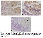
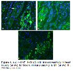
    Immunohistochemical and Immunfluorescence Results: A significant difference was detected between the groups in terms of immunoreactivity to 4-HNE, Anti 3,3 'Dityrosine and 8-OHdG in immunohistochemical and immunofluorescence staining findings (Table 1, P<0.05). Immunoreactivity for 4-HNE, Anti 3,3 'Dityrosine and 8-OHdG could not be detected at a significant level in the heart muscle tissues of the control group animals. While the 8-OHdG and 4-HNE immunoreactivity of the animals in the WMD group was found to be mild in the heart muscle, the immunoreactivity for Anti 3,3 'Dityrosine was found to be at a severe level (Fig 3-4 a-c).


    Büyütmek İçin Tıklayın
    Table 1: 4-HNE, 8-OHdG and DT immunoreactivities of control and WMD groups


    Büyütmek İçin Tıklayın
    Figure 3: a-c: 4-HNE, 8-OHdG mild immunoreactivity in heart muscle (arrow), b: Severe immunoreactivity to DT (arrow), IHC, Bar= 50 μm


    Büyütmek İçin Tıklayın
    Figure 4: a-c: 4-HNE, 8-OHdG mild immunoreactivity in heart muscle (arrow), b: Severe immunoreactivity to DT (arrow), IF, 20X Magnification

  • Başa Dön
  • Özet
  • Giriş
  • Materyal ve Metot
  • Bulgular
  • Tartışma
  • Kaynaklar
  • Tartışma
    WMD is seen as congenital form in newborns or adoptive form in 3-4 months old lambs and the clinical course of the disease varies as peracute, acute and subacute forms 1,6. The main clinical findings of the disease are as follows; inability to stand up, stiff-legged gait, shortness of breath, loss of appetite, weakness, unwillingness to move, difficulty in standing, stiff gait, curvature of the back, short and upright steps 4,8. In our study, in accordance with the literature data 1,6, the disease was detected mostly in animals up to 3 months (18 out of 20 animals, 90%). The remaining 2 animals were 1 week - 10 days old. In addition, many clinical symptoms such as anorexia, inability to get up from the ground, reluctance to move, hunched back, stiff gait, curvature of the back, short and upright steps, shortness of breath, inability to lift the head and limpness were noted in animals, as previously reported 4,8,28,29. In the present study, the diagnosis of WMD in lambs was made based on observed macroscopic and microscopic lesions (6, 28). The gross 2,7,13 and histopathological lesions 29-31 in the heart and gluteal muscles we detected in lambs were similar to those previously reported by different researchers.

    The most important factors in the etiology of WMD in lambs are Vit E and / or Se deficiency 5,7. Se enters the structure of the enzyme of glutathione peroxidase (GSH-Px) which works in the body's antioxidant protection and reduces hydrogen peroxide, super-oxide radicals and lipid peroxides to water 4,8. Vit E is a lipophilic antioxidant that reduces hydroperoxide formation and acts to scavenge free radicals at the extracellular or intracellular level 9,14. Vit E and Se-containing GSH-Px, are an important part of the antioxidant system found in all cells. In WMD, lipid peroxidation and hydrogen peroxide normally occurring in the organism cannot be scavenged from the muscles due to the decrease in GSH-Px activity caused by Se and antioxidant Vitamin E deficiencies 11. Free oxygen radicals that develop due to the decrease in antioxidant activity play a serious role in the pathogenesis of the disease by causing severe pathological changes such as lipid peroxidation in tissues, degradation of proteins, degeneration and finally necrosis of the myocardium 3,13. Therefore, an increase in oxidative stress and 4-HNE expression, which is an important active lipid peroxidation marker in heart tissue, is thought to be due to the decrease in antioxidant defense 17. As we expected, we found that 4-HNE expression increased statistically in the WMD group compared to the control group. In human medicine, there are findings that 4-HNE is produced in the heart in cardiovascular diseases such as atherosclerosis, hypertrophy, cardiomyopathy, myocardial ischemia-reperfusion injury and arrhythmias compared to healthy individuals in the control group of 4-HNE 32,33. In the literature review, no study was found in which lipid peroxidation in lambs with WMD was evaluated in terms of 4-HNE expressions by immunohistochemical methods. Consistent with our results, different researchers found that lipid peroxidation increased in WMD animals compared to healthy animals in terms of Malondialdehyde (MDA) levels 1,5,14. DT is a prominent marker for oxidative stress as protein oxidation and is formed by ROS attack on a wide range of proteins 20,22,23. DT is an irreversible and irreparable oxidative modification 21. We did not find any literature data in which DT expressions were evaluated by immunohistochemical methods in white muscle disease. However, it has been reported that DT levels increase statistically in disease group compared to the control group for chronic heart failure and acute myocardial infarction models 22,23,33. In the present study, we observed that DT expression in the heart of the animals in the WMD group was quite severe and increased significantly compared to the control group. We interpreted this increase in DT immunoreactivity as ROS-induced protein modifications may play an important role in the pathogenesis of WMD. ROS can cause specific oxidative DNA damage and 8-OHdG is the most used biomarker to detect this damage 1,24. 8-OHdG values have been evaluated in detail in human cardiovascular diseases such as coronary artery disease, non coronary artery disease, myocardial infarction, ischemic stroke, heart failure, atherosclerosis 25,26. In the literature reviews, it is seen that there is a positive relationship between 8-OHdG levels and cardiovascular diseases 25,26. However, there is only one study in which 8-OHdG levels were evaluated in immunohistochemical methods in lambs with WMD 1. We determined that the 8-OHdG expressions in the heart tissue of lambs with WMD increased statistically compared to control animals similar to that reported by Yildirim et al., 2019 1. We believe this increase in 8-OHdG immunoreactivity in lambs with WMD is likely a result of ROS-induced DNA damage. We think that ROS-induced lipid peroxidation and protein modifications as well as DNA damage play an important role in the pathogenesis of WMD.

    In conclusion, this study revealed that ROS, which is an important factor in the pathogenesis of WMD, causes lipid peroxidation, protein modification and DNA damage. There is no literature data in which important oxidative stress markers such as 4-HNE, DT and 8-OHDG were evaluated together with IHC and IF methods in lambs with WMD. In this respect, we believe that the data obtained from this study will contribute to the pathogenesis of the disease and the literature data.

  • Başa Dön
  • Özet
  • Giriş
  • Materyal ve Metot
  • Bulgular
  • Tartışma
  • Kaynaklar
  • Kaynaklar

    1) Yildirim S, Ozkan C, Huyut Z, Çınar A. Detection of Se, Vit E, Vit A, MDA, 8-OHdG, and CoQ10 levels and histopathological changes in heart tissue in sheep with white muscle disease. Biol Trace Elem Res 2019; 188: 419-423.

    2) Mert H, Yildirim S, Yoruk IH, et al. Retinol, α-tocopherol and vitamin D3 in white muscle disease. Med Weter 2018; 74: 441-444.

    3) Ataollahi F, Mohri M, Seifi HA, et al. Evaluation of copper concentration in subclinical cases of white muscle disease and its relationship with cardiac troponin I. PLoS One 2013; 8: e56163.

    4) Yavuz O. The pathological investigations on nutritional myopathy causing lamb deaths in neonatal period. Bahri Dağdaş Hayvancılık Araştırma Dergisi 2017; 6: 1-8.

    5) Beytut E, Erişir M, Aksakal M. Beyaz kas hastalıklı kuzuların kalp, iskelet kası ve karaciğerlerinde redükte glutatyon ve malondialdehit düzeyleri ile katalaz enzim aktivitesi. Kafkas Üniv Vet Fak Derg 2001; 7: 1-5.

    6) Loukopoulos P, Ioannou I, Kassinis N, et al. Congenital nutritional myodegeneration in a Cypriot mouflon (Ovis orientalis ophion) lamb. Revue Mèd Vèt 2012; 163: 402-404.

    7) Yildirim S, Irak K, Mert H, Dogan I, Mert N. Serum biochemical and histopathological examinations of some tissues of lambs with muscular dystrophy in Van. Sci Work Series C Veterinary Med 2016; 2: 52-55.

    8) Başbuğan Y, Yüksek N, Altuğ N, Ceylan E, et al. The diagnostic significance of serum troponin levels lambs with white muscle disease. Turkish Journal of Veterinary Research 2018; 2: 5-11.

    9) Değer Y, Mert H, Mert N, et al. Serum selenium, vitamin E, and sialic acids concentrations in lambs with white muscle disease. Biol Trace Elem Res 2008; 121: 39-43.

    10) Or ME, Dodurka HT, Kayar A. Beyaz kas hastalıklı kuzularda eritrosit glutasyon peroksidaz (GSH-Px) aktivitesi ve bazı serum parametrelerinin diagnostik önemi. Turk J Vet Anim Sci 2003; 27: 1-6.

    11) Kozat S, Gunduz H, Deger Y, et al. Studies on serum alpha-tocopherol, selenium levels and catalase activities in lambs with white muscle disease. Bull Vet Inst Pulawy 2007; 51: 281-284.

    12) Kozat S, Altuğ N, Yüksek N, Özkan C. Evaluation of the levels of homocysteine, troponin I, and nitric oxide in lambs with subclinical white muscle disease. Kafkas Univ Vet Fak Derg 2011; 17: 441-444.

    13) Yumusak N, Yigin A, Polat PF, Hitit M, Yilmaz R. Expression of ADAMTS-7 in myocardial dystrophy associated with white muscle disease in lambs. Pol J Vet Sci 2018; 21: 119-126.

    14) Bildik A, Yur F, Çamaş H, Dede S, Sekin S. Beyaz kas hastalıklı kuzularda hemoglobin tipleri ile lipid peroksidasyonu ve glutatyon düzeylerinin araştırılması. Yüzüncü Yıl Üniversitesi Veteriner Fakültesi 1996; 7: 95-98.

    15) Breitzig M, Bhimineni C, Lockey R, Kolliputi N. 4-Hydroxy-2-nonenal: A critical target in oxidative stress? Am J Physiol Cell Physiol 2016; 311: C537-C543.

    16) Smathers RL, Fritz KS, Galligan JJ, et al. Characterization of 4-HNE modified L-FABP reveals alterations in structural and functional dynamics. PLoS One 2012; 7: e38459.

    17) Ayala A, Muñoz MF, Argüelles S. Lipid peroxidation: production, metabolism, and signaling mechanisms of malondialdehyde and 4-hydroxy-2-nonenal. Oxid Med Cell Longev 2014; 2014: 360438.

    18) Lee WC, Wong HY, Chai YY, et al. Lipid peroxidation dysregulation in ischemic stroke: plasma 4-HNE as a potential biomarker? Biochem Biophys Res Commun 2012; 425: 842-847.

    19) Pillon NJ, Croze ML, Vella RE, et al. The lipid peroxidation by-product 4-hydroxy-2-nonenal (4-HNE) induces insulin resistance in skeletal muscle through both carbonyl and oxidative stress. Endocrinology 2012; 153: 2099-2111.

    20) Wu GR, Cheserek M, Shi YH, et al. Elevated plasma dityrosine in patients with hyperlipidemia compared to healthy individuals. Ann Nutr Metab 2015; 66: 44-50.

    21) Li B, Ge Y, Xu Y, et al. Spatial learning and memory impairment in growing mice induced by major oxidized tyrosine product dityrosine. J Agric Food Chem 2019; 67: 9039-9049.

    22) Mongirdienė A, Laukaitienė J, Skipskis V, Kašauskas A. The effect of oxidant hypochlorous acid on platelet aggregation and dityrosine concentration in chronic heart failure patients and healthy controls. Medicina (Kaunas) 2019; 55: 198.

    23) Mayer F, Falk M, Huhn R, Behmenburg F, Ritz-Timme S. Dityrosine as a marker of acute myocardial infarction? Experiments with the isolated Langendorff heart. Int J Legal Med 2016; 130: 1053-1060.

    24) Valavanidis A, Vlachogianni T, Fiotakis C. 8-hydroxy-2' -deoxyguanosine (8-OHdG): A critical biomarker of oxidative stress and carcinogenesis. J Environ Sci Health C Environ Carcinog Ecotoxicol Rev 2009; 27: 120-139.

    25) Kroese LJ, Scheffer PG. 8-hydroxy-2'-deoxyguanosine and cardiovascular disease: A systematic review. Curr Atheroscler Rep 2014; 16: 452.

    26) Di Minno A, Turnu L, Porro B, et al. 8-Hydroxy-2-Deoxyguanosine levels and cardiovascular disease: A systematic review and meta-analysis of the literature. Antioxid Redox Signal 2016; 24: 548-555.

    27) Alpar R. Spor, Sağlık ve Eğitimli Bilimlerinden Örnekle Uygulamalı İstatistik ve Geçerlik-Güvenlik SPSS’de Çözümleme Adımları ile Birlikte. 6. Baskı, Ankara: Detay Yayıncılık, 2020.

    28) Tunca R, Erdoğan HM, Sözmen M, et al. Evaluation of cardiac troponin I and inducible nitric oxide synthase expressions in lambs with white muscle disease. Turk J Vet Anim Sci 2009; 33: 53-59.

    29) Gunes V, Ozcan K, Citil M, Onmaz AC, Erdogan HM. Detection of myocardial degeneration with point-of-care cardiac troponin assays and histopathology in lambs with white muscle disease. Vet J 2010; 184: 376-378.

    30) Hanedan B, Erişir M, Kandemir FM, Özkaraca M. Changes in arginase activity and AST enzyme levels in the cardiac and skeletal muscle and liver of lambs with white muscle disease. Turk J Vet Anim Sci 2015; 39: 611-614.

    31) Beytut E, Karatas F, Beytut E. Lambs with white muscle disease and selenium content of soil and meadow hay in the region of Kars, Turkey. Vet J 2002; 163: 214-217.

    32) Mali VR, Palaniyandi SS. Regulation and therapeutic strategies of 4-hydroxy-2-nonenal metabolism in heart disease. Free Radic Res 2014; 48: 251-263.

    33) Nakamura K, Kusano KF, Matsubara H, et al. Relationship between oxidative stress and systolic dysfunction in patients with hypertrophic cardiomyopathy. J Card Fail 2005 11: 117-123.

  • Başa Dön
  • Özet
  • Giriş
  • Materyal ve Metot
  • Bulgular
  • Tartışma
  • Kaynaklar
  • [ Başa Dön ] [ Özet ] [ PDF ] [ Benzer Makaleler ] [ Yazara E-Posta ] [ Editöre E-Posta ]
    [ Ana Sayfa | Editörler | Danışma Kurulu | Dergi Hakkında | İçindekiler | Arşiv | Yayın Arama | Yazarlara Bilgi | E-Posta ]