[ Ana Sayfa | Editörler | Danışma Kurulu | Dergi Hakkında | İçindekiler | Arşiv | Yayın Arama | Yazarlara Bilgi | E-Posta ]
Fırat Üniversitesi Sağlık Bilimleri Veteriner Dergisi
2022, Cilt 36, Sayı 3, Sayfa(lar) 188-193
[ Özet ] [ PDF ] [ Benzer Makaleler ] [ Yazara E-Posta ] [ Editöre E-Posta ]
Deneysel Diyabetik Sıçan Böbreğinde Tarçın Ekstraktının TLR4 Ekspresyonu ve Mast Hücrelerinin Sayısal Dağılımı Üzerine Etkisinin Araştırılması
Hülya ŞEN, Tuğrul ERTUĞRUL
Ondokuz Mayıs University, Faculty of Veterinary Medicine, Department of Histology and Embryology, Samsun, TÜRKĠYE
Anahtar Kelimeler: Diyabet, tarçın, böbrek, mast hücre, TLR4
Özet
Objective: In this study, it was aimed to evaluate the effect of cinnamon extract on TLR4 expression and numerical distribution of mast cells in experimental diabetic rat kidneys and examine the relationships between TLR4 and mast cells.

Materials and Methods: Thirty-two Wistar albino male rats were used in this study. Control group, Diabetes group, Cinnamon group and Diabetes + cinnamon group. After confirming the occurrence of diabetes in rats, cinnamon at a dose of 0.5 mg/kg was administered by oral gavage to animals in the cinnamon group and diabetes + cinnamon group for 14 days. During the experiment, no application was made to the rats in the control group.

Results: When the cinnamon group was evaluated, there was an increase in mast cells compared to the control group. At the same time, there was a significant decrease in the number of mast cells in the diabetes + cinnamon group compared to the diabetes group. An increase in the intensity of TLR4 expression was determined between the control and experimental groups. The highest TLR4 expression was found in the diabetic group as a remarkable finding in our study. It was determined that cinnamon extract reduced TLR4 immunoreactivity that was increased in the diabetes group in diabetes + cinnamon group.

Conclusion: It was determined that cinnamon increases mast cell number and expression of TLR4 in healthy rats. At the same time, it caused a synergistic decrease in increased mast cell and TLR4 expression in rats with diabetes.

  • Başa Dön
  • Özet
  • Giriş
  • Materyal ve Metot
  • Bulgular
  • Tartışma
  • Kaynaklar
  • Giriş
    Diabetes mellitus is a metabolic disorder that can be caused by hereditary or environmental factors and increases a patient's risk of developing a variety of diseases 1. Diabetes is characterized by hyperglycemia, hyperlipidemia, and oxidative stress, which leads to chronic complications that affect various organs in the body 2.

    Despite advances in diabetes treatment, many patients seek alternatives for a variety of reasons. Because of the benefits it provides to patients, complementary and alternative medicine has grown in popularity 3. Cinnamon is a common spice that has been used by various cultures around the world for centuries. It is known to play a role in increasing the activity of the insulin receptor through autophosphorylation, promotes glucose uptake, stimulates glycogenesis and lowers blood sugar by inhibiting insulin activity 4. It has been suggested that cinnamon is effective in reducing fasting plasma glucose and oxidative stress, and therefore may prevent or delay progression to diabetes 5.

    Mast cells are found in a variety of tissues and are well known for their ability to cause allergic reactions. However, it also has several physiologically important functions, such as regulating angiogenesis and tissue homeostasis 6. Because mast cells are essential first defense cells in innate and adaptive immunity, they impact the outcome of many diseases. Depending on the location of the mast cell and the overall condition, mast cell activation might result in the activation of several immune effector mechanisms, cell differentiation, chemotaxis, or the inhibition of ongoing immunological processes 7.

    Toll-like receptors (TLRs) are a family of transmembrane-spanning proteins that recognize microbe-specific molecules, distinguish self from nonself antigens, act as tissue damage sentinels, and mediate inflammatory responses to aseptic tissue injury 8. TLR4 is a pattern recognition receptor that is activated by innate as well as adaptive immune cells. The activation of TLR4 by lipopolysaccharide (LPS) or damage-associated molecular models results in leads to the production of proinflammatory cytokines 9.

    In this study, it was aimed to evaluate the effect of cinnamon extract on TLR4 expression and numerical distribution of mast cells in experimental diabetic rat kidneys and examine the relationships between TLR4 and mast cells.

  • Başa Dön
  • Özet
  • Giriş
  • Materyal ve Metot
  • Bulgular
  • Tartışma
  • Kaynaklar
  • Materyal ve Metot
    Research and Publication Ethics: This study was approved by the Ondokuz Mayis University Animal Experiments Ethics Committee (11.03.2020, 2020/16).

    Animal Material: Thirty-two Wistar albino male rats weighing 250-300 g were used in this study. The rats were housed in conventional cages with 12 hours of light and 12 hours of darkness in a 21-23°C ambient temperature environment, and they were fed tap water and ad libitum food. The rats used in the study were randomly divided into four groups equal in number. Experimental groups: Group 1: Control group, Group 2: Diabetes group, Group 3: Cinnamon group and Group 4: Diabetes + cinnamon group.

    In our study, to form a diabetes model in animals in diabetes and diabetes + cinnamon group, animals received a single dose intraperitoneal (i.p.) injection of streptozotocin (STZ; 50 mg/kg). It was prepared by dissolving 450 mg of STZ (SO130; Sigma-Aldrich, USA) in 10 mL of distilled water 10.

    Determination of Blood Glucose Levels: A glucometer (PlusMED Accuro) was used to take blood from the hungry animals' tail vein 8 hours before the start of the trial to determine their blood glucose level preprandial. Animals involved in the study with a glucose level of 300 mg/dL had their preprandial blood glucose level measured for 8 hours on the 3rd day of STZ practice.

    After confirming the occurrence of diabetes in rats, cinnamon at a dose of 0.5 mg/kg 11 was administered by oral gavage to all animals in the cinnamon group and diabetes + cinnamon group for 14 days. During the experiment, no application was made to the rats in the control group.

    Following the experiment, the rats were sacrificed, and kidney tissue samples were taken. The kidney tissue samples were fixed in a 10% formaldehyde solution, then passed through standard histological tissue processing and blocked in paraffin.

    Mast Cell Histochemistry: To count the mast cells, ten serial cross-sections of 5 μm thickness were obtained from the prepared blocks at 30 μm intervals and stained with toluidine blue (92-31-9; Sigma-Aldrich, %0.5 and pH=0.5) 12. Cell counts were performed with a 100 square ocular micrometer to determine the numerical distribution of mast cells in the prepared serial crosssections. At a magnification of 40x, the mast cells in the ocular graticule were counted in per unit. For each piece of kidney tissue, the cells were counted in ten randomly selected regions. All these data were then converted to the number of mast cells per 1 mm2 unit area.

    Statistical Analysis: The SPSS package program was used to compare the mast cell numbers between the groups. For normality, Shapiro-Wilk test was used. Depending on the normality of the data, one-way analysis of variance (ANOVA) was used to evaluate the data, and Duncan's test was used to determine within-group differences. Obtained data are shown as mean ± standard error of the mean (mean ± SEM). A P-value of <0.05 was considered statistically significant 13.

    Immunohistochemical Staining: To determine the expression of TLR4 in 5 µm thick tissue sections taken from kidney tissue, one of the immunohistochemical methods, the "streptavidin-biotin-complex method," was used 14. The primary antibody utilized in immunohistochemistry was mouse monoclonal TLR4 (1/800 dilution, Santa Cruz Biotechnology, sc-293072). Antibody diluent reagent solution was used for reconstitution (Zymed 00-3118). As a secondary antibody, the Histostain® Plus kit was employed (Zymed kit: 85-6743). To reveal the antigen in the tissues, after the deparaffinization process, the sections were taken into a citrate buffer solution and heated in a microwave oven at 700 watts. The same procedure was performed three times, each for 5 minutes. At the end of the process, the sections in the citrate buffer solution were left to cool at room temperature for 20 minutes. Sections washed with Phosphate Buffered Saline (PBS) were incubated in 3% hydrogen peroxide solution for 10 minutes to block endogenous peroxidase activity. After the tissues removed from the PBS solution were thoroughly dried, the serum in the kit was dripped onto them to prevent non-specific protein binding. The blocking solution of the Histostain® Plus kit was used as protein blocking solution. The primary antibody was then dripped over the sections, which were then stored at +4 °C overnight. Only PBS solution was used in negative control group tissues. After washing, the sections were dripped with biotinylated secondary antibody and then incubated in streptavidin-horseradish peroxidase complex. In the last step, 3,3'-diaminobenzidine (DAB) was used as the chromogen (Zymed, 31079800) and the preparations were counterstained with hematoxylin and sealed with entellan.

    Immunohistochemical examination: The intensity of positive staining in the immunohistochemical examination was assessed semiquantitatively using a four-point scoring system being scored as negatively (-), weakly (+), moderately (++), or strongly (+++) 15.

  • Başa Dön
  • Özet
  • Giriş
  • Materyal ve Metot
  • Bulgular
  • Tartışma
  • Kaynaklar
  • Bulgular
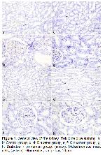
    Mast Cell Histochemical Findings: Mast cells stained with toluidine blue metachromatically were observed in the kidney tissues of both the control and experimental groups. While no morphological difference was observed between mast cells in all groups, they showed different sizes, round or oval shapes depending on their location. Mast cells were seen between the renal tubules, and especially around the renal corpuscle and the blood vessels. In the medulla, mast cells were found predominantly within the interstitium surrounding the tubules and collecting tubes, and in the connective tissue around.

    It was observed that the numbers of mast cells increased numerically in the experimental groups compared to the control group. When the mast cell numbers were compared, it was determined that there was a statistically significant difference between the mean number of mast cells in mm2 (P˂0.001) (Table 1). The highest increase in the mast cells was found in the diabetes group. When the cinnamon group was evaluated, there was an increase in mast cells compared to the control group. At the same time, there was a significant decrease in the number of mast cells in the diabetes + cinnamon group compared to the diabetes group (Figure 1).


    Büyütmek İçin Tıklayın
    Table 1: Mast cell counts after staining with toluidin blue in kidney tissue


    Büyütmek İçin Tıklayın
    Figure 1: General view of the kidney. Toluidine blue staining. a, b: Control group, c, d: Diabetes group, e, f: Cinnamon group, g, h: Diabetes + cinnamon group. (arrow): Metachromatic mast cells, (asterix): Glomerulus, range bar, 10 μm.

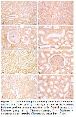
    TLR4 Immunohistochemical Findings: TLR4 immunoreactivity was observed in glomeruli, proximal, distal, loop of henle, and collecting tubules in kidney tissue of all groups. TLR4 expression was seen in the cells' cytoplasm and nucleus. Between the control and experimental groups, there was no difference in the location of intracellular TLR4 immunoreactivity. However, an increase in the intensity of TLR4 expression was determined between the control and experimental groups. Although it was observed that cinnamon increased TLR4 expression in kidney tissue, the highest TLR4 expression was found in the diabetic group as a remarkable finding in our study. It was determined that cinnamon extract reduced TLR4 immunoreactivity, which was increased in the diabetes group in diabetes + cinnamon group (Figure 2). Table 2 summarizes the semiquantitative analysis results of the intergroup for TLR4 immunohistochemistry staining.


    Büyütmek İçin Tıklayın
    Figure 2: Photomicrographs showing immunohistochemical labeling with TLR4 primary antibody in kidney. Brown staining indicates positive immune reactivity. a, b: Control group, c, d: Diabetes group, e, g: Cinnamon group, f, h: Diabetes + cinnamon group. (asterix): Glomerulus, range bar, 10 μm.


    Büyütmek İçin Tıklayın
    Table 2: Score of expression of TLR4 in the kidney tissue

  • Başa Dön
  • Özet
  • Giriş
  • Materyal ve Metot
  • Bulgular
  • Tartışma
  • Kaynaklar
  • Tartışma
    Mast cells are tissue-localized immune cells that play a key role in both innate and adaptive immunity as part of the physiological defense system 16. They have crucial roles in inflammation, autoimmunity, allergies, infectious diseases, and metabolic disorders 17. Chemokines, cytokines, growth factors, heparin, histamine, proteases, chymase, and tryptase, among other mast cell mediators, have a role in the pathophysiology of metabolic illnesses, including diabetes 18. In their study, Svensson et al. 19 showed that IgE levels increased in diabetes. At this point, this increase suggests that IgE-mediated mast cell activation may have an important effect on the pathogenesis of diabetes. Carlos et al. 20 reported that adaptive mast cell transfer before STZ administration confers resistance to diabetes and induces an increase in Treg cells in pancreatic lymph nodes. Also, they have stated that mast cells are associated with resistance to STZ-induced diabetes through an immunological tolerance mechanism mediated by Treg cells. In experimental studies, it was revealed that the number and activity of mast cells increased in the kidneys of rats with a diabetic model 21. In a study with diabetic rats, it was reported that the number of mast cells in the urinary bladder increased considerably. Furthermore, in the diabetic group in which Benfluorex - vitamin C treatment was investigated in the same study, fewer mast cells were found than in the diabetes group 22. Jones et al. 23 reported that the number of mast cell in the mesentery of diabetic rats increased statistically significantly compared to healthy rats. In the same study, it was revealed that antiallergic tranilast significantly reduced mast cell number in diabetic rats. It is well known that mast cells increase in diabetic rats, especially in the peripheral areas of the islets of Langerhans 24. Notedly, the previous study showed that a statistically significant increase in the number of mast cell in the ovarian and uterine tissues of experimental diabetic rats compared to the control group 25. In clinical and preclinical studies, it has been revealed that diabetes increases the density of mast cell and that agents that stabilize mast cells can improve diabetes experimentally 26. In addition, in our study, in which cinnamon extract also caused an increase in mast cells, it was observed that increased mast cells decreased in the diabetic group given cinnamon extract as a remarkable finding. This finding indicates that it might show an effective defense against possible damage to tissues and cells by diabetes through primary and secondary mediators in the granules of mast cells.

    It is known that TLR4 has critical importance in host defense against bacterial infections 27. TLR4 has also been found to protect against inflammatory tissue damage by promoting tissue repair and remodeling in tissue injury 28. Research has shown that metabolic disorders such as hyperglycemia will damage kidney cells and encourage cells to secrete and release inflammatory factors 29. Gulden et al. 30 reported that the development of diabetes was accelerated, and Treg cell activity decreased in parallel in TLR4-deficient mice. In terms of inflammatory factor expression, it is known that TLR4 endogenous and exogenous ligands stimulate renal tubular epithelial cells in the case of hyperglycemia 31. Therefore, it can be thought that the expression of TLR4 in hyperglycemia may induce the production of a rapid and strong inflammatory response by renal tubular epithelial cells 32. TLR4 activation has been shown in one study to partially prevent insulin-related insulin resistance caused by a high-fat diet 33. In addition, one study has shown that insulin treatment can reduce blood sugar and TLR4 protein expression 34. It has been shown that TLR4 expression was increased in experimentally diabetic rat kidneys. Moreover, this research indicated a significant difference in TLR4 expression in the glomerular basement membrane, proximal convoluted tubule, and renal interstitial area of the kidney in rats in the modeling group compared to the control group 35. In our study, in parallel with the above studies, an increase in TLR4 expression was observed in the kidney tissue in the diabetes groups and the cinnamon-treated groups. In addition, when compared to the diabetes group, TLR4 expression was lower in the diabetes + cinnamon group. Based on the current knowledge and the findings of this study, we suggest that cinnamon may contribute to strengthening the immune system and, in turn, indirectly treat diabetes.

    When mast cells are activated, they can release both newly synthesized lipid mediators, cytokines, chemokines, and preformed mediators like histamine from their granules. They then might cause an inflammatory response or participate in defensive responses due to this 36. The Fc receptors of mast cells express different receptors, including TLRs, chemokines, cytokines, as well as pathogen-associated molecular patterns of mast cell activation and immune responses 37. TLR4 receptors on the surface of human and mouse mast cells can regulate the FcRI-mediated mast cell signaling pathway in a synergistic manner, promoting mast cell degranulation and Th2 cytokine release 38. It has been demonstrated that TLR is expressed in both the cell membrane and cytosol of mast cells and can recognize viruses, bacteria, and fungi 39. A study reported that the number of mast cells and expression of TLR4 increased in the intestinal mucosa compared to healthy individuals with Crohn's disease 40. Huang et al. 41 stated that there were few scattered mast cells with TLR4 expression in healthy gingival tissues, but the number of mast cells and TLR4 positive mast cells increased significantly in the gingival tissues of the group with mild and severe periodontitis. In another study examining the relationship between mast cells and TLR4, it was stated that Giardia lamblia trophocytes, a parasite species, stimulated mast cells, and an increase in TLR4 expression was formed due to this stimulation 42. In a study using rats with necrotic enterocolitis in the ileum, it has been determined that breast milk oligosaccharides suppressed the mast cell number and TLR4 expression, and also reduced the damage in the ileum tissue 43. In our study, there was a parallel increase in the number of mast cells and expression of TLR4 in diabetes group and cinnamon group. Taken as a whole, this synergistic increase led us to suggest that mast cells might both secrete TLR4 and induce TLR4 expression.

    These findings showed that cinnamon increases mast cell number and expression of TLR4 in healthy rats. At the same time, it caused a synergistic decrease in increased mast cell and TLR4 expression in rats with diabetes. In conclusion, we think that cinnamon, by activating or stabilizing particular cells and cytokines, can help avoid cell and tissue damage that may arise due to metabolic problems.

  • Başa Dön
  • Özet
  • Giriş
  • Materyal ve Metot
  • Bulgular
  • Tartışma
  • Kaynaklar
  • Kaynaklar

    1) Hung HY, Qian K, Morris-Natschke SL, Hsu CS, Lee KH. Recent discovery of plant-derived anti-diabetic natural products. Nat Prod Rep 2012; 29: 580-606.

    2) Giovannini P, Howes MJ, Edwards SE. Medicinal plants used in the traditional management of diabetes and its sequelae in Central America: A review. J Ethnopharmacol 2016; 184: 58-71.

    3) Loren D, Roscoe R, Shack A. Complementary and alternative medicine for diabetes. Can J Diabetes 2018; 42: 154-161.

    4) Ranasinghe P, Pigera S, Premakumara G, et al. Medicinal properties of “true” cinnamon (cinnamomum zeylanicum): A systematic review. BMC Complement Altern Med 2013; 13: 1-10.

    5) Ziegenfuss TN, Hofheins JE, Mendel RW, Landis J, Anderson RA. Effects of a water-soluble cinnamon extract on body composition and features of the metabolic syndrome in pre-diabetic men and women. J Int Soc Sports Nutr 2006; 3: 45-53.

    6) Ragipoglu D, Dudeck A, Haffner-Luntzer M, et al. The role of mast cells in bone metabolism and bone disorders. Front Immunol 2020; 11: 1-16.

    7) Draberova L, Tumova M, Draber P. Molecular mechanisms of mast cell activation by cholesterol-dependent cytolysins. Front Immunol 2021; 12: 1-15.

    8) O’Neill LAJ. How Toll-like receptors signal: What we know and what we don’t know. Curr Opin Immunol 2006; 18: 3-9.

    9) Lee MB, Lee JH, Hong SH, et al. JQ1, a BET inhibitor, controls TLR4-induced IL-10 production in regulatory B cells by BRD4-NF-κB axis. BMB Rep 2017; 50: 640-646.

    10) Çetin A, Vardı N, Orman D. Deneysel diyabetin sıçan kalp dokusunda meydana getirdiği histolojik değişiklikler üzerine aminoguanidinin iyileştirici etkileri. İnönü Üniversitesi Sağlık Hizmetleri Meslek Yüksek Okulu Dergisi 2013; 1: 17-26.

    11) Hemayatkhah JV, Baqerzadeh P. The effect of cinnamon extract on ovary in adult rats treated lead acetate. Journal Of Anımal Biology 2017; 9: 1-9.

    12) Enerback L. Mast cells in rat gastrointestinal mucosa: II. Dye-binding and metachromatic properties. Acta Pathol Microbiol Scand 1966; 66: 303-312.

    13) John PWM. Statistical Design and Analysis of Experiments. 1st Edition, NewYork: Macmilllan, 1971.

    14) True LD. Principles of Immunohistochemistry. New York: Gower Medical Publishing, 1990.

    15) Chatterjee S, Gangula PR. Dong YL, Yallampalli C. Immunocytochemical localization of nitric oxide synthase- III in reproductive organs of female rats during the oestrous cycle. Histochem J 1996; 28: 715-723.

    16) Wernersson S, Pejler G. Mast cell secretory granules: Armed for battle. Nat Rev Immunol 2014; 14: 478-494.

    17) Huh JY, Park YJ, Ham M, Kim JB. Crosstalk between adipocytes and immune cells in adipose tissue inflammation and metabolic dysregulation in obesity. Mol Cells 2014; 37: 365-371.

    18) Zhang J, Shi GP. Mast cells and metabolic syndrome. Biochim Biophys Acta 2012; 1822: 14-20.

    19) Svensson J, Eising S, Mortensen HB, et al. High levels of immunoglobulin E and a continuous increase in immunoglobulin G and immunoglobulin M by age in children with newly diagnosed type 1 diabetes. Hum Immunol 2012; 73: 17-25.

    20) Carlos D, Juliana NUY, Rocha FA, et al. Mast cells control insulitis and increase Treg cells to confer protection against STZ-induced type 1 diabetes in mice. Eur J Immunol 2015; 45: 2873-2885.

    21) de Morais RB, do Couto Muniz VP, Costa EN, et al. Mast cell population in the development of diabetic nephropathy: Effects of renin angiotensin system inhibition. Biomed Pharmacother 2018; 107: 1115-1118.

    22) Özkorkmaz EG. Streptozotosin Diyabetik ve Benfluoreks-C Vitamini Tedavili Sıçanların Mesane Dokusunda Histolojik İncelemeler. Doktora Tezi, Ankara: Ankara Üniversitesi, Fen Bilimleri Enstitüsü, 2008.

    23) Jones SE, Gilbert RE, Kelly, DJ. Tranilast reduces mesenteric vascular collagen deposition and chymase-positive mast cells in experimental diabetes. J Diabetes Complications 2004; 18: 309-315.

    24) Abdel-Hamid AAM, Firgany AEL. Increased mast cell number is associated with a decrease in beta-cell mass and regeneration in type 2 diabetic rats. Acta Histochem 2019; 12: 508-515.

    25) Hayıroğlu AE, Karaca T, Demirtaş S. Streptozotosin ile deneysel diyabet oluşturulan sıçanlarda östrus siklusunun değişik evrelerinde ovaryum ve uterus dokularında mast hücrelerinin dağılımının histokimyasal ve immünohistokimyasal olarak incelenmesi. Kafkas Tıp Bilimleri Dergisi 2016; 6: 29-37.

    26) Pichler R, Afkarian M, Dieter BP, Tuttle KR. Immunity and inflammation in diabetic kidney disease: Translating mechanisms to biomarkers and treatment targets. Am J Physiol Renal Physiol 2017; 312: 716-731.

    27) Freudenberg MA, Tchaptchet S, Keck S, et al. Lipopolysaccharide sensing an important factor in the innate immune response to Gram-negative bacterial infections: Benefits and hazards of LPS hypersensitivity. Immunobiology 2008; 213: 193-203.

    28) Jiang D, Liang J, Noble PW. Regulation of non-infectious lung injury, inflammation, and repair by the extracellular matrix glycosaminoglycan hyaluronan. Anat Rec 2010; 293: 982-985.

    29) On'kin JBKL, Longo Mbenza B, Tchokonte Nana V, Okwe AN, Kabangu NK. Hyperbolic relation between beta cell function and insulin sensitivity for type 2 diabetes mellitus, malaria, influenza, Helicobacter pylori, Chlamydia pneumoniae, and hepatitis C virus infection induced inflammation/oxidative stress and temporary insulin resistance in Central Africans. Turk J Med Sci 2017; 19: 1834-1841.

    30) Gulden E, Ihira M, Ohashi A, et al. Toll-like receptor 4 deficiency accelerates the development of insulin-deficient diabetes in non-obese diabetic mice. PLoS ONE 2013; 89: e75385.

    31) Perry BD, Rahnert JA, Xie Y, et al. Palmitate‑induced ER stress and inhibition of protein synthesis in cultured myotubes does not require Toll‑like receptor 4. PLoS ONE 2018; 13: e0191313.

    32) Chen G, Sun W, Liang Y, et al. Maternal diabetes modulates offspring cell proliferation and apoptosis during odontogenesis via the TLR4/NF‑κB signalling pathway. Cell Prolif 2017; 50: e12324.

    33) Shi H, Kokoeva MV, Inouye K, et al. TLR4 links innate immunity and fatty acid-induced insulin resistance. Clin Invest 2006; 116: 3015-3025.

    34) Ghanim H, Mohanty P, Deopurkar R, et al. Acute modulation of toll-like receptors by insulin. Diabetes Care 2008; 319: 1827-1831.

    35) Liu S-Y, Nie X-Z, Zhou W-Y, Chen J. Expression and effect of TLR4 in rats with diabetic nephropathy. Asian Pac J Trop Med 2013; 68: 635-639.

    36) Shelburne CP, Abraham SN. The mast cell in innate and adaptive immunity. Adv Exp Med Biol 2011; 716: 162-185.

    37) Krystel-Whittemore M, Dileepan KN, Wood JG. Mast cell: A multi-functional master cell. Front Immunol 2016; 6: 620.

    38) Novak N, Bieber T, Peng WM. The immunoglobulin E-Toll-like receptor network. Int Arch Allergy Immunol 2010; 151: 1-7.

    39) Kılınç E, Baranoğlu Y. Mast cell stabilizers as a supportive therapy can contribute to alleviate fatal inflammatory responses and severity of pulmonary complications in COVID-19 infection. Anadolu Kliniği Tıp Bilimleri Dergisi 2020; (COVID 19 Özel Sayısı): 111-118.

    40) Kobayashi R, Okamura S, Ohno T, et al. Hyperexpression of FcgammaRI and Toll-like receptor 4 in the intestinal mast cells of Crohn's disease patients. Clin Immunol 2007; 125: 149-158.

    41) Huang B, Dai Q, Huang S-G. Expression of Toll‑like receptor 4 on mast cells in gingival tissues of human chronic periodontitis. Mol Med Rep 2018; 175: 6731-6735.

    42) Muñoz-Cruz S, Gomez-García A, Matadamas-Martínez F, et al. Giardia lamblia: Identification of molecules that contribute to direct mast cell activation. Parasitol Res 2018; 1178: 2555-2567.

    43) He-Yang J, Zhang W, Liu J, Xue P, Zhou X. Human breast milk oligosaccharides attenuate necrotizing enterocolitis in rats by suppressing mast cell accumulation, DPPI activity and TLR4 expression in ileum tissue, and regulating mitochondrial damage of Caco-2 cells. Int Immunopharmacol 2020; 88: 106881.

  • Başa Dön
  • Özet
  • Giriş
  • Materyal ve Metot
  • Bulgular
  • Tartışma
  • Kaynaklar
  • [ Başa Dön ] [ Özet ] [ PDF ] [ Benzer Makaleler ] [ Yazara E-Posta ] [ Editöre E-Posta ]
    [ Ana Sayfa | Editörler | Danışma Kurulu | Dergi Hakkında | İçindekiler | Arşiv | Yayın Arama | Yazarlara Bilgi | E-Posta ]