[ Ana Sayfa | Editörler | Danışma Kurulu | Dergi Hakkında | İçindekiler | Arşiv | Yayın Arama | Yazarlara Bilgi | E-Posta ]
Fırat Üniversitesi Sağlık Bilimleri Veteriner Dergisi
2023, Cilt 37, Sayı 1, Sayfa(lar) 026-031
[ Özet ] [ PDF ] [ Benzer Makaleler ] [ Yazara E-Posta ] [ Editöre E-Posta ]
Dermatofitozisli Sığırlarda 25-Hidroksivitamin D ve Paratiroid Hormon Konsantrasyonları
Şükrü DEĞİRMENÇAY1, M. Sertaç EROĞLU1, Akın KIRBAŞ2, M. Cemal ADIGÜZEL3
1Atatürk University, Faculty of Veterinary Medicine, Department of Internal Medicine, Erzurum, TÜRKİYE
2Yozgat Bozok University, Faculty of Veterinary Medicine, Department of Internal Medicine, Yozgat, TÜRKİYE
3Atatürk University, Department of Microbiology, Faculty of Veterinary Medicine, Erzurum, TÜRKİYE
Anahtar Kelimeler: Dermatofitozis, 25-hidroksivitamin D, paratiroid hormon, sığır
Özet
Bu çalışmada, dermatofitozisli sığırlarda serum 25-hidroksi vitamin D [25-(OH)D] ile paratiroid hormon (PTH), kalsiyum (Ca) ve fosfor (P) düzeylerindeki değişikliklerin araştırılması amaçlandı. Çalışma materyalini yaşları 6-8 aylık olan 28 dermatofitozisli (enfekte grup) ve 14 sağlıklı sığır (kontrol grubu) oluşturdu. Sonuçlarımıza göre kontrol grubuyla karşılaştırıldığında, enfekte grup anlamlı olarak daha düşük serum 25-(OH)D seviyelerine (P=0.001) sahipken, daha yüksek PTH, Ca ve P seviyelerine sahipti. Ayrıca serum 25-(OH)D düzeyleri lökosit (WBC) ve nötrofil (NEU) sayıları ile negatif korelasyona sahipti. Enfekte grup lezyonların dağılımına göre hafif ve şiddetli olmak üzere iki alt gruba ayrıldı. Kontrol ve hafif gruplarla karşılaştırıldığında, şiddetli grup en yüksek WBC (P=0,007) ve NEU (P=0,005) değerlerine ve en düşük 25-(OH)D düzeylerine (P=0,000) sahipti. Sonuç olarak dermatofitozisli sığırlarda D vitamini düzeylerinin önemli ölçüde düştüğü ve bu durumun bağışıklığı baskılayarak hastalığın kötüleşmesine yol açtığı sonucuna varıldı. PTH yüksekliği, D vitamini eksikliğini telafi etmek için vücudun enfeksiyona verdiği yanıt olarak değerlendirildi. Bu bulgular ışığında D vitamininin, hastalığın patogenezinde rol oynadığı ve tedavinin vazgeçilmez bir bileşeni olduğu düşünüldü.
  • Başa Dön
  • Özet
  • Giriş
  • Materyal ve Metot
  • Bulgular
  • Tartışma
  • Kaynaklar
  • Giriş
    Dermatophytosis, also known as Tinea or Ringworm, is a fungal infection in the cell layer of the epidermis 1. Domestic animals including cattle, cats, and dogs as well as laboratory animals like rabbits are frequently affected by this superficial zoonotic infection 2. In the etiology of the disease, Trichophyton spp., Epidermophyton spp. and Microsporum spp. types are included 1. However, especially Trichophyton verrucosum, Trichophyton mentagrophytes and Trichophyton megnini cause the disease 2. Lesions with the shape of a round, white-grey crust are common in keratinized tissues like the skin, nails, horns, and feet 3. In cattle, the lesions generally appear on the skin around the head and neck. Animals exhibit alopecia and thickening in response to autolysis of the stratum corneum and fiber structure 4.

    Cattle dermatophytosis outbreaks are most common in the fall and winter and are caused by contact with contaminated areas such as crowded cattle barns and mangers. Lesions ranging from moderate to severe may develop after infection in the host. Weak, young, and especially immunosuppressed animals are more prone to infection due to stress, such as transport or weaning, and more severe skin lesions occur 5. The disease has negative effects on animal growth, skin integrity, and the production of meat and milk, and it is more common in young animals than in adults 6. Vitamin D has a crucial role in several processes, including immune system regulation, keratinocyte turnover, and maintaining the integrity of the epidermal barrier 7. In vitamin D deficiency, the absorption of calcium (Ca) from the intestines decreases. Decreased Ca ion increases the release of parathyroid hormone(PTH). While PTH acts on bone, kidney and intestine, it allows the passage of Ca into the blood and reduces the reabsorption of phosphate 8. The body's primary source of vitamin D is the epidermis. Under normal conditions, the body may produce enough vitamin D (from particular precursors) when exposed to ultraviolet (UV)-B radiation 7. It has been stated that the vitamin D requirement of cattle can be met by nutrition and exposure to sunlight 9. The synthesis of vitamin D from the skin, however, declines throughout the winter 10.

    As is known, Dermatophytosis is a self-limiting disease, but several factors like immunosuppression, stress, overcrowding, illness, poor nutrition and age predispose animals to infection 11. Considering the important effects of vitamin D on the integrity of the epidermal barrier and the regulation of the immune system, we hypothesized that serum 25-hydroxy vitamin D [25-(OH)D] levels would be quite low in cattle with dermatophytosis. In addition, with this study, we aimed to investigate the changes in 25-(OH)D, PTH, Ca and phosphorus (P) levels, which are closely related to each other, in cattle with dermatophytosis.

  • Başa Dön
  • Özet
  • Giriş
  • Materyal ve Metot
  • Bulgular
  • Tartışma
  • Kaynaklar
  • Materyal ve Metot
    Research and Publication Ethics: This study was performed in accordance with the approved ethical rules of Atatürk University (protocol no. 2021/1, decision number: 30) and for each cattle written informed consent was obtained from the owner.

    Animals and Protocol Design: The material of the study consisted of 42 cattle of 6-8 months old, Simmental and Brown Swiss breeds, and both genders. According to clinical examination findings and blood test results, the cattle were divided into two groups: Dermatophytosis (infected, n=28), and healthy (control, n=14). Cattle with dermatophytosis were divided according to the distribution of their lesions as mild (n=14) and severe (n=14). Thus, a second grouping was made. Cattle with any disease symptoms other than dermatophytosis were not included in the study.

    Mycological Examination: Cattle with circular, white, topical, or dense alopecic, and bran lesions were included in the study. While sampling from the skin lesions of the animals, the conditions of asepsis and antisepsis were complied with by using alcohol. Skin scraping samples were taken using a sterile scalpel tip and a sterile plastic brush using the brushing technique 12 and cultured on Sabouraud Dextrose Agar containing 0.5% chloramphenicol (HiMedia, Mumbai, India). Cultures were incubated at 25±1 ºC for up to three weeks and controlled periodically for fungal growth. The identification of isolated colonies was made according to the dermatophytes identification scheme. The macroscopic examination of colonies was recorded then cultures were stained with lactophenol cotton blue for microscopic identification and then visualized under the microscope. In the microscopic examination, the presence of septa, shape, size of hyphae, and conidial cells were evaluated under a light microscope for examination 13.

    Blood Sampling: For haematological and biochemical investigations, blood samples from all the calves were taken from the vena jugularis externa and placed in tubes containing EDTA (Vacutainer, K2E 3.6 mg, BD, UK) and gel (Vacutainer, BD, UK). Blood samples were centrifuged at 3000 rpm for 10 minutes after waiting at room temperature in gel tubes. The sera were stored at -80°C until biochemical analysis.

    Hematological Analyses: A hematology analyser was used to measure the cattle's white blood cell (WBC), lymphocyte (LYM), monocyte (MON), neutrophil (NEU), eosinophil (EOS), red blood cell (RBC), and hemoglobin (HGB) counts, and hematocrit (HCT) and platelet (PLT) levels (Abacus Junior Vet5, Hungary). The haematological assays were completed immediately.

    Biochemical Analyses: Serum 25-(OH)D and PTH concentrations were determined by the electrochemiluminescence immunoassay (ECLIA) method using a chemistry analyzer (Cobas e801; Roche Diagnostics, Switzerland). A biochemistry auto analyser (Beckman Coulter, AU5800, USA) was used to analyse serum Ca and P levels using commercial kits.

    Statistical Analyses: For statistical analysis, SPSS software (Version 25.0, SPSS Inc., Chicago, IL, USA) was utilized 14. To evaluate the data distribution between the groups, the Kolmogorov-Smirnov test was utilized (Infected and Control groups). Parameter comparisons were made using the Independent-Samples t-test between healthy cattle and cattle with dermatophytosis. One-way analysis of variance (ANOVA) and the post hoc Duncan test were used for statistical analysis of the subgroups; control, mild, and severe groups. The Pearson Correlation test was used to measure the correlation between the parameters. All results were presented as mean ± standard deviation (SD), and P<0.05 was used for all statistical comparisons.

  • Başa Dön
  • Özet
  • Giriş
  • Materyal ve Metot
  • Bulgular
  • Tartışma
  • Kaynaklar
  • Bulgular
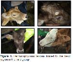
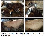
    Clinical Findings: Most cattle with dermatophytosis also had dandruff, topical alopecia, and circular, dense, white, and erythematous lesions that spread throughout the body, particularly in the head and neck region. Lesions in the mild group were typically limited to the head (Figure 1), whereas lesions in the severe group spread to the head, neck, and back (Figure 2).


    Büyütmek İçin Tıklayın
    Figure 1: Dermatophytosis lesions limited to the head region in the mild group


    Büyütmek İçin Tıklayın
    Figure 2: Widespread dermatophytosis lesions in the severe group

    Fungal Culture Findings: Trichophyton spp. colonies was visualized like a powdery to granular surface, was white to cream in color, flat, and reversed golden brown to reddish-brown in Sabouraud Dextrose Agar plate.

    Hematological Findings: According to the hematologic results, the infected cattle's MON (P=0.006) and EOS (P=0.016) values were lower while WBC, LYM, NEU, and PLT values were higher than those of the control group (Table 1). The highest WBC (P=0.007) and NEU (P=0.005) values were found in the severe group compared to the control and mild groups (Table 2).


    Büyütmek İçin Tıklayın
    Table 1: Comparison of haematological and biochemical results between healthy cattle and cattle with dermatophytosis


    Büyütmek İçin Tıklayın
    Table 2: Comparison of haematological and biochemical parameters of calves in control, mild and severe groups

    Biochemical Findings: Serum 25-(OH)D levels were significantly lower in cattle with dermatophytosis than in healthy cattle (P=0.001). On the other hand, PTH, Ca and P levels were high, but this increase was not statistically significant (Table 1). Compared to the control group, vitamin D levels were quite low in both the mild and severe groups, but the severe group had the lowest levels (P=0.000). Mild and severe groups had higher PTH, Ca, and P levels than the control group, but only the severe group's Ca value was statistically significant (P=0.041) (Table 2).

    Serum 25-(OH)D levels were moderately negatively correlated with P (r=-0.455, P=0.002) and NEU (r=-0.447, P=0.003) while weakly negatively correlated with WBC (r=-0.389, P=0.011). PTH levels were moderately positively correlated with P (r=0.432, P=0.004) (Table 3).


    Büyütmek İçin Tıklayın
    Table 3: Correlation results between 25-(OH)D and PTH, Ca, P, WBC, LYM, and NEU levels in dermatophytosis and healthy calves (Pearson correlation analysis)

  • Başa Dön
  • Özet
  • Giriş
  • Materyal ve Metot
  • Bulgular
  • Tartışma
  • Kaynaklar
  • Tartışma
    In this study, serum 25-(OH)D, PTH, Ca, and P levels were investigated in cattle with dermatophytosis. We determined that 25-(OH)D levels were significantly reduced in cattle with dermatophytosis. The current study results showed that increased Ca, P, and PTH levels were not significant statistically.

    Vitamin D has effects on keratinocyte proliferation and differentiation, apoptosis, barrier maintenance and immunomodulatory effects on the skin 15. Previous research reported that vitamin D levels are low in dermatophytosis cases in humans 16 and cattle 17. Similarly, in this study, we found very low serum 25-(OH)D levels in cattle with dermatophytosis. There was a negative correlation between serum 25-(OH)D levels and WBC and NEU levels. Furthermore, the lowest vitamin D levels and the highest WBC and NEU levels were found in the severe group. Thus, it is possible to argue that low vitamin D levels suppress the immune system of the skin and contribute to the development and progression of dermatophytosis lesions. In line with this, vitamin D has been shown to enhance cathelicidin synthesis in human keratinocytes 18. Cathelicidin, on the other hand, is a key effector molecule of innate immunity in the skin and increased epidermal cathelicidin levels could result in a reduction in cutaneous inflammasome activity 19. According to a study, cathelicidin expression in lesional skin was higher in serum vitamin D sufficient groups compared to serum vitamin D deficient groups 20. Topically applied vitamin D analogues like calcipotriol reduce inflammation and morphological changes in psoriatic lesions 21. At the same time, calcipotriol treatment reduces pro-inflammatory cytokines while significantly increasing cathelicidin expression 22. Based on these findings, it is possible to infer that low vitamin D levels in cattle with dermatophytosis will indirectly weaken the skin's immune system, thereby exacerbating dermatophytosis lesions. In the current study, which supports this inference, we found that the animals in the severe group had the lowest vitamin D levels. Of course, the use of vitamin D in treatment can be argued to be beneficial. In support of our view, the occurrence of fungal infections in young and immunocompromised cattle reveals the relationship between 25-(OH)D and the immune system 23. The use of vitamin D in the combat against infectious diseases has also been found to have therapeutic benefits 16.

    Mice with bacterial skin disease that are deficient in vitamin D in their diets have been found more susceptible to skin infections. It has been proposed that these mice respond to infection by producing more PTH to compensate for the lack of vitamin D because applying PTH to mouse skin reduced susceptibility to group A Streptococcus skin infection 24. Similarly, in this study, while vitamin D levels were lower in cattle with dermatophytosis compared to the control group, PTH levels were higher. Accordingly, this can be interpreted as these cattle respond to infection by increasing their PTH levels to compensate for insufficient vitamin D. Supporting our view, PTH has been shown to increase 1,25-D3 levels in keratinocytes 25, induce cathelicidin antimicrobial peptide response and enhance immune defense 26. This reveals that the secretion of PTH, which is normally known to be triggered by the low Ca levels caused by the decreased vitamin D 26, is also secreted in order to eliminate the disadvantage caused by low vitamin D in the skin's immune system. On the other hand, it has been shown that PTH-related peptides (PTHrP), expressed in the skin and other tissues, modulate intracellular calcium. Calcium has been implicated as an early signal in the differentiation of keratinocytes 27. Additionally, the expression of antimicrobial skin peptides is increased in keratinocytes under high calcium circumstances 28,29. Sezer et al. 30 found high blood Ca levels in weaned calves with dermatophytosis. Similarly, our study showed that the blood Ca levels of cattle with dermatophytosis were greater than those of healthy cattle. This situation can be evaluated as an immune response of the skin against dermatophytosis lesions.

    As a result, it was determined that vitamin D levels decreased and PTH levels increased in cattle with dermatophytosis. The vitamin D levels of cattle with extensive dermatophytosis lesions were found to be significantly lower. The conclusion was drawn that PTH is secreted at higher levels to compensate for low vitamin D levels, which is believed to have a negative effect on the skin's immune system. Due to the aforementioned beneficial effects, it was thought that the combined use of vitamin D and PTH in the treatment of cattle with dermatophytosis would be beneficial and this situation should be investigated with further studies.

  • Başa Dön
  • Özet
  • Giriş
  • Materyal ve Metot
  • Bulgular
  • Tartışma
  • Kaynaklar
  • Kaynaklar

    1) Guo Y, Ge S, Luo H, et al. Occurrence of Trichophyton verrucosum in cattle in the Ningxia Hui autonomous region, China. BMC Vet Res 2020; 16: 187.

    2) Agnetti F, Righi C, Scoccia E, et al. Trichophyton verrucosum infection in cattle farms of Umbria (Central Italy) and transmission to humans. Mycoses 2014; 57: 400-405.

    3) Al-Ani FK, Younes FA, Al-Rawashdeh OF. Ringworm infection of cattle and horses in Jordan. Acta Vet Brno 2002; 71: 55-60.

    4) Birdane FM, Kabu M. The use of two different vaccines in the treatment of Anatolian buffaloes with trichophytosis. Indian J Anim Res 2017; 52: 1350-1352.

    5) Yılmazer R, Aslan Ö. The investigation of efficacy of neguvon-whitfield’s ointment combinations for the treatment of ringworm in cattle. J Heal Sci 2010; 19: 175-183.

    6) Erel O. A novel automated direct measurement method for total antioxidant capacity using a new generation, more stable ABTS radical cation. Clin Biochem 2004; 37: 277-285.

    7) Mattozzi C, Paolino G, Richetta AG, Calvieri S. Psoriasis, vitamin D and the importance of the cutaneous barrier’s integrity: An update. J Dermatol 2016; 43: 507-514.

    8) Khundmiri SJ, Murray RD, Lederer E. PTH and vitamin D. Compr Physiol 2016; 6: 561-601.

    9) Huffman C, Duncan C, Lightfoot CC. Vitamin D studies in cattle. J Dairy Sci 1935; 18: 511-526.

    10) Hymøller L, Jensen SK, Lindqvist H, et al. Supplementing dairy steers and organically managed dairy cows with synthetic vitamin D3 is unnecessary at pasture during exposure to summer sunlight. J Dairy Res 2009; 76: 372-378.

    11) Samanta I. Cutaneous, Subcutaneous and Systemic Mycology. In: Samanta I. (Editor). Veterinary Mycology. 1st Edition, New Delhi: Springer 2015: 11-153.

    12) Mackenzie DW. “Hairbrush Diagnosis” in detection and eradication of non-fluorescent scalp ringworm. Br Med J 1963; 2: 363-365.

    13) Diren Sığırcı B, Metiner K, Çelik B, et al. Dermatophytes isolated from dogs and cats suspected dermatophytoses in Istanbul, Turkey within a 15-year-period: An updated report. Kocatepe Vet J 2019; 12: 116-121.

    14) IBM Corp. Released 2017. IBM SPSS Statistics for Windows, Version 25.0. Armonk, NY: IBM Corp.

    15) Bikle DD. Vitamin D and the skin: Physiology and pathophysiology. Rev Endocr Metab Disord 2012; 13: 3-19.

    16) Çifci N. Retrospective evaluation of effects of vitamin D levels on skin diseases. Kocaeli Med J 2018; 7: 47-54.

    17) Alıç Ural D, Erdoğan S. Determination of the relationship between dermatophytosis disease activity and serum 25 (OH) D3 vitamin levels in limited cattle population under field conditions. MAKÜ Sag Bil Enst Derg 2019; 7: 132-138.

    18) Schauber J, Gallo RL. The vitamin D pathway: A new target for control of the skin’s immune response? Exp Dermatol 2008; 17: 633-639.

    19) Reinholz M, Ruzicka T, Schauber J. Cathelicidin LL-37: an antimicrobial peptide with a role in inflammatory skin disease. Ann Dermatol 2012 ; 24: 126-135.

    20) Kim SK, Park S, Lee E-S. Toll-like receptors and antimicrobial peptides expressions of psoriasis: Correlation with serum vitamin D level. J Korean Med Sci 2010; 25: 1506-1512.

    21) Lebwohl M, Menter A, Koo J, Feldman SR. Combination therapy to treat moderate to severe psoriasis. J Am Acad Dermatol 2004; 50: 416-430.

    22) Peric M, Koglin S, Dombrowski Y, et al. Vitamin D analogs differentially control antimicrobial peptide/ “Alarmin” expression in psoriasis. PLoS One 2009; 4: e6340.

    23) Gudding R, Lund A. Immunoprophylaxis of bovine dermatophytosis. Can Vet J 1995; 36: 302-306.

    24) Muehleisen B, Bikle DD, Aguilera C, et al. PTH/PTHrP and vitamin D control antimicrobial peptide expression and susceptibility to bacterial skin infection. Sci Transl Med 2012; 4: 135ra66.

    25) Bikle DD, Nemanic MK, Gee E, Elias P. 1,25-dihydroxyvitamin D3 production by human keratinocytes. Kinetics and regulation. J Clin Invest 1986; 78: 557-566.

    26) Christakos S, Ajibade DV, Dhawan P, Fechner AJ, Mady LJ. Vitamin D: Metabolism. Endocrinol Metab Clin North Am 2010; 39: 243-253.

    27) Abou-Samra AB, Jüppner H, Force T, et al. Expression cloning of a common receptor for parathyroid hormone and parathyroid hormone-related peptide from rat osteoblast-like cells: A single receptor stimulates intracellular accumulation of both cAMP and inositol trisphosphates and increases intracel. Proc Natl Acad Sci U S A 1992; 89: 2732-2736.

    28) Liu AY, Destoumieux D, Wong AV, et al. Human beta-defensin-2 production in keratinocytes is regulated by interleukin-1, bacteria, and the state of differentiation. J Invest Dermatol 2002; 118: 275-281.

    29) Morizane S, Yamasaki K, Kabigting FD, Gallo RL. Kallikrein expression and cathelicidin processing are independently controlled in keratinocytes by calcium, vitamin D3, and retinoic acid. J Invest Dermatol 2010; 130: 1297-1306.

    30) Sezer K, Hanedan B, Ozcelik M, Kirbas A. Assessment of oxidative stress, trace elements, serum biochemistry, and hormones levels in weaned calves with dermatophytosis. J Hell Vet Med Soc 2021; 72: 2653-2660.

  • Başa Dön
  • Özet
  • Giriş
  • Materyal ve Metot
  • Bulgular
  • Tartışma
  • Kaynaklar
  • [ Başa Dön ] [ Özet ] [ PDF ] [ Benzer Makaleler ] [ Yazara E-Posta ] [ Editöre E-Posta ]
    [ Ana Sayfa | Editörler | Danışma Kurulu | Dergi Hakkında | İçindekiler | Arşiv | Yayın Arama | Yazarlara Bilgi | E-Posta ]