[ Ana Sayfa | Editörler | Danışma Kurulu | Dergi Hakkında | İçindekiler | Arşiv | Yayın Arama | Yazarlara Bilgi | E-Posta ]
Fırat Üniversitesi Sağlık Bilimleri Veteriner Dergisi
2023, Cilt 37, Sayı 1, Sayfa(lar) 049-053
[ Özet ] [ PDF ] [ Benzer Makaleler ] [ Yazara E-Posta ] [ Editöre E-Posta ]
Gebelik Toksemisi Olan Keçilerde Vitamin B12 ve Folik Asit Düzeylerinin Araştırılması
Pelin Fatoş POLAT DİNÇER1,Kenan Çağrı TÜMER2
1Dokuz Eylül University, Faculty of Veterinary Medicine, Department of Internal Medicine, İzmir, TÜRKİYE
2Kastamonu University, Faculty of Veterinary Medicine, Department of Internal Medicine, Kastamonu, TÜRKİYE
Anahtar Kelimeler: Gebelik toksemisi, halep keçisi, glukoz, folat, B12Pregnancy toxemia, aleppo goat, glucose, folate, B12
Özet
Bu çalışmada gebelik toksemili halep keçilerinde beta hidroksi butirik asit (BHBA), vitamin B12, folat ve glikoz seviyeleri belirlenip hastalık ile ilişkisinin ortaya konulması amaçlandı. Bu amaçla tahmini gebeliğin son 6 haftasında bulunan, 1-4 yaş aralığında toplam 54 Halep keçisi çalışmaya dahil edildi. Tüm keçilerin klinik muayeneleri ve BHBA ölçümleri yapıldı. Keçiler kan β-hidroksibütirat (BHBA) değerlerine göre ketonemik olmayan (<0.8 mmol/L, n= 15), orta derecede hiperketonemik (0.8-1.6 mmol/L, n= 13) ve şiddetli hiperketonemik (>1.6 mmol/L, n= 26) olarak sınıflandırıldı. BHBA değerinde ketonemik olmayan grup ile orta derecede hiperketonemik grup arasında anlamlı fark bulunamazken (P>0.05), ketonemik olmayan grup ile şiddetli hiperketonemik grup arasında anlamlı fark tespit edildi (P<0.001). Vitamin B12 konsantrasyonu üç grup arasındaki fark anlamlı tespit edildi (P<0.005). Folate ve glukoz konsantrasyonu ise 3 grup arasındaki fark anlamlı bulunmadı (P>0.05). Gebelik toksemisi olan keçilerde değişkenler arasındaki korelasyon grafiğinde BHBA konsantrasyonu ile vitamin B12 (r=0.392, P<0.05) ve glikoz (r=0.394, P<0.05) arasında pozitif korelasyon gözlendi. Ayrıca vitamin B12 konsantrasyonu ile folat (r=0.206, P<0.05) ve glikoz (r=0.323, P<0.05) arasında pozitif korelasyon gözlendi. Sonuç olarak, elde edilen bulgular hiperketoneminin serum B12 ve glukoz konsantrasyonları ile ilişkili olduğunu düşündürmektedir. Gebelik toksemisi olan keçilerde B12 yüksekliğinin altında yatan mekanizmayı anlamak için ilave çalışmalara ihtiyaç duyulmaktadır.
  • Başa Dön
  • Özet
  • Giriş
  • Materyal ve Metot
  • Bulgular
  • Tartışma
  • Kaynaklar
  • Giriş
    Pregnancy toxemia occurs in over-conditioned sheep and goats carrying twins, triplets, or large fetuses during last 4-6 weeks of pregnancy 1. Goats are socioeconomically significant animals because they can adapt to a wide range of climatic conditions, can be grazed even in stony places that cannot be appraised in any other manner, and are especially preferred by family companies 2. Due to the herd-based nature of pregnancy toxemia, it results in significant economic losses due to the loss of both goats and offspring when preventive and treatment are not carried out appropriately. In the early stages of the disease, the majority of animals are asymptomatic or exhibit anorexia, dry and mucous feces, ataxia, and blindness. Some animals may exhibit convulsions and respiratory distress in the last stages, along with an inability to stand or lateral recumbency. If timely and proper treatment is not provided, the mortality rate is around 80% 3,4. The underlying mechanism of pregnancy toxemia is the inability of the pregnant ewe to meet the increased energy demands of the growing fetus during the final stages of pregnancy. To meet energy imbalance, body fat stores are breakdown to produce sufficient energy by gluconeogenesis. However excessive lipolytic activity results in an increase of keton bodies in the circulation. Fetuses cannot synthesis glucose to meet their energy requirements and are therefore dependent on glucose given by the maternal circulation 4,5. The underlying mechanism of pregnancy toxemia is the inability of the pregnant ewe to meet the increased energy demands of the growing fetus during the final stages of pregnancy. Body fat stores are broken down to produce sufficient energy. However excessive lipolysis results in an increase of ketone bodies in the circulation 6-8.

    Vitamin B12, also known as cyanocobalamin, is a water-soluble vitamin and is an important part of enzyme systems involved in multiple metabolic pathways and energy production, mainly through rumen fermentation 9,10. This vitamin functions as a coenzyme and is involved in energy metabolism and cell replication 11. In particular, methylmalonyl CoA mutase is a cobalamine-dependent mitochondrial enzyme and plays a regulatory role in gluconeogenesis and fatty acid oxidation in ruminants 12. If the cobalt concentration in the ruminal fluid is below 0.5 mg/mL, the ruminal synthesis of vitamin B12 by ruminal flora is inhibited and its passage into blood and other tissues is reduced 10,13. When ration does not contain cobalt, B12 production in the rumen decreases within a few days, however, the vitamin B12 stored in the liver is enough for a few days 14. Synthesized cobalamin is absorbed from intestine into the the portal circulation and transported to the liver with folates. While some of the folates are utilized by liver cells, others are methylated and then released into the bloodstream for use by peripheral tissues 15. Similar to vitamin B12, folate is also a water-soluble vitamin, and has vital functions such as protein synthesis and red blood cell production 16. Unlike other water-soluble vitamins, approximately 60% of B12 is stored in the liver 17. In the case of B12 deficiency, it has been reported that folate concentration is depleted in liver cells 18. Excretion of absorbed vitamin B12 occurs via the urinary, biliary and fecal routes, but most is excreted in the urine 10,11,19. Serum B12 level can vary with factors such as feed, heredity, body reserve and herd management. In normal cobalt intake, serum B12 level is between 1-3 ng/mL. The most important factors for preventing vitamin B12 and folic acid deficiency are adequate and balanced feed intake as well as healthy rumen, liver, and kidney functions 20.

    The studies on serum vitamin B12 and folate status in goat are limited. In this study, it was aimed to determine the beta hydroxy butyric acid (BHBA), B12, folate and glucose levels of Aleppo goats with pregnancy toxemia and to reveal their relationship with the disease and thus to contribute to prophylaxis and treatment.

  • Başa Dön
  • Özet
  • Giriş
  • Materyal ve Metot
  • Bulgular
  • Tartışma
  • Kaynaklar
  • Materyal ve Metot
    Research and Publication Ethics: Before starting the study, ethical approval was obtained from Dokuz Eylul University Animal Experiments Local Ethics Committee, dated 30.11.2022, decision number 48/2022.

    Animal Selection: This study was conducted on 54 Aleppo goats raised in countryside in the city of Izmir. Goats are from different farms and their care and feeding vary. Clinical examinations and BHBA measurements of all goats were performed. BHBA measurements in blood were made using a ketone meter (FreeStyle Optium B-ketone, Abbott, ABD). Goats are divided into 3 different groups based on their blood β-hydroxybutyrate (BHBA) as follows: non-ketonemic (<0.8 mmol/L, n= 15), moderate hyperketonemic (0.8-1.6 mmol/L, n= 13) and severe hyperketonemic (>1.6 mmol/L, n= 26) 21,22.

    Collection of Serum Samples and Biochemical Analysis: 10 mL blood was collected from all goats from V. jugularis into tubes without anticoagulant. The samples were centrifuged at 3000 rpm for 10 minutes and the serum obtained was stored at –20 °C until analysis. Serum vitamin B12 (pg/mL), folate (ng/mL) and glucose (mg/dL) concentrations were measured using an automatic immunoassay analyzer (ADVIA Centaur XPT Immunoassay System, Siemens, Germany).

    Statistical Analysis: The conformity of the data to the normal distribution was evaluated with Histogram, Q-Q plots and Shapiro-Wilk test. The Kruskal Wallis test was used for comparisons between groups of more than two. Dunn-Bonferroni test was used for multiple comparisons. The relationship between quantitative variables was evaluated with Spearman correlation analysis. Analysis of the data was performed in an open source software (R 4.2.2, RStudio: Integrated Development for R. RStudio, PBC, Boston, MA.). Significance level was accepted as P<0.05.

  • Başa Dön
  • Özet
  • Giriş
  • Materyal ve Metot
  • Bulgular
  • Tartışma
  • Kaynaklar
  • Bulgular
    In the clinical examination, symptoms of anorexia, depression, muscle weakness, and ataxia were observed in the moderately hyperketonemic group. In the severe hyperketonemic group, muscle tremors, opisthotonos, grinding of the teeth, blindness, ataxia, and sternal recumbency were prominent symptoms. Table 1 shows the median, minimum and maximum values of serum BHBA, vitamin B12, folate and glucose concentrations for study groups as well as the statistical difference between groups. While no significant difference was found between the non-ketonemic (median= 0.4 mmol/L; range= 0.3-0.6 mmol/L) and moderate hyperketonemic group (median= 1.2 mmol/L; range= 0.9-1.6 mmol/L) in BHBA value, there was a significant difference between the non-ketonemic and severe hyperketonemic group (median= 6.8 mmol/L; range= 1.9-26.2 mmol/L) and between moderate hyperketonemic and severe hyperketonemic group (P<0.001). Serum vitamin B12 concentrations were higher in moderate hyperketonemic group (median= 416 pg/mL; range= 186-677 pg/mL) compared to non-ketonemic group (median= 266 pg/mL; range= 138-394 pg/mL). Serum folate and glucose levels did not differ between groups.


    Büyütmek İçin Tıklayın
    Tablo 1: Mean, minimum, maximum values of biochemical parameters in pregnancy toxemia and healthy goats

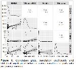
    Figure 1 illustrates correlation graphs, correlation coefficients and statistical significance between various variables in goats with pregnancy toxemia. Serum BHBA levels correlated positively with serum vitamin B12 (r=0.392, P<0.05) an glucose (r=0.394, P<0.05) levels. In addition, serum vitamin B12 levels showed a positive correlation with serum folate (r=0.206, P<0.05) and glucose (r=0.323, P<0.05) levels.


    Büyütmek İçin Tıklayın
    Figure 1: Correlation plots, correlation coefficients and statistical significance between several variables in goats with pregnancy toxemia

  • Başa Dön
  • Özet
  • Giriş
  • Materyal ve Metot
  • Bulgular
  • Tartışma
  • Kaynaklar
  • Tartışma
    Pregnancy toxemia is a common metabolic disease in sheep and goats. It has high mortality rate and causes substantial economic losses. In addition to clinical symptoms, ketonemia, ketonuria and hypoglycemia are important biochemical findings associated with the disease 23.

    Since BHBA level is more stable than acetone and acetoacetate, it is more preferred in the diagnosis of ketosis 24. In this study, study groups were formed based on blood BHBA levels. Serum BHBA levels were higher in the severe hyperketonemic group than in non-ketonemic group. The serum BHBA concentrations in moderate hyperketonemia group differ from those in non-ketonemic group and severe hyperketonemia group. The results obtained from the study were similar to those of the researchers 25-31.

    B12 is crucial for the energy metabolism of sheep because it acts as a cofactor in processes that support the production of propionic acid and is the main precursor of glucose synthesis in the liver. If there is a shortage of B12 in ruminants, energy efficiency has also decreased 32,33. Souto et al. 29 reported that serum vitamin B12 levels were within the reference intervals in sheep with pregnancy toxemia. In another study, researchers found that vitamin B12 levels associated with rumen microbiata status, the composition of the feed and rumen fermentation process 10. Similar to our results, Soeres et al. 34 also reported that sheep with pregnancy toxemia and fed with energy enriched ration had increased serum cobalamin and folate levels. They also argued that dietary energy content may hav an effect on serum cobalamin and folate levels.

    Folate plays an important role in DNA and methionine metabolisms and is synthesied by rumen microorganism in ruminants. Its circulating levels is significantly altered by metabolic and nutritional disorders. In addition circulating folate concentrations closely related with circulating cobalamin levels 10,35. Folic acid supplementation in pregnant sheep on certain days of gestation resulted in a dose dependent increases in serum folate and cobalamin 36. Studies of folic acid associated with pregnancy in the world 29,36-38 are limited, and the value of folic acid for pregnancy toxemia in Aleppo goats has been reported for the first time. In a study in sheep, Soeres et al. investigated the amount of folic acid in energy-poor, energy-rich, and pregnancy toxemia sheep. They reported that the difference between the three groups was significant and that the pregnancy toxemia group had the highest folic acid level 34. Soeres et al. 34, on the contrary, no significant relationship was found between serum folate levels between the control and pregnancy toxemia groups. Girard et al. reported 38, folic acid is important on its own during pregnancy, and it can be added to pregnant animals since it also causes an increase in B12 levels.

    Although hypoglycemia is frequently identified in pregnant toxemia, it is not a consistent finding 23,25,26,39. Considering the reference range for serum glucose concentrations in goats (50-63 mg/dL), the median of serum glucose concentrations in moderate and severe hyperketonemic goats in the current study were below the lower reference range in the present study. However, the percentage of hyperglycemic cases in moderate and severe hyperketonemia groups were 15.4% and 46.2%, respectively. Similar to our results, Souto et al. found that 51% of sheep pregnancy toxemia had hyperglycemia 29. Researchers hypothesized that glucose concentrations in pregnancy toxemia could indicate whether a fetus is viable 8. In 2012, Lima et al. 8 evaluated the chance of fetal survival in goats with pregnancy toxemia according to glycemic status and determined the fetal survival rate to be higher when cesarean section was performed in hypoglycemic goats. On the other hand, Jeffrey and Higgins reported in their study that hypoglycemia-induced nervous system damage may occur in pregnancy toxemia and these would be irreversible 40. Based on all these studies, it is thought that the glycemic status of pregnancy toxemia may change individually, and it is important to control the glycemic status of the patient before performing parenteral or oral glucose replacement.

    In conclusion, our results suggest that hyperketonemia associated with serum cobalamin and glucose concentrations. Future studies are needed to understand the underlying mechanism of B12 increase in goats with pregnancy toxemia.

  • Başa Dön
  • Özet
  • Giriş
  • Materyal ve Metot
  • Bulgular
  • Tartışma
  • Kaynaklar
  • Kaynaklar

    1) Radostits OM, Gay CC, Hinchcliff KW, et al. Veterinary Medicine: A textbook of the diseases of cattle, horses, sheep, pigs and goats. 10th Edition, St Louis: Elsevier, 2007.

    2) Khan JR, Ludri RS. Changes in blood glucose, plasman on-esterified fatty acids and insülin in pregnant and non-pregnant goats. Trop Anim Health Prod 2002; 34: 81-90.

    3) Smith MC, Sherman DM. Goat Medicine. 1th Edition, Oxford: Wiley-Blackwell, 2009. 4. Baştan İ, Salar S. Koyun ve keçilerde gebelik toksemisi. Harran Üniversitesi Veteriner Fakültesi Dergisi 2013; 1: 42-47.

    5) Gül Y. Geviş Getiren Hayvanların İç Hastalıkları. 2. Baskı, Malatya: Medipres, 2006.

    6) Hefnawy AE, Youssef S, Shousha S. Some immunohormonal changes in experimentally pregnant toxemic goats. Vet Med Int 2010; 4: 768438.

    7) González FHD, Hernández F, Madrid J, et al. Acid–base and electrolyte status during early induced pregnancy toxaemia in goats. Vet J 2012; 193: 598-599.

    8) Lima MS, Pascoal RA, Stilwell GT. Glycaemia as a sign of the viability of the foetuses in the last days of gestation in dairy goats with pregnancy toxaemia. Ir Vet 2012; 65: 1.

    9) Herdt TH, Hoff B. The use of blood analysis to evaluate trace mineral status in ruminant livestock. Vet Clin N Am Food A 2011; 27: 255-283.

    10) González-Montaña JR, Escalera-Valente F, Alonso AJ, et al. Relationship between vitamin B12 and cobalt metabolism in domestic ruminant: An update. Animals 2020; 10: 1855.

    11) Rizzo G, Laganà AS. A review of vitamin B12. Mol Nutr 2020; 105-129.

    12) Girard CL, Matte JJ. Effects of intramuscular injections of vitamin B12 on lactation performance of dairy cows fed dietary supplements of folic acid and rumen-protected methionine. J Dairy Sci 2005; 88: 671-676.

    13) Stemme K, Lebzien P, Flachowsky G, et al. The influence of an increased cobalt supply on ruminal parameters and microbial vitamin B12 synthesis in the rumen of dairy cows. Arch Anim Nutr 2008; 62: 207-218.

    14) Goff JP. Determining the mineral requirement of dairy cattle. In Proceedings of the 11th Annual Florida Ruminant Nutrition Symposium, University of Florida. 2000; 106-132.

    15) Watanabe F, Bito T. Vitamin B12 sources and microbial interaction. Exp Biol Med 2018; 243: 148-158.

    16) Olivares JL, Fernández R, Fleta J, et al. Vitamin B12 and folic acid in children with intestinal parasitic infection. J Am College Nutr 2002; 21: 109-113.

    17) Girard CL, Lapierre H, Desrochers A, et al. Net flux of folates and vitamin B12 through the gastrointestinal tract and the liver of lactating dairy cows. Br J Nutr 2001; 86: 707-715.

    18) Abou-Zeina HA, Zaghawa A, Nasr SM, et al. Effects of dietary cobalt deficiency on performance, blood and rumen metabolites and liver pathology in sheep. Metabol 2008; 4: 12.

    19) Kather S, Gruetzner N, Kook PH, et al. Review of cobalamin status and disorders of cobalamin metabolism in dogs. J Vet Intern Med 2020; 34: 13-28.

    20) Stelletta C, Gianesella M, Morgante M. Metabolic and nutritional diseases. 1th Edition, UK: Wallingford, 2008.

    21) Vijayanand V, Balagangatharathilagar M, Gnanaraj, et al. On field diagnostic indicators of sub clinical pregnancy toxaemia in goats and its therapeutic evaluation. J Entomol Zool Stud 2021; 9: 1110-1119.

    22) Rook JS. Pregnancy toxemia of ewes, does, and beef cows. Vet Clin North Am Food Anim Pract 2000; 16: 293-317.

    23) Batmaz H. Koyun ve Keçilerin İç Hastalıkları. Semptomdan Tanıya Tanıdan Sağaltıma. 1. Baskı, Bursa: Nobel, 2013.

    24) Duffield TF. Subclinical ketosis in lactating dairy cattle. Vet Clin North Am Food Anim 2000; 16: 231- 253.

    25) Darwish AA, El Ebissy IA. The diagnostic value of acute phase proteins in barki ewes with pregnancy toxemia. AJVS 2019; 62: 27-37.

    26) Prasannkumar R, Vasava RG, Jani H, et al. Studies on clinical signs and biochemical alteration in pregnancy toxemic goats. Vet World 2016; 8: 869- 874.

    27) Albay MK, Karakurum MC, Sahinduran S, et al. Selected serum biochemical parameters and acute phase protein levels in a herd of Saanen goats showing signs of pregnancy toxaemia. Vet Med 2014; 59: 336-342.

    28) Issi M, Gül Y, Başbuğ O. Evaluation of renal and hepatic functions in cattle with subclinical and clinical ketosis. J Vet Anim Sci 2016; 40: 47-52.

    29) Souto RJ, Afonso JA, Mendonça CL, et al. Biochemical, endocrine, and histopathological profile of liver and kidneys of sheep with pregnancy toxemia. Pesqui Vet Bras 2019; 39: 780-788.

    30) Balikci E, Yildiz A, Gurdogan F. Investigation on some biochemical and clinical parameters for pregnancy toxemia in Akkaraman ewes. J Anim Vet Adv 2009; 8: 1268-1273.

    31) Gurdogan F, Balikci E, Yildiz A. Some acute phase proteins, oxidative stress biomarkers and antioxidant enzyme activities in ewes with pregnancy toxemia (short paper). IJVR 2014; 15: 297-299.

    32) Strobel HJ. Vitamin B12-dependent propionate production by the ruminal bacterium Prevotella ruminicola 23. AEM 1992; 58: 2331-2333.

    33) Ertürküner AA, İssi M. Farklı ırklardaki koyun ve keçilerde serum B12 vitamini ve kobalt düzeylerinin araştırılması. FÜ Sağ Bil Vet Derg 2020; 34: 35-42.

    34) Soares PC, Carvalho CCD, da Cunha Mergulhão FC, et al. Serum concentrations of folic acid and cobalamin and energy metabolism of ewes as a function of the energy density of the diet, peripartum period, and pregnancy toxemia. Trop Anim Health Prod 2023; 55: 10.

    35) Ragaller V, Hüther L, Lebzien P. Folic acid in ruminant nutrition: A review. BJN 2008; 10: 153-164.

    36) Girard CL, Castonguay F, Matte JJ. Response of serum concentrations of folates to dietary supplements of folic acid given to ewes during gestation. Can J Anim Sci 1999; 79: 387-389.

    37) Washburn SE, Caudill MA, Malysheva O, et al. Formate metabolism in fetal and neonatal sheep. American Journal of Physiology-Endocrinology and Metabolism, 308, E921–E927. eep, and Suffolk ewes. JAS 2015; 74: 1711-1715.

    38) Girard CL, Castonguay F, Fahmy MH, et al. Serum and milk folates during the first two gestations and lactations in Romanov, Finnsheep, and Suffolk ewes. JAS 1996; 74: 1711-1715.

    39) Scott PR, Sargison ND, Penny CD, et al. Cerebrospinal fluid and plasma glucose concentrations of ovine pregnancy toxaemia cases, inappetant ewes and normal ewes during late gestation. Brit Vet J 1995; 151: 39-44.

    40) Jeffrey M, Higgins RJ. Brain lesions of naturally occurring pregnancy toxemia of sheep. Vet Pathol 1992; 29: 301-307.

  • Başa Dön
  • Özet
  • Giriş
  • Materyal ve Metot
  • Bulgular
  • Tartışma
  • Kaynaklar
  • [ Başa Dön ] [ Özet ] [ PDF ] [ Benzer Makaleler ] [ Yazara E-Posta ] [ Editöre E-Posta ]
    [ Ana Sayfa | Editörler | Danışma Kurulu | Dergi Hakkında | İçindekiler | Arşiv | Yayın Arama | Yazarlara Bilgi | E-Posta ]