[ Ana Sayfa | Editörler | Danışma Kurulu | Dergi Hakkında | İçindekiler | Arşiv | Yayın Arama | Yazarlara Bilgi | E-Posta ]
Fırat Üniversitesi Sağlık Bilimleri Veteriner Dergisi
2024, Cilt 38, Sayı 1, Sayfa(lar) 008-013
[ Özet ] [ PDF ] [ Benzer Makaleler ] [ Yazara E-Posta ] [ Editöre E-Posta ]
Sıçanlarda Fruktoz ile İndüklenmiş Metabolik Sendrom Modelinde Enalapril'in Böbrek Fonksiyonları Üzerindeki Koruyucu Etkisi
Bedrettin ORHAN1, Hasan GENÇOĞLU2, Özlem ÜÇER3, Necip İLHAN4, Kazım ŞAHİN5, Ayhan DOĞUKAN6
1Uludag University, Faculty of Medicine, Department of Internal Medicine, Division of Hematology Bursa, TÜRKİYE
2Firat University, Faculty of Science, Department of Molecular Biology, Elazığ, TÜRKİYE
3Fırat University, Faculty of Medicine, Department of Pathology, Elazığ, TÜRKİYE
4Firat University, Faculty of Medicine, Department of Biochemistry, Elazığ, TÜRKİYE
5Firat University, Faculty of Veterinary Medicine, Department of Animal Nutrition Elazığ, TÜRKİYE
6Firat University, Faculty of Medicine, Department of Internal Medicine, Division of Nephrology Elazığ, TÜRKİYE
Anahtar Kelimeler: Fructose, enalapril, rat
Özet
Metabolik sendrom, insülin direnci, hiperinsülinemi, dislipidemi, hipertansiyon ve obezite ile karakterize bir tablodur. Yüksek fruktoz tüketimiyle oluşturulmuş deneysel metabolik sendrom modellerinde ratlarda hipertansiyon, hipertrigliseridemi ve insülin direnci görülmüştür. Bir anjiotensin dönüştürücü enzim inhibitörü olan Enalapril, anjiyotensin-I’i hidrolize eden dönüştürücü enzim dipeptidil karboksipeptidazı inhibe eder. Bu çalışmanın amacı, ratlarda fruktoz ile oluşturulmuş deneysel metabolik sendrom modelinde, bir anjiotensin dönüştürücü enzim inhibitörü olan enalapril’in böbrek fonksiyonları, plasma lipid düzeyleri ve bazı hücre içi yolak belirteçleri üzerine potansiyel koruyucu etkilerini ortaya koymaktır. Çalışmaya 8 haftalık, 28 adet Wistar Albino ırkı erkek sıçanlar alındı. Bu sıçanlar rastgele 4 gruba ayrıldı ve 8 hafta sonunda sakrifiye edildi. Kan örnekleri; böbrek fonksiyon testleri, karaciğer fonksiyon testleri ve lipid düzeyleri için, böbrek doku örnekleri ise Western blot analizler için toplandı. Böbrek dokusunda transforme edici büyüme faktörü beta (TGF-β), tümör nekroze edici faktör alfa (TNF-α), nükleer faktör kappa B (NF-κB), interlökin-6 (IL-6), mothers against decapentaplegic-3 (SMAD-3) protein düzeyleri Western blot ile ölçüldü. Yüksek fruktozla beslenen sıçanlarda enalapril uygulaması serum glikoz, total kolesterol (TC), düşük yoğunluklu lipoprotein (LDL), trigliserid, aspartat aminotransferaz (AST), alanin aminotransferaz (ALT) ve kreatinin düzeyleri üzerinde olumlu etki gösterirken, HDL, BUN parametreleri üzerine anlamlı etki göstermemiştir. Enalapril uygulaması yüksek fruktoz ile beslenen sıçanlarda sadece fruktoz ile beslenen sıçanlara göre TGF-β, IL-6, SMAD-3 protein düzeylerini anlamlı şekilde düşürürken, NF-κB, TNF-α protein düzeylerinde anlamlı değişikliğe yol açmamıştır. Sonuç olarak yüksek fruktozla beslenme karaciğer ve böbrek fonksiyon testleri üzerinde kötüleştirici ve yıkıcı etkiler gösterirken enalapril uygulmasının bu etkileri iyileştirdiği görülmüştür.
  • Başa Dön
  • Özet
  • Giriş
  • Materyal ve Metot
  • Bulgular
  • Tartışma
  • Kaynaklar
  • Giriş
    Metabolic syndrome is an entity characterized by insulin resistance, hyperinsulinemia, hypertension, dyslipidemia, and central obesity 1. Metabolic syndrome (MS) has reached epidemic proportions world-wide including cardiovascular damage, nonalcoholic fatty liver disease, and increased incidence of chronic kidney disease (CKD)2.

    Fructose is commonly found in added sugar in our food, in the form of high-fructose corn syrup and sucrose. Dietary fructose consumption has been predicted as an enviromental factor which causes obesity and anomalies caused by metabolic syndrome. In experimental models of fructose induced metabolic syndrome hypertension, hypertriglyceridemia, hyperinsulinemia and insulin resistance were seen in rats3. In addition to insulin resistance and hypertriglyceridemia, hypertension is induced in rats fed with a high fructose diet, and this was described by Hwang et al. for the first time4. It was found that plasma angiotensin-2 (AT-2) levels and systolic blood pressure increased significantly in rats fed the fructose diet. In these animal models, the use of angiotensin-converting enzyme (ACE) inhibitors or angiotensin receptor antagonists (ARB) has been shown to prevent the development of hypertension5.

    Nuclear factor kappa B (NF-κB), an accepted intracellular pathway, has been shown to be an obesity-dependent pathway in metabolic syndrome6. Activation of the NF-κB pathway can lead to increased AT-2 and the release of TGF-β (transforming growth factor beta) and inflammatory cytokines (Interleukin-6 [IL-6], Tumor necrosis factor-alpha [TNF-α]) and increased intracellular pathways (Mothers against decapentaplegic-3 [SMAD-3]), thereby causing fibrosis7-9. Activation of the TGF-β signaling pathway, which proceeds in a cascade, begins with the binding of the ligand to its receptors. There are three types of receptors on the cell membrane: TGF-β type I (TβRI), TGF-β type II (TβRII), and TGF-β type III (TβRIII). TβRI and TβRII receptors have serine/threonine kinase properties. Binding of TGFβ to the type II receptor causes the kinase activity of this receptor to emerge and phosphorylation of the glycine-serine region in the structure of the type I receptor. In this way, activated TβRI phosphorylates SMAD proteins in the cytosol. SMAD 2 and 3 TGF-β, called receptor regulatory SMAD; SMAD 1, 5 and 8 are involved in the signaling pathway of bone morphogenic protein and anti-mullerian hormone and are activated by phosphorylation by ligand/receptor complexes 10,11. As a result of these pathways, TGF-β induces the formation of fibrosis in the lung, kidney and liver, and understanding TGF-β and its family pathways is important for clinical studies to be conducted for the regulation of the signaling activities of serine/threonine kinase receptors and SMADs12.

    Enalapril, an angiotensin-converting enzyme (ACE) inhibitor, reduces RAS activity in the body, and has both cardioprotective and renal protective effects1. The protective effects of enalapril treatment on kidney are hemodynamic (ameliorating glomerular capillary and intraglomerular pressure) as well as a non-hemodynamic (reduction of mesangial proliferation, gene expression upregulating, inflammatory cell infiltration and ameliorating renal fibrotic changes) mechanism13.

    The aim of this study was to determine the potential protective roles on kidney functions, plasma lipid levels and some intracellular pathway markers of enalapril in an experimental model of metabolic syndrome induced by fructose in rats.

  • Başa Dön
  • Özet
  • Giriş
  • Materyal ve Metot
  • Bulgular
  • Tartışma
  • Kaynaklar
  • Materyal ve Metot
    Research and Publication Ethics: All animal procedures were approved by the Animal Experimentation Ethics Committee of Fırat University (Decision No: 114-2015/59 Elazığ, Türkiye). All procedures involving rats were conducted in strict compliance with the relevant laws, the Animal Welfare Act, Public Health Services Policy, and guidelines established by the Institutional Animal Care and Use Committee of the Institute.

    Animals and Study Design: Twenty-eight (8 week-old; 200-220 g body weight) male Wistar-Albino rats were used from the Fırat University Laboratory Animal Research Center (Elazığ, Türkiye). The rats were randomly divided according to body weight, which was similar, into four equal groups containing seven rats each. The rats were fed either i): a standard diet (rat chow) as control (Control), ii): a fructose diet (Fructose) (containing 60% fructose), iii): a standard diet with/ administrated Enapril into drinking water (10 mg/kg body weight per day) (Enapril), iv): a combination of fructose and enapril (Fructose+Enapril) for 8 weeks.

    Laboratory Analyses: At the end of the experiment, all rats were killed by cervical dislocation. Blood samples and tissues from kidney were taken and processed for biochemical and Western blot examination.

    Serum glucose, total cholesterol (TC), high density lipoprotein cholesterol (HDL-C), LDL cholesterol (LDL-C), triglyceride (TG), aspartate aminotransferase (AST), alanine aminotransferase (ALT), blood urea nitrogen (BUN) and creatinine concentrations were measured by an automatic analyzer (Samsung LABGEO PT10, Samsung Electronics Co, Suwon, Korea). Repeatability and device/method precision of LABGEOPT10 was established according to the IVR-PT06 Guideline.

    Western Blot Analyses: The protein levels of IL-6, NF-κB, TGF-β, TNF-α and SMAD-3 in kidney tissue were determined by Western blotting according to the previously described method 14.

    To determine levels of the proteins in kidney Western blot analysis, the samples were homogenized in in 1:10 (w/v) in 10 mM Tris-HCl buffer at pH 7.4, comprising 0.1 mM NaCl, 0.1 mM phenylmethylsulfonyl fluoride, and 5 μM soybean (soluble powder; Sigma, St. Louis, MO) as trypsin inhibitor. The sample (20 μg of protein per lane) was mixed with sample buffer, boiled for 5 min, and separated by sodium dodecyl sulfate-polyacrylamide (12%) gel electrophoresis under denaturing conditions, and then electroblotted onto a nitrocellulose membrane (Schleicher and Schuell Inc., Keene, NH). Nitrocellulose blots were washed in PBS and blocked with 1% bovine serum albumin in PBS for 1 h prior to application of the primary antibodies (IL-6, NF-κB, TGF-β, TNF-α and SMAD-3 Abcam, Cambridge, UK). Primary antibody was previously diluted (1:1000 or 1:5000) in the same buffer containing 0.05% Tween-20. The nitrocellulose membrane was incubated overnight at 4°C with protein antibody. The blots were washed and incubated with horseradish peroxidase-conjugated goat antimouse IgG, the secondary antibody (Abcam). Specific binding was detected using diaminobenzidine and hydrogen peroxide as substrates. Protein load was controlled using a monoclonal mouse antibody against β-actin antibody (Sigma, St. Louis, MO). Protein levels were quantified densitometrically using an image analysis system (Image J; National Institute of Health, Bethesda, MD).

    Statistical Analysis: Data were analyzed using the IBM SPSS version 22 software. The changes among groups were analyzed using one-way analysis of variance (ANOVA) followed by the Tukey post hoc test, and P<0.05 was considered statistically significant. The data were presented as mean ± standard error.

  • Başa Dön
  • Özet
  • Giriş
  • Materyal ve Metot
  • Bulgular
  • Tartışma
  • Kaynaklar
  • Bulgular
    The effects of enalapril administration on biochemical parameters in rats with metabolic syndrome induced by a diet with high levels of fructose are presented in Table 1. While enalapril administration in rats fed with high fructose affected serum glucose, total cholesterol, LDL, triglyceride, AST, ALT, and creatinine levels, it did not significantly affect HDL, BUN parameters.


    Büyütmek İçin Tıklayın
    Table 1: The effect of Enalapril on weight and biochemical parameters in rats fed with fructose

    The mean body weight of the animals at the end of the 8th week were as follows: control group 397.1±40.2 grams, fructose group 404.1±31.6 grams, enalapril group 382.7±28.3 grams, fructose+enalapril group was 370.7±37.9 grams (Figure 1). No statistical significans was found between body weight of the rats at the end of 8 weeks despite the mean weight of the fructose group was higher (P>0.05). Serum glucose levels in rats fed with high doses of fructose were found to increase by 30% compared to the control group (P˂0.0001). Although enalapril administration caused a decrease in blood glucose levels in rats fed with high fructose, this decrease was not statistically significant (P˃0.05). While the serum triglyceride level in rats fed with high doses of fructose increased by 38% compared to the control group (P˂0.05), enalapril administration caused a decrease in blood triglyceride levels in rats fed with high fructose, and this decrease was statistically significant (P<0.05). While the serum creatinine level in rats fed with high doses of fructose increased by 130% compared to the control group (P˂0.0001), enalapril administration caused a decrease in blood creatinine level in rats fed with high fructose and this decrease was statistically significant (P˂0.0001). In addition, although creatinine in the group fed with high fructose and given enalapril was lower in than the control group, this decrease was not statistically significant (P>0.05). Although enalapril administration caused a decrease in blood total cholesterol, ALT, AST, BUN, and LDL levels in rats fed high fructose, this decrease was not statistically significant (P>0.05).


    Büyütmek İçin Tıklayın
    Figure 1: Initial and final weight of the rats at the end of eight weeks

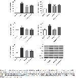
    The studied IL-6, NF-κB, TGF-β, TNF-α, and SMAD-3 protein levels were higher in the fructose group than the control group. Enalapril administration caused a significant decrease in IL-6, TGF-β, SMAD-3 levels in the high fructose group compared to the group given only fructose (P<0.01). The group receiving high fructose displayed 97.3%, 43.0%, 115.9% in IL-6, TGF-β and SMAD-3 protein levels compared to control, respectively. Enalapril administration to fructose fed group resulted in a significant decrease in IL-6, TGF-β, and SMAD-3 protein levels (24.6%, 32.9%, 35.0%, respectively). There was no noteworthy difference observed when comparing TNF-α and NF-κB protein levels between groups (Figure 2; P>0.05).


    Büyütmek İçin Tıklayın
    Figure 2: The effect of Enalapril on protein levels of IL-6 (Panel A), NF-κB (Panel B), TGF-β (Panel C), TNF-α (Panel D) and SMAD-3 (Panel E) in rats fed with fructose. The intensity of the bands was quantified by densitometric analysis and β-actin was included to ensure equal protein loading (Panel F). Data are expressed as percent of control value. Each bar represents the mean and standard error. Blots were repeated at least 3 times * P<0.05, ** P<0.01, *** P<0.001 as compared to control group; # P<0.05, ## P<0.01, ### P<0.001 as compared to fructose group

  • Başa Dön
  • Özet
  • Giriş
  • Materyal ve Metot
  • Bulgular
  • Tartışma
  • Kaynaklar
  • Tartışma
    In human studies and animal models, consumption of a high fructose diet seemed to be related to obesity, higher triglyceride levels, and insulin resistance15. It has been shown that Wistar rats fed with 60% fructose diet for 6-8 weeks is sufficient to cause MS16-18. In the light of these findings and studies, the model created with high fructose (60%) diet was deemed appropriate for the research.

    At the end of 8 weeks, an increase in the body weights of the rats was observed in all four groups. However, there was no significant difference among the four groups regarding body weight. In one of the two different metabolic syndrome models, no statistical difference were detected between the groups in the weights of rats fed with 60% fructose after 12 weeks, while in another study, rats fed with 35% fructose were found to be heavier than the control group at the end of 16 weeks2,18. Our study and other studies show that the duration of fructose feeding, rather than the fructose content, causes a significant increase in body weight.

    In a metabolic syndrome model performed in Wistar albino rats containing a 60% fructose diet, after eight weeks, an ARB application of telmisartan showed a significant decrease in AST, ALT, T-K, LDL-C, VLDL-C levels and a significant increase in HDL-C levels 15. In our study, obtaining similar results at the end of 8 weeks supports that ACEi and ARBs show the same effects on liver enzymes and serum lipids, albeit with a different mechanism. However, the fact that HDL-C levels did not change with enalapril administration in our study may be attributed to the small sample size in the study.

    In another metabolic syndrome model created with a high-fat diet in 20 weeks in rats, losartan administration, an ARB, showed an increase in T-C levels, which was expected to decrease, and no significant changes were observed in creatinine clearance19. The fact that the metabolic syndrome model was made with a high-fat diet and losartan, an ARB, in the mentioned study may explain the differences with our study. In our study, it is a surprising result that serum creatinine values were found at such high levels in the fructose group. Despite the elevated creatinine levels, BUN did not increase in our study. This might be hypothesized that the creatinine elevation might be related to the decrease in tubular secretion rather than renal failure. It is also an interesting result that creatinine values were lower in the fructose + enalapril group than the fructose group in our study. The fact that there was no difference in creatinine levels in the group given enalapril alone compared to the control group suggests that the creatinine change associated with enalapril in rats may not be related to changes in intraglomerular pressure, so called RAS activation. The variable levels of creatinine, which increased in fructose, and decreased in the enalapril group, may be related to fructose effects on creatinine by mechanisms that we cannot explain.

    In a study conducted in ZSF1 rats, which are genetically suitable for the metabolic syndrome model, enalapril applied at a dose of 60 mg/kg/day into the drinking water of rats for 32 weeks showed significant improvement in serum cholesterol and TG levels but no effect on serum glucose concentration20. In our study, even enalapril administration given at a dose of 10 mg/kg/day for only eight weeks caused an improvement in serum glucose concentrations. A high dose of enalapril in the previously mentioned study may suppress rats' energy consumption via ACE inhibition21. This suppression is thought to be related to the inhibition of adipocyte growth via AT-222.

    The studied IL-6, NF-κB, TGF-β, TNF-α, and SMAD-3 protein levels of rat kidneys were higher in the fructose given group than the control group. While enalapril administration caused a significant decrease in TGF-β, SMAD-3, IL-6 levels in the high fructose given group compared to the group given only fructose. However, enalapril administration caused a decrease in TNF-α and NF-κB levels; this decrease was not found statistically significant. In a metabolic syndrome model created with 60% fructose-fed rats for 12 weeks, significant increases in TNF-α, TGF-β, and NF-κB levels were observed in Western blot analyzes of liver tissues, and the levels of these inflammatory markers were found to be low in the group has given telmisartan, an ARB18. In our study, although significant increases were observed in the levels of TGF-β, IL-6, SMAD-3, NF-κB, TNF-α in kidney tissues in the fructose-administered group, NF-κB, and TNF-α levels were not significantly decreased with enalapril administration. This can be attributed to the relatively short duration of our experiment. In a study conducted with Sprague-Dawley rats, the administration of benazepril, an ACEi, significantly decreased TNF-α, TGF-β, NF-κB, and SMAD- 3/4 expressions in the heart tissue of rats compared to the control group23. The similar results obtained in our study can be explained by the fact that ACE inhibition stops the inflammatory process by inhibiting the intracellular pathways with AT-2 blockade24.

    Metabolic syndrome is a chronic disease with various components and many complications caused by these components. MS causes severe morbidity and mortality all over the world. It harms the national economy by causing injuries and loss of workforce and the care and treatment expenditures created by complications. The positive steps to be taken in the treatment of metabolic syndrome will contribute to society and the state economy. Therefore, metabolic syndrome and related complications can be prevented with treatment modalities targeting inflammatory markers involved in pathogenesis. Our study showed that enalapril, an ACEi, shows promising results in treating complications such as diabetes, kidney failure, and obesity caused by metabolic syndrome. However, further experimental and clinical studies are needed.

  • Başa Dön
  • Özet
  • Giriş
  • Materyal ve Metot
  • Bulgular
  • Tartışma
  • Kaynaklar
  • Kaynaklar

    1) Aygen B, Kucuksu M, Aydin S, Ozercan IH. Effect of enalapril maleate on ghrelin levels in metabolic syndrome in rats. Peptides 2015; 67: 39-44.

    2) Bratoeva K, Stoyanov GS, Merdzhanova A, Radanova M. Manifestations of renal impairment in fructose-induced metabolic syndrome. Cureus 2017; 9: e1826.

    3) Ng HY, Lee YT, Kuo WH, et al. Alterations of renal epithelial glucose and uric acid transporters in fructose induced metabolic syndrome. Kidney Blood Press Res 2018; 43: 1822-1831.

    4) Hwang IS, Ho H, Hoffman BB, Reaven GM. Fructose-induced insulin resistance and hypertension in rats. Hypertension 1987; 10: 512-516.

    5) Tran LT, Yuen VG, McNeill JH. The fructose-fed rat: A review on the mechanisms of fructose-induced insulin resistance and hypertension. Mol Cell Biochem 2009; 332: 145-159.

    6) Wang T, Zhang X, Li JJ. The role of NF-kappaB in the regulation of cell stress responses. Int Immunopharmacol 2002; 2: 1509-1520.

    7) Yamamoto Y, Gaynor RB. IkappaB kinases: Key regulators of the NF-kappaB pathway. Trends Biochem Sci 2004; 29: 72-79.

    8) Hirschberg R. Kindlin-2: a new player in renal fibrogenesis. J Am Soc Nephrol 2013; 24: 1339-1340.

    9) Flanders KC. Smad3 as a mediator of the fibrotic response. Int J Exp Pathol 2004; 85: 47-64.

    10) Shi Y, Massagué J. Mechanisms of TGF-beta signaling from cell membrane to the nucleus. Cell 2003;113: 685-700.

    11) Massagué J, Seoane J, Wotton D. Smad transcription factors. Genes Dev 2005; 19: 2783-810.

    12) Miyazawa K, Shinozaki M, Hara T, Furuya T, Miyazono K. Two major smad pathways in TGF-beta superfamily signalling. Genes Cells 2002; 7: 1191-204.

    13) Taal MW, Brenner BM. Renoprotective benefits of RAS inhibition: From ACEI to angiotensin II antagonists. Kidney Int 2000; 57: 1803-1817.

    14) Sahin K, Tuzcu M, Orhan C, et al. Anti-diabetic activity of chromium picolinate and biotin in rats with type 2 diabetes induced by high-fat diet and streptozotocin. Br J Nutr 2013; 110: 197-205.

    15) Basciano H, Federico L, Adeli K. Fructose, insulin resistance, and metabolic dyslipidemia. Nutr Metab (Lond) 2005; 2: 5.

    16) Sánchez-Lozada LG, Tapia E, Jiménez A, et al. Fructose-induced metabolic syndrome is associated with glomerular hypertension and renal microvascular damage in rats. Am J Physiol Renal Physiol 2007; 292: F423-429.

    17) Mamikutty N, Thent ZC, Haji Suhaimi F. Fructose-drinking water ınduced nonalcoholic fatty liver disease and ultrastructural alteration of hepatocyte mitochondria in male wistar rat. Biomed Res Int 2015; 2015: 895961.

    18) Rabie EM, Heeba GH, Abouzied MM, Khalifa MM. Comparative effects of Aliskiren and Telmisartan in high fructose diet-induced metabolic syndrome in rats. Eur J Pharmacol 2015; 760: 145-153.

    19) Machado H, Pinheiro HS, Terra MM, et al. Dissociation of antihypertensive and metabolic response to losartan and spironolactone in experimental rats with metabolic sindrome. J Bras Nefrol 2012; 34: 328-336.

    20) Bilan VP, Salah EM, Bastacky S, et al. Diabetic nephropathy and long-term treatment effects of rosiglitazone and enalapril in obese ZSF1 rats. J Endocrinol 2011; 210: 293-308.

    21) Santos EL, de Picoli Souza K, Guimarães PB, et al. Effect of angiotensin converting enzyme inhibitor enalapril on body weight and composition in young rats. Int Immunopharmacol 2008; 8: 247-253.

    22) Engeli S, Negrel R, Sharma AM. Physiology and pathophysiology of the adipose tissue renin-angiotensin system. Hypertension 2000; 35: 1270-1277.

    23) Yan SH, Zhao NW, Zhu XX, et al. Benazepril inhibited the NF-κB and TGF-β networking on LV hypertrophy in rats. Immunol Lett 2013; 152: 126-134.

    24) Ruiz-Ortega M, Rodríguez-Vita J, Sanchez-Lopez E, Carvajal G, Egido J. TGF-beta signaling in vascular fibrosis. Cardiovasc Res 2007; 74: 196-206.

  • Başa Dön
  • Özet
  • Giriş
  • Materyal ve Metot
  • Bulgular
  • Tartışma
  • Kaynaklar
  • [ Başa Dön ] [ Özet ] [ PDF ] [ Benzer Makaleler ] [ Yazara E-Posta ] [ Editöre E-Posta ]
    [ Ana Sayfa | Editörler | Danışma Kurulu | Dergi Hakkında | İçindekiler | Arşiv | Yayın Arama | Yazarlara Bilgi | E-Posta ]