[ Ana Sayfa | Editörler | Danışma Kurulu | Dergi Hakkında | İçindekiler | Arşiv | Yayın Arama | Yazarlara Bilgi | E-Posta ]
Fırat Üniversitesi Sağlık Bilimleri Veteriner Dergisi
2024, Cilt 38, Sayı 1, Sayfa(lar) 078-081
[ Özet ] [ PDF ] [ Benzer Makaleler ] [ Yazara E-Posta ] [ Editöre E-Posta ]
Türkiye’nin Bingöl İlindeki Kıl Keçilerinde Gebelik Toksemisinin Yaygınlığı
Murat UZTİMÜR, Cennet Nur ÜNAL
Bingöl University, Faculty of Veterinary Medicine, Department of Internal Medicine, Bingöl, TÜRKİYE
Anahtar Kelimeler: Goat, BHBA, prevalence, pregnancy toxemia
Özet
Bu çalışmada Bingöl iline bağlı 9 farklı köyde (Garip, Yamaç, Alibir, Yeşil, Sarıçiçek, Gökçeli, Çavuşlar, Ardıçtepe, Balpınar) gebelik toksemisinin prevalansının araştırılması amaçlanmıştır. Çalışmadaki hayvanlarda gebelik toksemisinin belirlenmesi için kan beta hidroksibütirik asit (BHBA) konsantrasyonu ölçümü kullanılmıştır. Çalışma materyalini gebeliğin son 3 haftalık döneminde olan 398 adet kıl keçisi oluşturmaktadır. Kan BHBA konsantrasyonu <0.80 mmol/L Sağlıklı (S), 8-1.6 mmol/L subklinik gebelik toksemili (SGT), >1.6 mmol/L klinik gebelik toksemili (KGT) olarak kabul edildi. Keçilerin 70 tanesi SGT, 25 tanesinin KGT ve 303 tanesinin ise S olduğu belirlenmiştir. Keçilerin vücut kondisyon skorunun ortalama 2.8±0.7 ve ortalama 4.5±0.8 yaşında olduğu belirlendi. Kan BHBA konsantrasyonları FreeStyle Optium Neo H (Abbott Diabetes Care Ltd, Witney, UK) cihazı kullanılarak analiz gerçekleştirildi. Bingöl’ün Garip, Yamaç, Alibir, Yeşil, Sarıçiçek, Gökçeli, Çavuşlar, Ardıçtepe, Balpınar köylerinde 303 (%76.13) adet S, 95 (%23.87) adet gebelik toksemili keçi olduğu belirlendi. Gebelik toksemili keçilerin 70 adedi SPT (%73.68)’den oluşurken 25 adedinin ise KGT’li (%26.32) keçilerden oluştuğu saptandı. Sonuç olarak Bingöl ilinin farklı yerlerindeki keçi işletmelerinde gebelik toksemisinin yüksek olduğu ve hayvancılık işletmelerinde önemli beslenme ve yönetim hatalarının olduğunu göstermektedir.
  • Başa Dön
  • Özet
  • Giriş
  • Materyal ve Metot
  • Bulgular
  • Tartışma
  • Kaynaklar
  • Giriş
    Pregnancy toxemia is a metabolic disease that occurs in the last 2-3 weeks of pregnancy in goats and sheep1,2. In the latter trimester of pregnancy, the fetus grows by 60-80%, and dry matter intake declines, resulting in a negative energy balance (NED) in animals2. Furthermore, pregnancy with multiple offspring, functional insufficiency of the liver, excessive fat, weakness, irregular feeding, parasitic infestations, sudden weather changes, stress, and the uterus narrowing the area of the rumen due to advanced pregnancy all contribute to the formation of pregnancy toxemia3-5. The pathophysiological mechanisms behind maternal toxemia remain incompletely understood; nevertheless, it is believed to arise from an imbalance in glucose homeostasis, leading to disruptions in the metabolism of proteins, lipids, carbohydrates, and other nutrients1,6,7. If the fetus and mother's glucose requirements are unsatisfied, the body tries to make up the energy shortfall by breaking down fat stores. Non-esterified fatty acids (NEFA) released as a result of lipolysis are oxidized through the tricarboxylic acid cycle in the liver to produce energy8. Pregnancy toxaemia is caused by lipid accumulation in the liver from oxidizing fatty acids to ketone bodies (beta hidroksibütirik asit (BHBA), acetic acid, and acetone), which raises the concentrations of ketone bodies in blood, urine, and milk when increased lipid mobilization surpasses the liver's capacity6,9.

    There are two forms of pregnancy toxemia: Clinical pregnancy toxemia (CPT) and subclinical pregnancy toxemia (SPT). As the disease progresses, if it is not treated in the terminal phase, where nervous symptoms such as the inability to stand, leaning the head on something, muscle tremors, teeth grinding, opisthotonus, and blindness are observed, mortality usually occurs within 3-4 days1,5. While Khan et al.10 determined that the prevalence of pregnancy toxemia was 35% in their study, Scott et al.11 revealed that this rate was 88.9% in their study. It is stated that if pregnancy toxemia is not treated, the mortality rate can reach up to 80%1. The disease's extensive prevalence results in severe losses for cattle enterprises and national economies. Gestational toxemia results in significant economic losses, such as the mortality of pregnant goats, veterinary care and treatment expenses, reduced milk production, and progeny loss12. Consequently, preventative measures rather than treatment for pregnant toxemia are required1,5,13. The incidence of pregnancy toxemia may vary based on nutritional status and environmental factors1. As a result of this, determining the disease's prevalence in a certain area is essential for creating preventative plans. However, there is not enough information about the prevalence of pregnancy toxemia in goats in our country. Thus, this study aims to investigate the prevalence of pregnancy toxemia in hair goats in Bingöl province.

  • Başa Dön
  • Özet
  • Giriş
  • Materyal ve Metot
  • Bulgular
  • Tartışma
  • Kaynaklar
  • Materyal ve Metot
    Research and Publication Ethics: The local ethics committee for animal experiments at Bingöl University provided the permission documents needed for the study plan. Work began following the approval of these permits (BÜ HADYEK Committee Number: 2023/02, Decision Number 02/03).

    Animals: The study was conducted in 9 different villages (Garip, Yamaç, Alibir, Yeşil, Sarıçiçek, Gökçeli, Çavuşlar, Ardıçtepe, Balpınar) of Bingöl province, located between 41º 20 and 39º - 56º eastern longitudes and 39º - 31 and 36º - 28º northern latitudes of Turkey. The goats body condition score in the study was assessed using the criteria defined by Villaquiran et al.14. The body condition score of the goats was determined to be 2.8±0.7 on average and 4.5±0.8 years on average. In this study, all of the animals for which BHBA analysis was performed come from family businesses. According to the information provided by the owners of the animals, they feed the animals hay, barley straw, barley silage, and maize silage in barns during the winter and on pastures in the summer.

    BHBA Analysis: Blood BHBA concentrations were analyzed using the FreeStyle Optium Neo H (Abbott Diabetes Care Ltd, Witney, UK) device and disposable β-ketone test strips (FreeStyle Optimum β-Ketone, Abbott Diabetes Care Ltd, UK). Before starting the analysis, the device was calibrated by attaching a special calibrator kit to the FreeStyle Optium Neo H device. After the calibration process, 1 drop of blood was taken from the jugular vein in accordance with the technique and scanned into the device, and the results obtained after 10 seconds were recorded. Blood BHBA concentration was accepted as <0.80 mmol/L H, 8-1.6 mmol/L SPT, >1.6 mmol/L CPT1,7. In this study, animals with protozoan diseases (such as theileria, babesia, anaplasma), metabolic diseases and animals with any drug application were not included in the study.

    Statistical Analysis: The data underwent rigorous statistical analysis using SPSS 26 software (IBM SPSS Statistics for Windows, Version 22.0. Armonk, NY: IBM Corp.). All results were presented as mean value ± standard deviation. Whether the data showed normal distribution or not was analyzed with the Shapiro-Wilk test. Statistical differences between groups were analyzed with the Kruskall Wallis test. Paired group comparisons were made with the Mann Whitney U test. The one-way ANOVA was followed up with the post hoc Tukey multiple comparisons test. The statistical significance level between groups was accepted as p value <0.05.

  • Başa Dön
  • Özet
  • Giriş
  • Materyal ve Metot
  • Bulgular
  • Tartışma
  • Kaynaklar
  • Bulgular
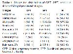
    According to the blood BHBA concentrations, 303 (76.13%) of the goats in the villages of Garip, Yamaç, Alibir, Yeşil, Sarıçiçek, Gökçeli, Çavuşlar, Ardıçtepe, and Balpınar in Bingöl were found to be healthy, whereas 95 (23.87%) had pregnant toxemia. These goats were determined to be 4.5±0.8 years old on average. Seventy of the goats with pregnant toxemia were found to be SPT (73.68%), while twenty-five of them were found to be CPT (26.32%). The distribution of CPT, SPT and H in different villages of Bingöl province is presented in Table 1. Goat populations in various villages were found to be: 5 out of 36 in Garip village, 6 out of 52 in Yamaç village, 15 out of 65 in Alibir village, 9 out of 38 in Yeşil village, 8 out of 26 in Sarıçiçek village, and 26 out of 48 in Gökçeli village. After investigation, it was found that 15 out of 48 goats, 65 goats in the village of Çavuşlar, 7 out of 56 goats in the village of Ardıçtepe, and 5 out of 37 goats in the village of Balpınar were pregnant toxemia.


    Büyütmek İçin Tıklayın
    Table 1: Shows the distribution of CPT, SPT, and H in different Bingöl provincial villages

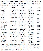
    Blood BHBA concentrations according to different parts of Bingöl province and their statistical significance between groups are shown in Table 2. In the villages of Garip, Yamaç, Ardıçtepe, and Balpınar, it was found that there was a statistically significant difference (P<0.001) between H-SPT and H-CPT. In the villages of Alibir, Yeşil, Sarıçiçek, Gökçeli, and Çavuşlar, it was found that there was a statistically significant difference (P<0.001) between H-SPT, H-CPT, and SPT-CPT. The goats possessed an average body condition score of 2.8±0.7 and an average age of 4.5±0.8.


    Büyütmek İçin Tıklayın
    Table 2: BHBA concentrations (mmol/L) according to different villages of Bingöl province and their statistical significance between the groups

  • Başa Dön
  • Özet
  • Giriş
  • Materyal ve Metot
  • Bulgular
  • Tartışma
  • Kaynaklar
  • Tartışma
    Pregnancy toxemia is a metabolic disorder in which pregnant goats suffer hyperketonemia. This condition is typically triggered by a decrease in energy balance during the last two to three weeks of pregnancy2,15. The prevalence of pregnancy toxemia in the world has been researched more in sheep than in goats, and there are a limited number of studies investigating the prevalence of pregnancy toxemia in goats10,16. However, no studies investigating pregnancy toxemia in goats have been found in Türkiye. In this study, it was aimed to determine the prevalence of pregnancy toxemia in hair goats in Bingöl province by blood BHBA measurement, and the presented study is the first research conducted in this context in Bingöl province. Animals that develop NED deplete their fat stores. The body consequently produces acetone, acetoacetic acid, and BHBA also referred to as ketone bodies. BHBA is widely preferred in the diagnosis of ketosis because it is found more stable in the blood among the ketone bodies mentioned1,11. While the blood BHBA cut-off value for the diagnosis of SPT in goats was determined as 0.8-1.6 mmol/L1,17, a value of >1.6 mmol/L was accepted for the diagnosis of CPT18. In this study, blood BHBA level was preferred in the diagnosis of pregnancy toxemia. In addition, the above-mentioned BHBA levels were used as a reference in this study to group pregnancy toxemia. In this study, it was determined that BHBA levels in the CPT groups were significantly higher than in the H and SPT groups. Previous published literature findings also support the ΒHBA results of the CPT and SPT groups in this investigation15,17.

    In both developing and undeveloped nations, goats play a vital role in the economy due to their high productivity of meat, milk, and wool. This is especially true in regions that are too rocky, mountainous, or shrubby for agriculture7. Pregnancy toxemia is a disease for which prevention strategies need to be developed due to maternal and offspring losses, high treatment costs, and the predisposition of these animals to transitional diseases (retention secundinarum, mastitis, metritis)15,19. To take comprehensive measures to prevent the occurrence of pregnancy toxemia, the prevalence of the disease in the region must be known. Furthermore, it is mentioned that regional variations in administrative practices, racial predispositions, and environmental factors may all affect the occurrence of pregnant toxemia16. In the study conducted by Sathish16, the prevalence of SPT in sheep was found to be 15.33%, while Gupta et al.20 found it to be 14.86% in their study. In another study, it was stated that the prevalence of the disease in sheep was approximately 5-20%6. Pregnancy toxemia prevalence was reported as 13.3% by Ismail et al.21, 36.7% by Murugeswari and Mathialagan2, and 35% by Khan et al. 10. In this study, the prevalence of pregnancy toxemia was found to be 23.87%, while SPT was 73.68% and CPT was 26.32%. The results of this study are similar to those of Ji et al.6, Sathish,16 and Gupta et al.20 data, while Murugeswari and Mathialagan2 and Khan et al.10 were found to be lower than the results. The possible reason for this may be related to regional differences, economic situation and seasonal conditions.

    Many different risk factors play important roles in the occurence of pregnancy toxemia10,21. In order to prevent pregnancy toxemia and increase production efficiency, the interaction between animals and the environment must be taken into account 10,22. Goats biological systems are impacted by physiological stressors, such as seasonal fluctuations22,23. In Bingöl province, goats are kept in pastures in summer and in barns in winter due to harsh climatic conditions and snowfall. It is well recognized that the inability to fulfill the rising energy needs of winter and the modifications and adaption process to a new environment have a negative impact on the metabolism 5,23,24. Khan et al.10 found the prevalence of pregnancy toxemia to be higher in winter than in summer. Pregnancy toxemia is more common in the winter, and one reason for this is because in some regions, the months of January and February are the birth seasons and correspond with the final two to three weeks of pregnancy21. Pregnancy toxemia has been determined to be 23.87% prevalent in Bingöl province. The reason for this is believed to be a combination of the winter season, physiological stress factors, lack of grazing, and the fact that births take place around this time in reproductive planning.

    Like many metabolic illnesses, pregnancy toxemia is known to be significantly impacted by age25. Older animals with pregnant toxemia have been shown to meet the energy deficit less quickly and insufficiently than younger animals10. In addition, this situation leads to disruption of metabolic adaptation mechanisms and an increase in NEFA and BHBA concentrations25. The average age of the goats in this study was 4.5±0.8 years old, which is considered to be an important risk factor that will affect the prevalence of pregnancy toxemia in goats10,25.

    As a result, the animals included in thisstudy consist of 398 hair goats that were in the last 3 weeks of their pregnancy. It was found that 303 of the goats were healthy (H), 25 were CPT, and 70 were SPT. In light of the findings of this study, it is believed that pregnancy toxemia significantly damages the financial structure of family-run companies in the area and that season and advanced age play a major role in the risk of pregnancy toxemia in hair goats. In further studies, it would be useful to analyze all risk factors affecting prevalence in detail and evaluate a larger study population.

  • Başa Dön
  • Özet
  • Giriş
  • Materyal ve Metot
  • Bulgular
  • Tartışma
  • Kaynaklar
  • Kaynaklar

    1) Rook JS. Pregnancy toxemia of ewes, does, and beef cows. Vet Clin North Am Food Anim Pract 2000; 16: 293-317.

    2) İ ssi M, Gül Y, Başbuğ O. Evaluation of renal and hepatic functions in cattle with subclinical and clinical ketosis. Turkish Journal of Veterinary Animal Sciences 2016; 40: 47-52.

    3) A ndrews A. Pregnancy toxaemia in the ewe. In Pract 1997; 19: 306-314.

    4) İ ssi M, Gül Y, Kandemir FM, Başbuğ O. Primer ketozisli süt ineklerinin tedavisinden önce subkutan insülin uygulamasının kan glikoz düzeyleri üzerine etkileri. Yüzüncü Yıl Üniversitesi Veteriner Fakültesi Dergisi 2009; 20: 13-16.

    5) K elay A, Assefa A. Causes, control and prevention methods of pregnancy toxemia in ewe. J Life Sci Biomed 2018; 8: 69-76.

    6) J i X, Liu N, Wang Y, et al. Pregnancy toxemia in ewes: A review of molecular metabolic mechanisms and management strategies. Metabolites 2023; 13: 149.

    7) U ztimür M. Gazioğlu A. Yilmaz Ö. Changes in free amino acid profile in goats with pregnancy toxemia. Vet Res Commun 2023; 1-9.

    8) S argison ND. Pregnancy toxaemia. Diseases of sheep, 2007: 7; 359-362.

    9) B aştan İ, Salar S. Koyun ve keçilerde gebelik toksemisi. Harran Üniversitesi Veteriner Fakültesi Dergisi 2013; 2: 42-47.

    10) K han YR, Durrani AZ, Muhammad IJAZ, et al. Determination of hemato-biochemical biomarkers, associated risk factors and therapeutic protocols for pregnancy toxemia in beetal goats. Kafkas Üniv Vet Fak Derg 2021; 27: 525-532.

    11) Scott PR, Sargison ND, Penny CD. Evaluation of recombinant bovine somatotropin in the treatment of ovine pregnancy toxaemia. Vet J 1998; 155: 197-199.

    12) Abdelaal A, Zaher H, Elgaml SA, Abdallah H. Prognostic value of serum cardiac troponin t and nitric oxide as cardiac biomarkers in pregnancy toxemic goats. Glob Vet 2013; 11: 817-823.

    13) Temizel EM, Batmaz H, Keskin A, et al. Butaphosphan and cyanocobalamin treatment of pregnant ewes: Metabolic effects and potential prophylactic effect for pregnancy toxaemia. Small Rumin Res 2015; 125: 163-172.

    14) Villaquiran M, Gipson TA, Merkel RC, Goetsch AL, Sahlu T. Body condition scores in goats. American Institute for Goat Research, Langston University 2004: 1-8.

    15) Polat Dinçer PF, Tümer KÇ. Investigation of Vitamin B12 and Folic Acid Levels in Goats with Pregnancy Toxemia. Firat Universitesi Saglik Bilimleri Veteriner Dergisi 2023; 37: 49-53.

    16) Sathish KB. Incidence, diagnosis and treatment of pregnancy toxaemia in Hassan sheep. J Pharm Innov 2023; 12: 3708-3713

    17) Bani Ismail ZA, Al-Majali AM, Amireh F, Al-Rawashdeh OF. Metabolic profiles in goat does in late pregnancy with and without subclinical pregnancy toxemia. Vet clin pathol 2008; 37: 434-437.

    18) Firat A, Ozpinar A. Metabolic profile of pre-pregnancy, pregnancy and early lactation in multiple lambing Sakiz ewes. Ann Nutr Metab 2002; 46: 57.

    19) Barbagianni MS, Spanos SA, Ioannidi KS, et al. Increased incidence of peri-parturient problems in ewes with pregnancy toxaemia. Small Rumin Res 2015; 132: 111-114.

    20) Gupta VK, Kumar A, Vihan VS, Sharma SD. Studies on haemogram in sub-clinical ketosis in goats and sheep in organized farming system. IJSR 2008; 14: 114-117.

    21) Ismail ZB, Al-Rawashdeh O, Al-Majali AM, Amireh F. Prevalence and risk factors for pregnancy toxemia of goats in Jordan. Anim Biol Anim Husb 2015; 7: 53-59.

    22) Fatet A, Pellicer-Rubio MT, Leboeuf B. Reproductive cycle of goats. Anim Reprod Sci 2011; 124: 211-219.

    23) Ribeiro NL, Germano Costa R, Pimenta Filho EC, Ribeiro MN, Bozzi R. Effects of the dry and the rainy season on endocrine and physiologic profiles of goats in the Brazilian semi-arid region. Ital J Anim Sci 2018; 17: 454-461.

    24) Sharma K, Saini AL, Singh N, Ogra JL. Seasonal variations in grazing behaviour and forage nutrient utilization by goats on a semi-arid reconstituted silvipasture. Small Rum Res 1998; 27; 47-54.

    25) Radin L, Šimpraga M, Vince S, Kostelić A, Milinković-Tur S. Metabolic and oxidative status of Saanen goats of different parity during the peripartum period. J Dairy Res 2015; 82: 426-433.

  • Başa Dön
  • Özet
  • Giriş
  • Materyal ve Metot
  • Bulgular
  • Tartışma
  • Kaynaklar
  • [ Başa Dön ] [ Özet ] [ PDF ] [ Benzer Makaleler ] [ Yazara E-Posta ] [ Editöre E-Posta ]
    [ Ana Sayfa | Editörler | Danışma Kurulu | Dergi Hakkında | İçindekiler | Arşiv | Yayın Arama | Yazarlara Bilgi | E-Posta ]