[ Ana Sayfa | Editörler | Danışma Kurulu | Dergi Hakkında | İçindekiler | Arşiv | Yayın Arama | Yazarlara Bilgi | E-Posta ]
Fırat Üniversitesi Sağlık Bilimleri Veteriner Dergisi
2005, Cilt 19, Sayı 1, Sayfa(lar) 007-011
[ Özet ] [ PDF ] [ Benzer Makaleler ] [ Yazara E-Posta ] [ Editöre E-Posta ]
PURIFICATION AND PROKARYOTIC CLONING OF NUCLEOPROTEIN (NP) GENE OF RINDERPEST VIRUS (RPV) RBOK VACCINE STRAIN
Şükrü TONBAK1, Aykut ÖZDARENDELİ1, Mehmet Ziya DOYMAZ2, Yusuf BOLAT1
1Fırat Üniversitesi, Veteriner Fakültesi, Viroloji Anabilim Dalı, Elazığ-TÜRKİYE
2Fırat Üniversitesi, Tıp Fakültesi, Mikrobiyoloji Anabilim Dalı, Elazığ-TÜRKİYE
Keywords: Rinderpest virus, Nucleoprotein gene, Cloning, Plasmid
Summary
In this study, it was aimed to expres the NP protein of rinderpest virus (RPV) RBOK vaccine strain in a prokaryotic system. Vero cells were infected with RBOK vaccine strain. Total RNA was isolated from infected cells and reverse transcription was carried out and cDNA of NP gene was obtained. The NP gene was amplified by polymerase chain reaction (PCR) with the primers specific to the NP gene. Nucleoprotein gene of RPV-RBOK vaccine strain was cloned into vector PinPointTM Xa-3 by using the HindIII and KpnI restriction enzyme sites. Recombinant NP protein synthesized by E.coli was purified. To determine the recombinant NP protein which is approximately 80kDa, western blotting was performed.
  • Top
  • Summary
  • Introduction
  • Methods
  • Results
  • Disscussion
  • References
  • Introduction
    Rinderpest virus (RPV) infection is an acute, usually fatal, highly contagious viral disease of cloven-hoofed animals, particularly cattle and buffaloes. The disease causes necrosis and erosions on the mucosa of respiratory and digestion systems 1-3.

    RPV belongs to the morbillivirus genus of paramyxoviridae family, and has single stranded, negative polarity, non-segmented RNA genome 4,5. It has six structural proteins, namely nucleoprotein (NP), phosphoprotein (P), matrix (M), fusion (F), hemagglutinin (H), large (L), and two nonstructural V and C proteins. Using transcription mapping method, the sequences of the genes on the viral genome was found to be as N-P-M-F-H-L from 3' direction to 5' direction 6,7.

    The NP protein constitutes the major component of the nucleocapsid core and is known to play a major role in transcription and replication of the virus 8. The sequence analysis reveals an open reading frame (ORF) 1575 nucleotid in lenght. The ORF encodes NP protein of 525 amino acids with a moleculer weight 65 kDa. The NP gene 3' non-coding region consists of 53 nucleotides, and 5' non-coding region consists of 40 nucleotides 9.

    The aim of this study was to express the NP protein of RPV-RBOK vaccine strain in a prokaryotic system.

  • Top
  • Summary
  • Introduction
  • Methods
  • Results
  • Disscussion
  • References
  • Methods
    Cells and Virus: African green monkey kidney (Vero) cells (Foot and Mouth Disease Institute, Ankara, Turkey) were cultured in Dulbecco’s Modified Eagle’s Medium (DMEM, Sigma Chemical Co. St. Louis, MO, USA) containing 10% fetal bovine serum (Sigma), penicilin (100 IU/ml) and streptomycin (100µg/ml). RPV-RBOK vaccine strain adapted to cell culture as produced in the Etlik Research Institute was used in this study.

    Total RNA Isolation: Approximately 65-75% of confluent Vero cells were infected with the RPV-RBOK vaccine strain. When cytopathological effect (CPE) was observed in approximately 80% of the cells, total RNA isolation was performed with TRI-Reagant (Sigma) as described by the manufacturer.

    Reverse Transcription (RT) and Polymerase Chain Reaction (PCR): To design the NP gene spesific primers, EMBL gene bank X68311 RPV-RBOK vaccine strain gene sequence was utilized. According to the gene sequence, the forward primer (NP1: 5' GG AAG CTT ATG GCT TCT CTC TTG A 3') and reverse primer (NP2: 5' GG GGT ACC TCA GTT GAG AAT ATC 3') were created. HindIII enzyme cut site was added to the forward primer, and KpnI enzyme cut site was added to reverse primer. The reverse primer was used to make cDNAs of the nucleoprotein gene by using RT. Using these cDNAs as templates, 10pmol reverse and forward primers, 1.25mM dNTP (Promega), 25mM MgCl2 (Promega), 10X PCR buffer (Promega), 2U Taq DNA Polymerase (Promega), and 50µl PCR mixture were prepared. After preheating at 95ºC for 2 minutes, the PCR was performed containing 32 cycles with denaturation 94ºC for 1 minute, annealing at 44ºC for 1 minute, extension at 72ºC for 5 minutes and final extension at 72ºC for 15 minutes. PCR products were visualized on 1.5% agarose gel stained with ethidium bromide 10.

    Construction of Recombinant pin3-NP Plasmid: Both RPV-RBOK vaccine strain NP gene and prokaryotic expression vector PinPointTMXa-3 (Promega) were cut with HindIII and KpnI restriction enzymes and purified from the agarose gel using the Wizard PCR Preps DNA Purification System (Promega, A7170) in accordance with the protocol of the manufacturer. The ligation reaction using T4 DNA Ligase enzyme was carried out. Using alkaline lysis method, the recombinant plasmid DNA was obtained from the transformed competent E.coli JM109 cells. The recombinant plasmid pin3-NP was identified XbaI, HindIII and KpnI digestion. Furthermore, PCR was performed using recombinant plasmid DNA as a template with the primers specific to the NP gene 11.

    Production of Recombinant Biotinylated NP Protein: Procedures for the expression of recombinant biotinylated NP protein were as described by technical manufacturer. Recombinant plasmid pin3-NP and control vector were transformed to JM109 cells of E.coli. Transformed cells were inoculated to LB agar containing 100µg/ml ampicillin and kept at 37ºC for 16 hours. E.coli strain JM109 carrying the recombinant plasmid and control plasmid was grown for overnight in LB medium containing 100µg/ml ampicillin and 2µM biotin at 37ºC. Overnight cultures were diluted 1/5 in 25ml LB medium with 100µg/ml ampicillin and 2µM biotin. After four hours of culture at 37ºC, the expressions of recombinant biotinylated NP protein and CAT protein were induced by addition of 200µM IPTG and cultures further incubated at 37ºC for 5 hours with shaking.

    Cell Lysis and Affinity Purification of Recombinant NP Protein: The cells were centrifuged at 8000 rpm for 10 minutes and the supernatant was removed. The cells were incubated with 1ml of ice cold cell lysis buffer (50mM Tris-HCl pH 7.5, 50mM NaCl, 5% glycerol) placed on ice and cells were resuspended. Then lysozyme was added to a final concentration of 1mg/ml and mixture was stirred at 4ºC for 1 hours. After adding sodium deoxycholate (DOC) to a final concentration of 0.1%, stirring was continued for an additional 15 minutes. Finally, 200U DNase I was added to reduce viscosity of the solution, then stirred for 20 minutes at 4ºC. The cell lysate was centrifuged at 10000 rpm for 10 minutes at 4ºC, so that remove cell debris. Then the supernatant was transferred to a clean tube. After the supernatant was mixed with 75µl SoftLinkTMResin and stirred gently for 2 hours at 4ºC. The resin was washed throughly with cell lysis buffer twice and was added to the resin suspension up to concentration of 5mM in order to elute the resin bound protein fraction. Then, allow the resin to settle and purified protein to a clean tube transferred.

    SDS-PAGE and Western Blotting: To determine the production of recombinant protein in expressed E.coli SDS-PAGE was carried out. Firstly, pour off 10% resolving gel on electrophesis apparatus, when polymerization is complete and pour off stacking gel solution directly onto the surface of the polymerized resolving gel. While the stacking gel is polymerizing, the samples prepared by heating to 100ºC for 3 minutes in 1XSDS gel loading buffer (50mM TrisCl pH6.8, 100mM dithiothreitol [DTT], 2% SDS, 0.1% bromophenol blue, 10% glycerol) for denature proteins, after the samples were load up into the bottom of the wells. The samples were run at 150V current for 4 hours. Then, proteins were transferred onto nitrocellulose membran at 10mA current for 1 hours. After transfer process, the membran was blocked with 5% nonfat dried milk at 37ºC for 1 hours. The membran was washed with TBST buffer (10mM TrisCl pH8.0, 150mM NaCl, 0.05% Tween20) five times, the membran was incubated with 1/500 dilution streptavidin (Kpl, USA) at 37°C for 1 hour. The membran was washed with TBST buffer. Then, the membran were left into a chromogen substrate solution containing 0.1% diamino benzidine tetrachloride “DAB” (Sigma, USA) and 0.02% H2O2 for a period of time for monitoring.

  • Top
  • Summary
  • Introduction
  • Methods
  • Results
  • Disscussion
  • References
  • Results
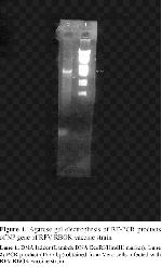
    The pathological changes in the cells infected with RPV-RBOK vaccine strain on the 5th day of the infection were observed. Following the PCR, the NP gene with a lenght of approximately 1575 nucleotides was shown in 1.5% agarose gel (Figure 1, lane 2).


    Click Here to Zoom
    Figure 1: Agarose gel electrophesis of RT-PCR products of NP gene of RPV RBOK vaccine strain.
    Lane 1: DNA ladder (Lambda DNA EcoRI/HindIII marker). Lane 2: PCR product (1575 bp) obtained from Vero cells infected with RPV RBOK vaccine strain.

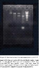
    By cutting the recombinant pin3-NP plasmid DNA with HindIII and KpnI, a piece of 1575 nucleotides was obtained (Figure 2, lane 3). Furthermore, following the cutting with the XbaI enzyme which does not recognize any site on the prokaryotic expression vector, it was observed that the recombinant plasmid was opened in a linear structure as expected (Figure 2, line 4). The primers specific to the NP gene were used and the presence of the NP gene in the recombinant plasmid was shown. Accordingly, an amplification product of approximately 1575 base pair long was shown in 1.5% agarose gel stained with ethidium bromide (Figure 2, lane 2).


    Click Here to Zoom
    Figure 2: Demonstration of recombinant plasmid pin3-NP.
    Lane 1: DNA ladder (Lambda DNA EcoRI/HindIII marker). Lane 2: pin3-NP plasmid DNA was used as template in PCR assay where RPV-NP gene spesific primers were used. Lane 3: HindIII/KpnI cut pin3-NP plasmid DNA. Lane 4: XbaI cut pin3-NP plasmid DNA. Lane 5: Uncut pin3-NP plasmid DNA.

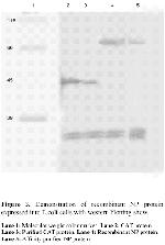
    Recombinant plasmid pin3-NP and control vector were transformed into JM109 cells of E.coli and grown overnight, by taking single colony inoculated into 5ml LB medium containing 100µg/ml ampicillin and 2µM biotin at 37ºC for 16 hours. Then 25ml of the same fresh LB medium was inoculated with the overnight cultures and under same conditions. After 4 hours of culture, the expression of fusion protein was induced by addition 200µM IPTG. The recombinant protein was expressed at 37ºC for 4 hours incubation. The CAT protein and the recombinant NP protein were detected on western blotting 40kDa, 80kDa respectively (Figure 3, lane 2 and 4). We tried to purify to the recombinant NP protein using avidin-biotin purification system. The recombinant CAT and NP proteins were produced in E.coli, cell lysis and purification of proteins were accomplished. Affinity purified CAT and NP proteins were determined by western blotting (Figure 3, lane 3 and 5).


    Click Here to Zoom
    Figure 3: Demonstration of recombinant NP protein expressed into E.coli cells with western blotting show.
    Lane 1: Molecular weight color marker. Lane 2: CAT protein.
    Lane 3: Purified CAT protein. Lane 4: Recombinant NP protein.
    Lane 5: Affinity purified NP protein

  • Top
  • Summary
  • Introduction
  • Methods
  • Results
  • Disscussion
  • References
  • Discussion
    In this study, affinity purification and expression of NP protein of RPV-RBOK vaccine strain were reported. In general, nucleoprotein is an internal protein of viruses, and it is important to develop vaccines using the viruses or viral proteins, particularly to develop cellular immunity 9,12,13. Furthermore, the detection of the NP protein is used in the diagnosis of viral diseases. For the effective control and elimination of RPV disease, it is a significant step to distinguish serologically the infected and vaccinated animals. Recently, the utilization of recombinant vaccines for effectively struggle with RPV has become dominant. However, it is impossible to distinguish the vaccinated and infected animals concerning conventional vaccination 14. However, using the diagnostic kits prepared from nucleoprotein it is sharply possible to distinguish the animals vaccinated with recombinant vaccine from the infected animals 3,9,12.

    The purification of recombinant proteins is typically performed by a variety of methods. But the advantage of this expression system is based on principle the biotin to avidin interaction. The PinPointTM Xa-3 expression vector which allows that transformed E.coli produces a biotinylated recombinant fusion protein, and thus makes the purification easier. This method is short quick and does not require too many laboratory equipment 15-18.

    In the present study, expression of NP protein of RPV-RBOK vaccine strain in E.coli, and purification and detection of NP protein with western blotting was showed. It has been reported by Kumar and his collegues 14 that, prokaryotically expressed NP protein can directly be used as positive antigen in ELISA assays. In the future we plan to obtain more protein, and use this protein in both immunization and diagnostic purpose.

  • Top
  • Summary
  • Introduction
  • Methods
  • Results
  • Discussion
  • References
  • References

    1) Bolat Y, Doymaz MZ. Paramyxoviridae Ailesi. Fırat Üniv.Vet Fak. Viroloji Ders Notları. Fırat Üniv. Yayınları, Elazığ. 1998; 302-304.

    2) Diallo A, Libeau G, Couacy-Hymann E, et al. Recent developments in the diagnosis of rinderpest and peste des petits ruminants. Vet Microbiol 1995; 44: 307-317.

    3) Ohishi K, Inui K, Barrett T, et al. Long-term protective immunity to rinderpest in cattle following a single vaccination with a recombinant vaccinia virus expressing the virus hemagglutinin protein. J Gen Virol 2000; 81: 1439-1446.

    4) Blixenkrone-Möller M, Sharma B, Varsanyi TM, et al. Sequence analysis of the genes encoding the nucleocapsid protein and phosphoprotein (P) of phocid distemper virus, and editing of the P gene transcript. J Gen Virol 1992; 73: 885-893.

    5) Shaji D, Shaila MS. Domains of rinderpest virus phosphoprotein involved in interaction with itself and the nucleocapsid protein. Virology 1999; 258: 415-424.

    6) Ghosh A, Joshi VD, Shaila MS. Characterization of an in vitro transcription system from rinderpest virus. Vet Microbiol 1995; 44: 165-173.

    7) Kamata H, Tsukiyama K, Sugiyama M, et al. Nucleotide sequence of cDNA to the rinderpest virus mRNA encoding the nucleocapsid protein. Virus genes 1991; 5: 5-15.

    8) Conzelmann KK. Nonsegmented negative-strand RNA viruses: Genetics and manipulation of viral genomes. Annu Rev Genet 1998; 32: 123-162.

    9) Ismail T, Ahmad S, D’souza-ault M, et al. Cloning and expression of the nucleocapsid gene of virulent Kabete O strain of rinderpest virus in baculovirus:

    10) Use in differential diagnosis between vaccinated and infected animals. Virology 1994; 198: 138-147.

    11) Tonbak Ş, Özdarendeli A, Bolat Y. Sığır vebası virusu RBOK aşı suşu nükleoprotein (NP) geninin açıklanması. Fırat Üniversitesi Sağlık Bilimleri Dergisi 2003; 17(1): 81-85.

    12) Sambrook J, Fritsch EF, Maniatis T. Molecular cloning: A Laboratory Manual. Cold Spring Harbor Laboratory Press, Newyork. 2001.

    13) Kamata H, Ohkubo S, Sugiyama M, et al. Expression in baculovirus vector system of the nucleocapsid protein gene of rinderpest virus. J Virol Methods 1993; 43: 159-166.

    14) Nakamura K, Ohishi K, Ohkubo S, et al. Immunizing effect of vaccinia virus expressing the nucleoprotein of rinderpest virus on systemic rinderpest virus infection in rabbits. Comp Immunol Microbiol Infect Dis 1998; 21: 91-99.

    15) Kumar SS, Renji R, Saini M, et al. Use of prokaryotically expressed nucleocapsid protein as a positive antigen in Elisa. Biochem Mol Biol Int 1998; 46: 1093-1100.

    16) Kosovsky J, Durmanova V, Kudelova M, et al. A simple procedure for expression and purification of selected non-structural (a and b) herpes simplex virus 1 (HSV-1) proteins. J Virol Methods 2001; 92: 121-129.

    17) Lesley SA, Groskreutz DJ. Simple affinity purification of antibodies using in vivo biotinylation of a fusion protein. J Immunol Methods 1997; 207: 147-155.

    18) Winkler IG, Lochelt M, Levesque JP, et al. A rapid streptavidin-capture Elisa spesific for the detection of antibodies to feline foamy virus. J Immunol Methods 1997; 207: 69-77.

  • Top
  • Summary
  • Introduction
  • Methods
  • Results
  • Discussion
  • References
  • [ Başa Dön ] [ Özet ] [ PDF ] [ Benzer Makaleler ] [ Yazara E-Posta ] [ Editöre E-Posta ]
    [ Ana Sayfa | Editörler | Danışma Kurulu | Dergi Hakkında | İçindekiler | Arşiv | Yayın Arama | Yazarlara Bilgi | E-Posta ]