[ Ana Sayfa | Editörler | Danışma Kurulu | Dergi Hakkında | İçindekiler | Arşiv | Yayın Arama | Yazarlara Bilgi | E-Posta ]
Fırat Üniversitesi Sağlık Bilimleri Tıp Dergisi
2004, Cilt 18, Sayı 4, Sayfa(lar) 239-241
[ Özet ] [ PDF ] [ Benzer Makaleler ] [ Editöre E-Posta ]
THE ROLE OF EMERGENT SURGICAL DRAINAGE IN THE TREATMENT OF SUBACUTE SUBDURAL HEMATOMAS PRESENTING WITH TRANSIENT NEUROLOGICAL DEFICITS: CASE REPORT
Fatih S. EROL, Metin KAPLAN, Cahide TOPSAKAL, M. Faik ÖZVEREN, Ebru GERCEK
Firat University School of Medicine Department of Neurosurgery, Elazig – TÜRKİYE
Keywords: Subdural hematoma, surgical treatment, transient ischemic
Summary
Presentation with transient ischemic attacks in subacute subdural hematomas is quite rare. It is a clinical phenomeon in which hematoma mass effect with its own capability of producing local edema formation and the variations in cerebral vascular structures take place in the pathophysiology. Diseases such as atrial fibrillation or tromboembolic disorders of carotid artery, which may lead to transient ischemic attacks should be considered in the differential diagnosis. For treatment, emergent surgical drainage in order to eliminate the mass effect should be performed. In this study, a case of subdural hematoma presenting with motor aphasia and hemiparesis is reported of which neurological symptoms became permanent due to delay in the hematoma evacuation.

  • Top
  • Summary
  • Introduction
  • Case Presentation
  • Disscussion
  • References
  • Introduction
    Subacute subdural hematomas are almost encountered in the elderly, particularly following a minor head trauma 1,3. It is still on debate today due to controversies in the physiopathology, clinical progress and surgical treatment modalities in use 5. These hematomas may manifest with various symptoms such as headache, paresis, dementia, and epilepsy. An infrequent presentation with transient ischemic attacks has been reported 6. In this study, a case of subacute subdural hematoma in which transient ischemic attack (TIA) became permanent is reported and discussed in the context of the literature.

  • Top
  • Summary
  • Introduction
  • Case Presentation
  • Disscussion
  • References
  • Case Presentation
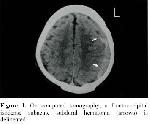
    72-year-old-man was admitted to our department with the complaints of aphasia and right hemiparesis, which developed suddenly and continued for twenty minutes. The patient had history of trauma. He had not nausea or epileptic seizure, nor had the symptoms of hypertension, diabetes, heart failure or other types of systemic diseases. On neurological examination, Glasgow Coma Scale was found as 15 points. He had motor aphasia and right hemiparesis dominating on the lower extremity. Plantar reflexes were extensor on both sides. Other systemic and laboratory findings were unremarkable. Electrocardiographic and echocardioagraphic findings were interpreted as normal. Carotid pulsations were symmetrical bilaterally and no bruits were noted. On computerized tomography (CT), a subacute subdural hematoma in the frontoparietal region was identified (Fig. 1). A diagnosis of chronic subdural hematoma presenting with transient ischemic attack was made considering the progression of symptoms and the absence of any coexistent lesion on tomography.

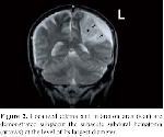
    Hematoma evacuation by burr-hole craniostomy and irrigatio drainage in the emergent setting was planned. However, no family consent was provided, therefore no surgical intervention could be made. The patient suffered from an ischemic attack manifested with motor aphasia and right hemiparesis on the 2nd day of follow-up period. The control magnetic resonance imaging (MR), in conjunction with the subacute subdural hematoma in the left frontoparietal region with the largest diameter being 1cm, an infarct lesion on the left parietal lobe with a significant mass effect due to localized edema was identified subjacent the hematoma (Fig 2).


    Click Here to Zoom
    Figure 1: On computed tomography, a frontooccipital isodense subacute subdural hematoma (arrows) is delineated.


    Click Here to Zoom
    Figure 2: Localized edema and infarction area (star) are demonstrated subjacent the subacute subdural hematoma (arrows) at the level of its largest diameter.

    Anti edema therapy by mannitol, and furosemide was implemented. On 24 hour follow-up examination after the beginning of the medical treatment, a partial recovery in the paresis, and a full recovery in the motor aphasia were noted. No complication developed during hospitalization and on 1st month follow-up, there was not increase thicking of subdural hemorrhage. Right hemiparesis was found to be persisting at that time.

  • Top
  • Summary
  • Introduction
  • Case Presentation
  • Disscussion
  • References
  • Discussion
    Coexistence of subacute subdural hematomas with TIA is seldom encountered and the real incidence is not known 8. Clinical progression of subacute subdural hematoma with the association of sudden developing transient ischemic neurological symptoms, display a different manifestation as to be a phenomenon. In this phenomenon, the most frequent symptoms are motor aphasia and paresis 4. Likewise, in our case, motor aphasia was associated with right hemiparesis.

    The mechanism in the development of TIA in conjunction with chronic subdural hematomas is controversial 8. Welsh EJ reported two cases of chronic subdural hematomas presenting with transient neurological deficits in which cardiac or carotid artery bruits were identified 10. In another case of Kaminski et al, atrial fibrillation was found. Likewise, atrial fibrillation again was found in a similar study of Guptha and Puthrasingham 2. In the etiology of transient ischemic attacks, heart originated emboli are the most frequent etiological factor. Carotid athero-embolus is known as another etiological factor 4,9. Besides, development of chronic subdural hematomas has been reported following anti-aggregation therapy used in order to reduce the incidence of cerebral emboli in atrial fibrillation or carotid atheroma cases 1. Therefore, those transient ischemic attacks reported before are not due to chronic subdural hematomas, but rather due to emboli originating from heart or carotid artery, therefore it is not appropriate to mention under this phenomenon since it leads some pathophysiological controversies. Therefore, it is crucial to make differential diagnosis between these diseases causing transient ischemic attacks.

    There are two basic factors in the pathophysiology of this phenomenon 2,6,8,10. 1) Mass effect of the chronic hemorrhage in the subdural space 2) Local cerebral edema caused by hematoma, without necessarily enlargement in the diameter. Furthermore, additionally mechanical stimulation to cortical mantle and even more, the changes in the head position could lead to the development of these symptoms intermittently 6,8. It is obvious that mass effect of the hematoma has a great role in the development of this phenomenon, since recovery in the attacks were achieved after the evacuation of the hematoma 4,7,10. Since no similar phenomenon is observed in most of the chronic subdural hematomas with significant mass effect, anatomical variations in the cerebral vascular structures adjacent to hematoma likely have the main effect in defining the physiopathology.

    TIA is a focal neurological deficit lasting 15-20 minutes not extending beyond 24 hours. The neurologic deficit was completely resolved after the first attack in our case. However, discrete from the cases reported before, in our case, hemiparesis was partially permanent despite anti-edema therapy after the second attack. Ischemia progressing to infarction in the region adjacent to hematoma was delineated on 48 hour follow-up MR imaging. Neurological deficits in our cases were transient initially, which became permanent because of the delay in the hematoma evacuation, therefore, we recommend emergent surgical evacuation in these kind of chronic subdural hematomas associated with this phenomenon. The previous cases reported in the literature with a good recovery in TIA after evacuation of coexisting hematoma, all support our hypothesis.

  • Top
  • Summary
  • Introduction
  • Case Presentation
  • Discussion
  • References
  • References

    1) Chen JCT, Levy M. Causes, epidemiology, and risk factors of chronic subdural hematoma. N Clin of N Am 2000;11:399-406.

    2) Guptha SH, Puthrasingam S. A case of chronic subdural haematoma presenting as a transient ischaemic attack. Age and Ageing 2001;30:172-173.

    3) Hamilton MG, Frizzell JB, Tranmer BI. Chronic subdural hematoma: The role for craniotomy reevaluated. Neurosurgery 1993;33: 67-72.

    4) Kaminski HJ, Hlavin ML, Likavec MJ, Schmidley JW. Transient neurologic deficit caused by chronic subdural hematoma. Am J Med 1992;92:698-700.

    5) Kudo H, Kuwamura K, Izava I. Chronic subdural hematoma in elderly people: present status on awaji island and epidemiological prospect. Neurol Med Chir (Tokyo) 1992;32:207-209.

    6) Moster ML, Johnston DE, Reinmuth OM. Chronic subdural hematoma with transient neurological deficits: A review of 15 cases. Ann Neurol 1983;14:539-542.

    7) Ribo M, Montaner J, Molina C, Abilleira S, Arenillas J, Sabin JA. Chronic subdural hematoma simulating a TIA. Implications for the management transient neurological deficit. Neurologia 2002;17:342-344.

    8) Russell NA, Goumnerova L, Atack EA, Atack DM, Benoit BG. Chronic subdural hematoma mimicking transient ischemic attacks. J Trauma 1985;25:1113-1114.

    9) Sherman DG, Easton JD. Clinical syndromes of brain ischemia. In: Wilkins RH, Rengachary SS, eds. Neurosurgery. New York. McGraw-Hill Co, 1996. 2053-2065.

    10) Welsh JE, Tyson GW, Winn HR, Jane JA. Chronic subdural hematoma presenting as transient neurologic deficits. Stroke 1979;10:564-567.

  • Top
  • Summary
  • Introduction
  • Case Presentation
  • Discussion
  • References
  • [ Başa Dön ] [ Özet ] [ PDF ] [ Benzer Makaleler ] [ Editöre E-Posta ]
    [ Ana Sayfa | Editörler | Danışma Kurulu | Dergi Hakkında | İçindekiler | Arşiv | Yayın Arama | Yazarlara Bilgi | E-Posta ]