[ Ana Sayfa | Editörler | Danışma Kurulu | Dergi Hakkında | İçindekiler | Arşiv | Yayın Arama | Yazarlara Bilgi | E-Posta ]
Fırat Üniversitesi Sağlık Bilimleri Tıp Dergisi
2006, Cilt 20, Sayı 6, Sayfa(lar) 415-421
[ Özet ] [ PDF ] [ Benzer Makaleler ] [ Yazara E-Posta ] [ Editöre E-Posta ]
Total Antioxidant Response and Oxidative Stress in Patients with Behçet's Disease
Ahmet IŞIK, Süleyman Serdar KOCA
Firat University Faculty of Medicine Department of Rheumatology Elazığ-TÜRKİYE
Keywords: Behçet's Disease, Total Antioxidant Response, Total Peroxide, Oxidative Stress Index
Summary
It was shown that many antioxidants decrease in Behçet’s disease (BD). Total antioxidant response (TAR), whose measurement method has been recently specified and developed, represents the total antioxidant properties of all antioxidants. This study aims to measure the antioxidant capacity in BD with TAR and to determine its relation with disease activity.

The study included 21 BD cases (10 active and 11 inactive) and 23 healthy volunteers as the control group. TAR and total peroxide (TP) were studied using appropriate methods and oxidative stress index (OSI) was calculated.

TAR level was 1,04±0,07 and 1,16±0,08 mmol Trolox eq./L in the BD group and in the control group, respectively; TP level was 13,4±4,5 and 7,72±1,86 µmol H202/L in the BD group and in the control group, respectively; OSI value was 1,45±0,52 and 0,67±0,18 Arbitrary Units in the BD group and in the control group, respectively. When compared to the control group, TAR level was significantly lower and TP and OSI levels were significantly higher in the BD group (p<0.001, for each). TAR level was negatively correlated with TP and OSI levels both in the BD group (r=-0.578, p<0.01 and r=-0.552, p<0.01, respectively) and the control group (r=-0.469, p<0.05 and r=-0.391, p<0.05, respectively). There was not any significant difference between active and inactive BD groups in terms of TAR, TP and OSI levels (p>0.05, for each).

TAR is an appropriate method that can be used to determine the antioxidant capacity in BD.

  • Top
  • Summary
  • Introduction
  • Methods
  • Results
  • Disscussion
  • References
  • Introduction
    Behcet’s disease (BD) is a chronic relapsing inflammatory disease. It is characterized by recurrent oral aphthous ulcer, genital ulcer/scar, ocular inflammation, skin and joint signs 1. The etiology and pathogenesis of BD has not been known fully yet, but genetic, environmental and autoimmune factors are thought to be involved 2. The interest to reactive oxygen species (ROS), which can be produced as a result of normal aerobic metabolism and whose production by active neutrophils increases during inflammation, has been recently grown 3.

    Activated neutrophils cause the production of non-radical ROS like hydrogen peroxide (H2O2) and singlet oxygen (1O2), radical ROS like hydroxyl anion-radical (OH•) and superoxide radical (O2•), and nitrogen-containing radicals like peroxynitrite (ONOO-) 3-7.

    Polymorphonuclear leukocytes increase the production of hpochlorous acid (HOCl) from H2O2 via myeloperoxidase enzyme 5-8. HOCl, in turn, causes tissue injury via sulphydryl oxidation and protein decarboxylation, leading to oxidative modification of macromolecules in the tissues 7,8.

    ROS lead to cell damage through its detrimental effects on peroxidation of double-chain fatty acids, protein and DNA, as well as, increase oxidative stress 3,4,7. Oxidants damage the antioxidant system, and thereby reduce the antioxidants levels 5-8.

    In BD, it has been reported that antioxidants decrease as a result of increased ROS production and oxidative stress 3,6,9,10. Impaired oxidant/antioxidant balance is held responsible for the tissue damage in BD 11,12. Furthermore, oxidative stress and lipid peroxidation (LPO) products have active part in the endothelial dysfunction and development of atherosclerosis 13,14. Chambers et al. 4 have demonstrated the presence of endothelial dysfunction in BD and have noted that endothelial dysfunction, a precursor of atherosclerosis, is associated with oxidative stress and LPO products.

    Blood contains many antioxidant molecules which prevent and/or inhibit harmful free radical reactions 15. Exogenous antioxidants, like vitamin C and vitamin E, and endogenous antioxidant like albumin, bilirubin, uric acid and superoxide dismutase and glutathione peroxidase as the scavenger enzymes can protect the cell against the potentially harmful effects of oxidant agents 5,7,15. Concentrations of these antioxidants in the plasma can be measured one by one, but this procedure is time-consuming, labour-intensive and costly, and requires complicated techniques 16,17. On the other hand, total antioxidant response (TAR) whose measurement method has been recently specified and developed can reflect the total antioxidative state of the plasma 16,17. In this method, TAR of the plasma against especially potent free radical reactions, which strongly lead to oxidative damage of biomolecules such as lipids, proteins and DNA, is measured. In this study, we aimed to measure both the levels of some individual antioxidant components and the TAR levels in plasma

    samples from active and inactive BD to evaluate their antioxidant status using a novel automated method 16,17. As a reciprocal measure, the total peroxide (TP) levels of the same plasma samples were also measured. The ratio of the plasma TP level to the TAR level was regarded as the oxidative stress index (OSI) 18.

  • Top
  • Summary
  • Introduction
  • Methods
  • Results
  • Disscussion
  • References
  • Methods
    The study included 21 BD cases who applied to outpatient clinic of Rheumatology Department of Internal Medicine of Medical Center of Firat University and 23 healthy individuals who age and sex matched. The diagnosis of BD was made according to the International Study Group’s criteria for BD 19. The patients were divided into two groups, as active and inactive, depending on the activity of their disease identified in the light of clinical and laboratory findings. The patients were interpreted as active if the patients with oral ulcer had at least two of below pathologies; genital ulcer, skin lesion, recent eye involvement, recent vascular involvement, recent neurological involvement, active arthritis, positive pathergy test and with high erythrocyte sedimentation rate (ESR) and/or C-reactive protein (CRP) 7,20,21. Histories of all individuals in the study were obtained, and systemic and rheumatological physical examinations were performed. Pathergy test was applied to all of the BD group, and 24-48 hours later, the patients were evaluated in terms of papulopustular lesions. All patients were assessed by an ophthalmologist for eye involvement. Approval of the local ethics committee and informed consent of the patients and healty individuals were obtained. Patients with dislipidemia, cardiovascular diseases, diabetes mellitus, renal failure, chronic infection, alcohol abuse, and who used antilipidemic and antioxidant drugs were excluded from the study.

    Blood samples were obtained at 0800-0900 a.m. after 8-12 hours of fasting. The samples were centrifuged at 3000 rpm for 10 minutes to obtain serums. Ordinary biochemical assays were carried out in the serum samples in an Olympus AU 600 Autoanalyzer using Olympus kits (Olympus Corp. Tokyo-Japan). Erythrocyte sedimentation rate was determined by classical Westergren method immediately in whole blood with 1 mg/mL ethylene diamine tetraacetic acid. The level of CRP was determined by immunoturbidimetric technique (Schiapparelli Biosystems, the Netherlands).

    The levels of serum total protein, albumin, bilirubin and uric acid were measured by using commercial kits (Olympus AU Autoanalyzer). TAR and TP, which are specific parameters, were measured using appropriate methods in the plasma samples obtained by centrifugation of blood put in heparinized tubes. Plasma samples were stored at -80 ºC until the day of analysis.

    Measurement of the total antioxidant status of plasma: The total antioxidant status of the plasma was measured using a novel automated colorimetric measurement method for the TAR developed by Erel (16,17). In this method the hydroxyl radical, the most potent biological radical, is produced by the Fenton reaction, and reacts with the colourless substrate Odianisidine to produce the dianisyl radical, which is bright yellowish-brown in colour. Upon the addition of a plasma sample, the oxidative reactions initiated by the hydroxyl radicals present in the reaction mix are suppressed by the antioxidant components of the plasma, preventing the colour change and thereby providing an effective measure of the total antioxidant capacity of the plasma. The assay results are expressed as mmol Trolox eq./L, and the precision of this assay is excellent, being lower than 3% 22.

    Measurement of total plasma peroxide (TP) concentration: The total plasma peroxide concentration were determined using the FOX2 method 23 with minor modifications 18. The FOX2 test system is based on the oxidation of ferrous iron to ferric iron by the various types of peroxides contained in the plasma samples, in the presence of xylenol orange which produces a coloured ferric-xylenol orange complex whose absorbance can be measured. The FOX2 reagent was prepared by dissolving ammonium ferrous sulphate (9.8 mg) in 250 mM H2SO4 (10 ml) to give a final concentration of 250 µM ferrous iron in acid. This solution was then added to 90 ml HPLC-grade methanol containing 79.2 mg butylated hydroxytoluene (BHT). Finally, 7.6 mg xylenol orange was added, with stirring, to make the working reagent (250 µM ammoniumferrous sulphate, 100 µM xylenol orange, 25 mM H2SO4, and 4 nM BHT, in 90% (v/v) methanol in a final volume of 100 ml). The blank reagent contained all the components of the solution except ferrous sulphate.

    Aliquots (200 µL) of plasma were mixed with 1.8 ml FOX2 reagent. After incubation at room temperature for 30 min, the vials were centrifuged at 12,000 g for 10 min. The absorbance of the supernatant was then determined at 560 nm. The total peroxide content of the plasma samples was determined as a function of the difference in absorbance between the test and blank samples using a solution of H2O2 as standard. The coefficient of variation for individual plasma samples was less than 5%.

    Oxidative stress index (OSI): The ratio of the TP to the total antioxidant potential gave the oxidative stress index, an indicator of the degree of oxidative stress (18).

    Statistics: All results were expressed as means ±SD. Student’s t-test and Spearman’s correlation analysis were performed using SPSS for Windows Release 11.0 (SPSS Inc. Chicago, Illinois, USA). The P value less than 0.05 was considered to be significant.

  • Top
  • Summary
  • Introduction
  • Methods
  • Results
  • Disscussion
  • References
  • Results
    The patient group comprised 8 females and 13 males, whose mean age was 34,4±7,4 years and mean duration of disease was 3,7±2,9 years, while the control group consisted of 9 females and 14 males, whose mean age was 36,7±4,2 years. Demographical characteristics and laboratory findings of the BD group and the control group are presented in Table 1. There was not significant difference between patient and control groups in terms of age, gender, hemoglobin, white blood cell count (WBC), fasting blood glucose, total cholesterol, LDL, HDL and triglyceride values (p>0.05, for each). The comparison between the patient group and the control group showed that CRP level was significantly higher in the former (p<0.05), and the ESR was higher, too, but the difference was not significant (p>0.05).


    Click Here to Zoom
    Table 1: Demographical characteristics and laboratory findings of the BD group and the control group.

    Total protein level in the BD group was significantly higher when compared with the control group (p<0.05). However, there was not any significant difference between the patient group and the control group with regard to individual antioxidants, which are albumin, bilirubin and uric acid (p>0.05, for each, Table 2).


    Click Here to Zoom
    Table 2: Antioxidant levels in the BD group and the control group.

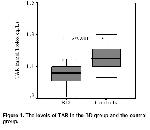
    TAR level was 1,04±0,07 and 1,16±0,08 mmol Trolox eq./L in the BD group and in the control group, respectively; TP level was 13,4±4,5 and 7,72±1,86 µmol H202/L in the BD group and in the control group, respectively; OSI value was 1,45±0,52 and 0,67±0,18 Arbitrary Units in the BD group and in the control group, respectively. The comparison between the BD group and the control group revealed that the level of TAR was significantly lower and TP and OSI levels were significantly higher in the former (p<0.001, for each, Table 2, Figures 1 and 2).


    Click Here to Zoom
    Figure 1: The levels of TAR in the BD group and the control group.


    Click Here to Zoom
    Figure 2: TP and OSI values in the BD group and the control group.

    The level of TAR was negatively correlated with TP (Figure 3) and OSI levels both in the BD group (r=-0.578, p<0.01 and r=-0.552, p<0.01, respectively) and the control group (r=-0.469, p<0.05 and r=-0.391, p<0.05, respectively). No significant correlation was found among duration of disease, and oxidant or antioxidant markers in the BD group (p>0.05, for each).


    Click Here to Zoom
    Figure 3: Correlation graphics between the level of TAR and the level of TP in BD group (a) and control group (b).

    Of the cases in the BD group, 100% (n:21) had oral ulcer, 57,1% (n:12) genital ulcer or scar, 90,4% (n:19) skin lesion, 28,5% (n:6) ocular involvement, 9,5% (n:2) vascular involvement, 4,7% (n:1) neurological involvement and 42,8% (n:9) positive pathergy test (Table 3).


    Click Here to Zoom
    Table 3: Clinical characteristics of patients with BD.

    The comparison between the cases who had positive pathergy test and negative pathergy test and between the cases with and without ocular involvement did not show significant difference in terms of the laboratory findings evaluated (p>0.05, for each). ESR was 28,3±22,5 and 7,2±5,4 mm/h and CRP level was 40,3±23,6 and 4,2±1,5 mg/L in the active (n:10) and inactive (n:11) BD groups, respectively. The increase in ESR and CRP in the active BD group was significant (p<0.01 and p<0.05, respectively). However, the difference between active and inactive BD groups in terms of albumin, bilirubin, uric acid, TAR, TP and OSI levels was not significant (p>0.05, for each).

  • Top
  • Summary
  • Introduction
  • Methods
  • Results
  • Disscussion
  • References
  • Discussion
    It was shown that ROS and other oxidants could be also formed in the normal physiological process 2,3. It is known that activated inflammatory cells cause ROS production in BD a systemic, autoimmune disease 8. Increased ROS, in turn, enhance LPO products, thus, lead to tissue injury 2-7. H2O2 and other derivatives of peroxides increase in some conditions, diffuse into plasma. Here, antioxidant components of plasma overwhelm them, and they are simultaneously consumed 15.

    When TP is measured, it means that the sum of many peroxides like protein peroxide, lipid peroxide and H2O2 are measured 24. Although it is known that H2O2 and lipid peroxides increase in BD 3,5-7, oxidative stress has not been evaluated through TP in BD. However, it has been reported that TP level increases in passive smokers 25, preeclampsia cases 26 and cutaneous leishmaniasis 27. It was shown in the present study that TP level also increased in BD. Possible reasons for this increase in TP might be the inevitable increase in lipid peroxides and ROS including H2O2 in BD.

    Many antioxidant molecules found in blood prevent or inhibit the harmful effects of free radicals 15. Whenever there is a decrease in antioxidants and/or an increase in oxidants, oxidant/antioxidant balance is impaired in favor of oxidants and this is known as oxidative stress 24,28. It is known that oxidative stress is responsible for tissue injury in many diseases and contributes to the development of atherosclerosis 13,14. Antioxidant activity indicates the antioxidant characteristics of only one antioxidant, whereas total antioxidant capacity (TAC) represents the total antioxidant characteristics of all antioxidants found in the plasma. TAR and total antioxidant status (TAS) are used synonymously with TAC 28. It is doubtlessly more advantageous to evaluate TAR, instead of individual antioxidant activities. Many methods have been developed recently for this aim. Total radical trapping antioxidant parameter (TRAP), oxygen radical absorbance capacity (ORAC) and ferric reducing antioxidant power (FRAP) are colorimetric methods previously developed to assess TAC 16,17,28. It has been reported that TAR, a new measurement method developed by Erel 16,17 correlated with data obtained by other measurement methods and has had some extra advantages 16,17.

    Blood has an important role in the oxidant/antioxidant balance, as it carries and distributes antioxidants through the body 28. Plasma has various antioxidant molecules. Albumin, uric acid, bilirubin and ascorbic acid are the major antioxidant components of plasma 16,17,28. TAR represents practically all of them 16,17. Albumin consists of about half of the TAC of the plasma 16,17. Albumin has several biological functions, particular as a ligand binder 29. Plasma thiol contents originate from albumin. Thiol groups, on the surface of albumin, bind oxidants 29. Low level of albumin can cause oxidative stress via leading to increase oxidants like homocystein 29. In chronic inflammation albumin is reduced. Bilirubin, a powerful endogenous antioxidant, is one of the catabolites of heme oxygenases 30. However, Harma et al. 26 have reported that bilirubin did not correlated with TAR, in their clinical study. Uric acid is another well-known low molecular weight water-soluble plasma antioxidant 16,17. Uric acid has a strong antioxidant activity and its concentration in the plasma is about 10 fold than antioxidants like vitamin C and vitamin E 28. In the present study, although there was a significant increase in total protein levels in the BD group, there was not any significant difference between the BD group and the control group in terms of the levels of such individual antioxidants as albumin, bilirubin and uric acid. It has been reported that total protein, bilirubin and uric acid levels correlated positively with TAC level 28. However, uric acid concentrations are influenced by age, diet, heavy exercise, renal failure and some metabolic diseases 28,31. Therefore, uric acid level may not appropriately reflect the TAC. However, it has been also reported that uric acid was not a strong antioxidant and might not protect against free radicals 32. Noyan et al. 21 have reported that while vitamin C levels reduced in BD oxidant MDA levels to be elevated, on the other hand, there is no changes in the levels of uric acid.

    Orem et al. 6 have reported a decrease in TAR level in BD. Similarly, TAR level was found low in the BD group in the present study. The increase in TP and OSI levels and the negative correlation between these increases and TAR level in the present study suggest that a possible cause of the decrease in TAR level may be increased oxidative stress. Since albumin, bilirubin and uric acid levels were not significantly different between the BD and control groups in the present study, the decrease in TAR is not possible to attribute to only these individual antioxidants. Therefore, the decrease in TAR must have resulted from the decrease in other antioxidants. It has been reported that activity of PON1 33 and level of SH 34, both of which are antioxidants, decreased in BD. Besides, Ece et al. 35 have noted that PON1 was positively correlated with TAR and negatively correlated with TP and OSI in cases with nephrotic syndrome. In the light of these data, a possible reason for the decrease in TAR in BD may be the decrease in other antioxidants, like PON1.

    Plasma TAR level has been reported to be lower in those with CVD, compared with those without CVD, in smokers, compared with non-smokers, in diabetic cases, compared with non-diabetic cases, and in hyperlipidemic cases, compared with those who have a normal lipid profile 36. Additionally, children who are exposed to passive cigarette smoking have been found to have a decrease in TAR level, and an increase in TP and OSI levels 25. It is known that oxidative stress is responsible for pathogenesis of atherosclerosis 37,38. As TAR is a fairly good representative of antioxidant capacity, and TP and OSI are representatives of oxidant capacity, decreased TAR and/or increased TP levels indicate oxidative stress 16,17,28. In the present study, there was a decrease in TAR level and an increase in TP and OSI levels in the BD group. In the light of these data, it is necessary to consider the possibility of development of atherosclerosis while evaluating BD cases.

    In consideration of the fact that increased ROS and LPO products as a result of inflammation can be responsible for the impairment in oxidant/antioxidant balance, the increase in ROS and LPO products and the decrease in antioxidant capacity are expected to be more marked in the active BD group, in comparison to the inactive BD group, due to marked inflammation. However, no significant difference was found between active and inactive BD groups with respect to TAR, TP, OSI, and levels of individual antioxidants, such as albumin, bilirubin and uric acid. This lack may be explained by the activity criteria used to determine the disease activity. It is known that currently there are no agreed activity criteria for BD. It is also possible that the oxidant/antioxidant balance is impaired at the onset of the disease and continues. In this study presented here, the fact that there is no relation between disease age and oxidant and antioxidant markers in the BD group may be supporting this theory.

    In conclusion, TAR is an appropriate measurement method demonstrating oxidant/antioxidant balance. Oxidant/antioxidant balance seems to be impaired at all stages of BD. In future, we think that this argument should be confirmed by controlled, multi-centered, prospective studies, which shall include large case series and employ reviewed disease activity criteria.

  • Top
  • Summary
  • Introduction
  • Methods
  • Results
  • Discussion
  • References
  • References

    1) Sakane T, Takeno M, Suzuki N, Inaba G. Behcet's disease. N Engl J Med. 1999; 341(17):1284-91.

    2) Onder M, Gurer MA. The multiple faces of Behcet's disease and its aetiological factors. J Eur Acad Dermatol Venereol. 2001; 15(2):126-36.

    3) Orem A, Efe H, Deger O, Cimsit G, Uydu HA, Vanizor B. Relationship between lipid peroxidation and disease activity in patients with Behcet's disease. J Dermatol Sci. 1997;16(1):11-6.

    4) Chambers JC, Haskard DO, Kooner JS. Vascular endothelial function and oxidative stress mechanisms in patients with Behcet's syndrome. J Am Coll Cardiol. 2001; 37(2):517-20.

    5) Kokcam I, Naziroglu M. Effects of vitamin E supplementation on blood antioxidants levels in patients with Behcet's disease. Clin Biochem. 2002; 35(8):633-9.

    6) Orem A, Yandi YE, Vanizor B, Cimsit G, Uydu HA, Malkoc M. The evaluation of autoantibodies against oxidatively modified low-density lipoprotein (LDL), susceptibility of LDL to oxidation, serum lipids and lipid hydroperoxide levels, total antioxidant status, antioxidant enzyme activities, and endothelial dysfunction in patients with Behcet's disease. Clin Biochem. 2002;35(3):217-24.

    7) Yazici C, Kose K, Calis M, Demir M, Kirnap M, Ates F. Increased advanced oxidation protein products in Behcet's disease: a new activity marker. Br J Dermatol. 2004; 151(1):105-11.

    8) Weiss SJ. Tissue destruction by neutrophils. N Engl J Med. 1989;320(6):365-76.

    9) Saglam K, Serce AF, Yilmaz MI, et al. Trace elements and antioxidant enzymes in Behcet's disease. Rheumatol Int. 2002; 22(3):93-6.

    10) Kose K, Yazici C, Cambay N, Ascioglu O, Dogan P. Lipid peroxidation and erythrocyte antioxidant enzymes in patients with Behcet's disease. Tohoku J Exp Med. 2002; 197(1):9-16.

    11) Dogan P, Tanrikulu G, Soyuer U, Kose K. Oxidative enzymes of polymorphonuclear leucocytes and plasma fibrinogen, ceruloplasmin, and copper levels in Behcet's disease. Clin Biochem. 1994; 27(5):413-8.

    12) Niwa Y, Miyake S, Sakane T, Shingu M, Yokoyama M. Auto-oxidative damage in Behcet's disease--endothelial cell damage following the elevated oxygen radicals generated by stimulated neutrophils. Clin Exp Immunol. 1982; 49(1):247-55.

    13) Witztum JL, Steinberg D. Role of oxidized low density lipoprotein in atherogenesis. J Clin Invest. 1991; 88(6):1785-92.

    14) Yla-Herttuala S, Palinski W, Butler SW, Picard S, Steinberg D, Witztum JL. Rabbit and human atherosclerotic lesions contain IgG that recognizes epitopes of oxidized LDL. Arterioscler Thromb. 1994; 14(1):32-40.

    15) Young IS, Woodside JV. Antioxidants in health and disease. J Clin Pathol. 2001; 54(3):176-86.

    16) Erel O. A novel automated direct measurement method for total antioxidant capacity using a new generation, more stable ABTS radical cation. Clin Biochem. 2004; 37(4):277-

    85)

    17) Erel O. A novel automated method to measure total antioxidant response against potent free radical reactions. Clin Biochem. 2004; 37(2):112-9.

    18) Harma M, Harma M, Erel O. Increased oxidative stress in patients with hydatidiform mole. Swiss Med Wkly. 2003; 133(41-42):563-6.

    19) The International Study Group for Behcet's disease. Evaluation of diagnostic ('classification') criteria in Behcet's disease--towards internationally agreed criteria. Br J Rheumatol. 1992; 31(5):299-308.

    20) Bekpinar S, Kilic N, Unlucerci Y, Akdag-Kose A, Azizlerli G, Ozbek-Kir Z. Evaluation of nitrosative and oxidative stress in Behcet disease. J Eur Acad Dermatol Venereol. 2005; 19(2):167-71.

    21) Noyan T, Sahin I, Sekeroglu MR, Dulger H. The serum vitamin C levels in Behcet's disease. Yonsei Med J. 2003; 44(5):771-8.

    22) Cao G, Prior RL. Comparison of different analytical methods for assessing total antioxidant capacity of human serum. Clin Chem. 1998; 44(6 Pt 1):1309-15.

    23) Miyazawa T, Saeki R, Inaba H. Detection of chemiluminescence in lipid peroxidation of biological systems and its application to HPLC. J Biolumin Chemilumin. 1989; 4(1):475-8.

    24) Abuja PM, Albertini R. Methods for monitoring oxidative stress, lipid peroxidation and oxidation resistance of lipoproteins. Clin Chim Acta. 2001; 306(1-2):1-17.

    25) Kosecik M, Erel O, Sevinc E, Selek S. Increased oxidative stress in children exposed to passive smoking. Int J Cardiol. 2005; 100(1):61-4.

    26) Harma M, Harma M, Erel O. Measurement of the total antioxidant response in preeclampsia with a novel automated method. Eur J Obstet Gynecol Reprod Biol. 2005; 118(1):47-51.

    27) Kocyigit A, Keles H, Selek S, Guzel S, Celik H, Erel O. Increased DNA damage and oxidative stress in patients with cutaneous leishmaniasis. Mutat Res. 2005; 585:71-8.

    28) Ghiselli A, Serafini M, Natella F, Scaccini C. Total antioxidant capacity as a tool to assess redox status: critical view and experimental data. Free Radic Biol Med. 2000; 29(11):1106-14.

    29) Sengupta S, Wehbe C, Majors AK, Ketterer ME, DiBello PM, Jacobsen DW. Relative roles of albumin and ceruloplasmin in the formation of homocystine, homocysteine-cysteine-mixed disulfide, and cystine in circulation. J Biol Chem. 2001; 276(50):46896-904.

    30) Dohi K, Satoh K, Ohtaki H, et al. Elevated plasma levels of bilirubin in patients with neurotrauma reflect its pathophysiological role in free radical scavenging. In Vivo. 2005;19(5):855-60.

    31) Puig JG, Mateos FA, Ramos TH, Capitan CF, Michan AA, Mantilla JM. Sex differences in uric acid metabolism in adults: evidence for a lack of influence of estradiol-17 beta (E2). Adv Exp Med Biol. 1986; 195:317-23.

    32) Smolenska Z, Kaznowska Z, Zarowny D, Simmonds HA, Smolenski RT. Effect of methotrexate on blood purine and pyrimidine levels in patients with rheumatoid arthritis. Rheumatology (Oxford). 1999; 38(10):997-1002.

    33) Karakucuk S, Baskol G, Oner AO, Baskol M, Mirza E, Ustdal M. Serum paraoxonase activity is decreased in the active stage of Behcet's disease. Br J Ophthalmol. 2004; 88(10):1256-8.

    34) Kose K, Dogan P, Ascioglu M, Erkilic K, Ascioglu O. Oxidative stress and antioxidant defenses in plasma of patients with Behcet's disease. Tohoku J Exp Med. 1995; 176(4):239-48.

    35) Ece A, Atamer Y, Gurkan F, Davutoglu M, Kocyigit Y, Tutanc M. Paraoxonase, total antioxidant response, and peroxide levels in children with steroid-sensitive nephrotic syndrome. Pediatr Nephrol. 2005; 20(9):1279-84.

    36) Demirbag R, Yilmaz R, Kocyigit A. Relationship between DNA damage, total antioxidant capacity and coronary artery disease. Mutat Res. 2005; 570(2):197-203.

    37) Madamanchi NR, Hakim ZS, Runge MS. Oxidative stress in atherogenesis and arterial thrombosis: the disconnect between cellular studies and clinical outcomes. J Thromb Haemost. 2005; 3(2):254-67.

    38) Violi F, Cangemi R. Antioxidants and cardiovascular disease. Curr Opin Investig Drugs. 2005; 6(9): 895-900.

  • Top
  • Summary
  • Introduction
  • Methods
  • Results
  • Discussion
  • References
  • [ Başa Dön ] [ Özet ] [ PDF ] [ Benzer Makaleler ] [ Yazara E-Posta ] [ Editöre E-Posta ]
    [ Ana Sayfa | Editörler | Danışma Kurulu | Dergi Hakkında | İçindekiler | Arşiv | Yayın Arama | Yazarlara Bilgi | E-Posta ]