 |
 |
| [ Ana Sayfa | Editörler | Danışma Kurulu | Dergi Hakkında | İçindekiler | Arşiv | Yayın Arama | Yazarlara Bilgi | E-Posta ] | |
| Fırat Üniversitesi Sağlık Bilimleri Veteriner Dergisi | |||||||||||||||||
| 2007, Cilt 21, Sayı 4, Sayfa(lar) 163-166 | |||||||||||||||||
| [ Özet ] [ PDF ] [ Benzer Makaleler ] [ Yazara E-Posta ] [ Editöre E-Posta ] | |||||||||||||||||
| Morphological And Morphometric Approach To The Bones Of The Wings İn The Long-Legged Buzzard (Buteo Rufinus) | |||||||||||||||||
| Ömer ATALAR1, İbrahim KÜRTÜL2, Derviş ÖZDEMİR3 | |||||||||||||||||
| 1Fırat Üniversitesi Veteriner Fakültesi, Anatomi Anabilim Dalı, Elazığ-TÜRKİYE 2Kafkas Üniversitesi Veteriner Fakültesi, Anatomi Anabilim Dalı, Kars-TÜRKİYE 3Atatürk Üniversitesi Veteriner Fakültesi, Anatomi Anabilim Dalı, Erzurum-TÜRKİYE |
|||||||||||||||||
| Keywords: Bones of the wings, long-legged buzzard | |||||||||||||||||
| Summary | |||||||||||||||||
In this study it was investigated the morphometric and morphological features of the bones of the wings in two long-legged buzzards by maceration. The findings on the humerus complied well with the literature reports except the smaller dorsal condyle on the humeral trochlea and ventral supracondylar tubercle at the distal end of the humerus. The ventral remigal papillae on the ulna were hardly observable and well less developed even though the long-legged buzzard is in the class of very strong flyers. The features of the bones of the manus skeleton were also similar with the general pattern of avian anatomy. This study aimed to identify the data using these bones in order to separete different bird species. |
|||||||||||||||||
| Introduction | |||||||||||||||||
Forelimbs of the avian species have been adapted to function as wings. This long time evolution has had dramatic morphological changes on the bones of the forelimbs in avian species as compared to those of other tetrapoda. Characteristics of the bones of the forelimbs in domestic avian species have long been observed by the researchers and documented in literature reports and textbooks 1-4. On the other hand, research on wild avian species is very limited. This has recently raised a question on what the similarities and dissimilarities of the gross anatomical structures of wild avian species are, if compared to those of domestic birds. Furthermore, recent researches on gross morphological observations have focused particularly on wild birds 5. Likewise, this study aimed at documenting the morphometric and morphological features of the bones of the wings in the long-legged buzzard, Buteo rufinus, whose live climates extend from North Africa through Mid Asia 6. Results of the present study may well contribute to the veterinary gross anatomy, which might be a valuable contribution to the veterinary gross anatomy literature. |
|||||||||||||||||
| Methods | |||||||||||||||||
The forelimbs of the two dead long-legged buzzard were macerated in boiling water. The bones were cleaned and dried. Morphometric data were obtained by a digital oculometer (Mitutoyo, Japan). Nomina Anatomica Avium,1993 7 was followed for the anatomical nomenclature. |
|||||||||||||||||
| Results | |||||||||||||||||
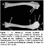
The humerus (Fig. 1), a highly pneumatic and strong one among the bones of the wing, composed of prominently developed dorsal and ventral tubercles.
Well developed capital groove was present, located between the articular surface of the head of the humerus and ventral tubercle. The deltopectoral crest extended down to one third of the bone and was stronger than bicipital crest. The transversal groove resembled a deep fovea. The prominent transverse groove for the coracobrachial nerve was present at the distal aspect of the intertubercular plane of the humerus. The pneumotricipital fossa was a highly large and deeply developed excavation. The pneumatic foramen was single and wide. Caudally located fossa for the dorsal and ventral crura, and cranially bounded coracobrachial impression were highly distinct structures. At the distal end of the humerus, a well developed ventral condyle and a smaller dorsal condyle were present on the humeral trochlea. Separating the dorsal and ventral condyles of the humerus, the intercondylar notch was very deep. The dorsal supracondylar tubercle was distinct while the ventral one was relatively less developed. A deep fossa for the brachial muscle was present on the ventral aspect of the distal extremity of the humerus. On the other hand, a small and shallow scapulotricipital groove and a deep humerotricipital groove were observed on the dorsal aspect of the distal end of the humerus. Some measurements of the humerus are displayed in table 1.
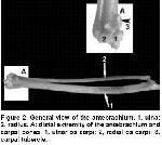
Antebrachium (Fig. 2) was the longest bone of the wing, possessing the longer and stronger ulna (Fig.2/1) and smaller and medially located radius (Fig. 2/2). The spatium between the two was proximally wider. The olecranon was less developed while the radial incisura was prominent. The ventral remigal papillae for attachment for the ligaments of the follicles of the secondary flight feathers were hardly observable. The carpal tubercle on the ventral aspect of the distal end of the ulna was a well developed structure and the radial depression on the dorsal aspect of the ulna showed a wide articular surface. Some measurements of the ulna and radius are shown in table 2 and 3, respectively.
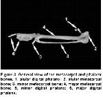
The proximal extremity of the radius was narrower than the ventral one. The humeral cotyla completely covered the dorsal surface of the head of the radius. The bicipital tubercle on the proximal extremity of the radius was well developed. The intermuscular lineae on the body of the radius were also highly distinct features. On the distal extremity of the radius, the tubercle for the ventral aponeurosis was apparent and a single deep tendinous groove was present on the dorsal aspect of the distal extremity of the radius. There was a larger and prominent ulnar os carpi (Fig. 2A/1) and a smaller and less developed radial os carpi (Fig. 2A/2). The major metacarpal bone (Fig. 3/4) was a strong and smooth bone while the minor metacarpal one (Fig. 3/3) was weak, possessing a slightly concave structure in nature. The intermetacarpal space was very narrow. The alular metacarpal bone (Fig. 3/2) was also present and well developed. Some measurements of the major and minor metacarpal bones are depicted in Tables 4 and 5, respectively.
The major digital phalanx (Fig. 3/6) possessing two distinct bones was the largest bone among the ossa digiti manus. The minor digital phalanx (Fig. 3/5) and bones of the alular digital phalanx (Fig. 3/1) were also present. |
|||||||||||||||||
| Discussion | |||||||||||||||||
The process of the evolution on the fact that the forelimbs of the avian species have been adapted to function as wings is indicated by the literature reports to differ greatly in accordance with the motion of species either flying or walking 3,4,7. These researches have pointed out that the bony features for attachment for the flight muscles specifically on the humerus are very compact and developed in long flyers. The findings in this observation comply well with the literature reports; that is, the features are eminent and highly developed except the smaller dorsal condyle on the humeral trochlea and ventral supracondylar tubercle at the distal end of the humerus. Likewise, literatures 3,4,7 reported that the ventral remigal papillae for attachment for the ligaments of the follicles of the secondary flight feathers should be very prominent and greatly developed in strong flyers and reduced in non-flyers. These features in the present study on the long-legged buzzard which is even though in the class of very strong flyers were, on the other hand, hardly observable and well less developed, resembling those of walking birds. The features of the bones of the manus skeleton in the long-legged buzzard observed hereby complied at gross anatomical aspect with the general pattern of avian anatomy. An example of that is the width of the intermetacarpal space. Literature reports 4,5,8,9 have indicated that it is narrow in flying birds and very wide in walking ones. This example again rendered the long-legged buzzard as a strong flyer. Numerical data obtained in this study was not statistically analyzed; thus, average values of each feature are shown in tables since the number of the animal used here was only two already dead animals. This is for the fact that some of the wild animals such as the long-legged buzzards are not easily obtainable since their populations have been decreasing and they are endangered species. As a consequence, the findings on the humerus were in parallel with the literature reports except the smaller dorsal condyle on the humeral trochlea and ventral supracondylar tubercle at the distal end of the humerus. The ventral remigal papillae for attachment for the ligaments of the follicles of the secondary flight feathers were hardly observable and well less developed even though the long-legged buzzard is in the class of very strong flyers. The features of the bones of the manus skeleton were also similar with the general pattern of avian anatomy. |
|||||||||||||||||
| References | |||||||||||||||||
1) Nickel R, Schummer A, Seiferle E. The Skin Anatomy of the Domestic Birds. Berlin: Verlag Paul Parey, 1977: 14-15.
2) King AS, Mclelland J. Birds, Their Structure and Function, 2nd ed. London: Bailliere Tindall, 1984: 58-60.
3) Hifny A, Abdalla KEH, Alam El-Din MA. Relation of the length of the main bones of the wing and pelvic limb to the mode of locomotion in certain birds. Accepted and discussed. Proc. of XVII Kongress. European Association of Veterinary anatomists, 1988; 28: 8-19.
4) Abdel-Moneim ME. Role of the bones of the wing and pelvic limb of quails in its mode of locomotion. Assuit Vet Med J 1992; 27: 1-11.
5) Bozkurt EU, Duzler A, Ozgel O, Kurtul I. Morphometric and morphological features of the bones of the wing in bald ibis. Indian Vet J 2002; 79: 470-476.
6) Demirsoy A. Basic Rules of Life, Vertebrates/Amniota (Reptiles, Birds, Mammals), Part: III/II. Ankara: Meteksan, 1992: 345-346.
7) Nomina Anatomica Avium.: International Committee on Avian Anatomical Nomenclature. A Committee of the World Association of Veterinary Anatomists, A Handbook of Avian Anatomy, 2nd ed., No.23 Cambridge MA: The Nuttall Ornithological Club, 1993.
|
|||||||||||||||||
| [ Başa Dön ] [ Özet ] [ PDF ] [ Benzer Makaleler ] [ Yazara E-Posta ] [ Editöre E-Posta ] | |||||||||||||||||
 |
| [ Ana Sayfa | Editörler | Danışma Kurulu | Dergi Hakkında | İçindekiler | Arşiv | Yayın Arama | Yazarlara Bilgi | E-Posta ] |


