[ Ana Sayfa | Editörler | Danışma Kurulu | Dergi Hakkında | İçindekiler | Arşiv | Yayın Arama | Yazarlara Bilgi | E-Posta ]
Fırat Üniversitesi Sağlık Bilimleri Veteriner Dergisi
2007, Cilt 21, Sayı 6, Sayfa(lar) 281-284
[ Özet ] [ PDF ] [ Benzer Makaleler ] [ Yazara E-Posta ] [ Editöre E-Posta ]
Pulmonary Sequestration and Bronchogenic Cyst in a Calf
İbrahim CANPOLAT1, Yesari ERÖKSÜZ2
1Fırat Üniversitesi Veteriner Fakültesi, Cerrahi Anabilim Dalı, Elazığ -TÜRKİYE
2Fırat Üniversitesi Veteriner Fakültesi, Patoloji Anabilim Dalı, Elazığ -TÜRKİYE
Keywords: Pulmonary sequestration, bronchogenic cyst, calf
Summary
Two-day-old, Simmental calf was presented with a pedinculated subcutaneous mass arising from midline of lower cervical region. The mass covered by the skin was removed surgically. Histological features of the mass were of underdeveloped lung tissue containing bronchogenic cyst. The bronchioles had excessive in number and the bronchi were hypoplastic and had no cartilagonous ring. The concurrent occurrence of a subcutaneous pulmonary sequestration and a bronchogenic cyst was noted as bronchopulmonary foregut malformation in a calf. As a result, this case report indicated that pulmonary sequestration and bronchonic cyst have common embryonic pathogenesis.
  • Top
  • Summary
  • Introduction
  • Case Presentation
  • Disscussion
  • References
  • Introduction
    The prevalence of congenital bronchopulmanory malformations is unknown in animal species, but they generally are thought to occur rarely. Probably, the most commonly reported bovine bronchopulmonary abnormality is abnormal mass of nonfunctioning lung tissue development apart from the original site of lung including abdominal, thoracic or subcutaneous locations. The term of ‘ectopic or accessoric lung' has been used to describe to this lesion in standard veterinary pathology textbooks 1,2,3, however various terms such as heterotopic lung, ectopic lung, bronchopulmonary foregut malformation or pulmonary sequestration have been used interchangebaly to describe this abnormality in separate bovine case reports 4,5,6,7.

    In human medical literature, the same term ‘accessory lung or accessory lobe' was renamed as pulmonary sequestration to define abnormal mass of nonfunctioning development of lung tissue anywhere in the body 8. Bronchogenic cysts are another congenital anomalies resulting from anomalous development of the ventral foregut; they are usually single but may be multiple. They have been found all along the tracheoesophageal course, pulmonary parenchyma, neck, abdomen, and lumbar regions 4. To the best of authors' knowledge, as a lung bud malformation, no concurrent occurrence of pulmonary sequestration and bronchogenic cyst is available in veterinary literature, whereas in human medicine, concurrent occurrence of bronchogenic cyst and pulmonary sequestration was reported in only 3 among the approximately 900 cases of pulmonary sequestrations 9. This case report describes morphologically a subcutaneous lung sequestration enmeshed with bronchogenic cyst in a calf.

  • Top
  • Summary
  • Introduction
  • Case Presentation
  • Disscussion
  • References
  • Case Presentation
    Two-day-old, Simmental crossbred calf was presented with a pedinculated subcutaneous mass situated on the midline of lower cervical region at aperture thoracis cranialis. The calf was fully developed female and born at full term.

    The mass which had initially been noted on the day of parturition was not associated with dysphagie, airway obstruction or previous trauma. It was ovoid appearance with 5-6 cm in diameter and freely movable and was covered and well protected by the skin. Cytological examination of the needle aspiration revealed the presence of scattered macrophages and a few neutrophil leucocytes. Ventro-dorsal and latero-lateral chest radiographs showed that the mass located in sternal manibrium. After injection of contrast medium, the mass did not showed enchantment. On request of surgical removal of the mass by the owner, the operation site on the lower neck was prepared and after premedication and local infiltration anesthesia, a 15 cm straight incision was made centered over the mass, attached to apertura thoracis cranialis by a fibrovascular pedicle containing an artery and a ligamenteous structure.

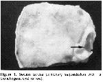
    The artery was approximately 5-6 mm in diameter. There was apperently no communication with gastrointestinal tract. Following the proper ligation, the mass was completely removed. It weighed 350 g, had fleshy and edematous appereance and measured 5x6x6 cm. Serial sectioning of the specimen revealed a multiocalar cyst, approximately 1 cm in diameter, it's inner surface was separeted and white, containing serous and clear fluid (Fig-1). Formalin fixed tissue samples were processed routinely and paraffin embedded tissue samples were stained with hematoxyline-eosin.


    Click Here to Zoom
    Figure 1: Section across pulmonary sequestration with a bronchogenic cyst (arrow).

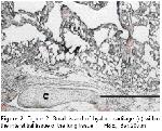
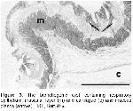
    Histological examination of the tissue specimens revealed that the mass was underdeveloped lung tissue which was rich in loose connective tissue along with brochioles and alveolar structures. The lung tissue was covered by both pleura and the skin. Microscopically, the bronchioles were present in excessive number and dilated apparence containing simple columnar or cuboidal epithelium. The brochi were hypoplastic and had no cartilagonous ring. The alveoli were also not fully developed and some alveoli and respiratory bronchioles were dilated. Alveolar spaces contained focally hemosiderin laden macrophages. Focal irregular lymphoid aggregates in paranchymal tissue were also present. Mild to moderate peribronchialar fibrosis and isolated cartiloganeous tissue were the additional findings detected (Figure-2 ). The cyst wall was composed of ciliated pseudostatified columnar epithelium, hyaline cartilagae and mucous glands (Fig-3).


    Click Here to Zoom
    Figure 2: Figure 2. Small island of hyaline cartilage (c) within the interstitial tissue of the lung tissue, H&E, Bar:20m.m


    Click Here to Zoom
    Figure 3: The bronchogenic cyst containing respiratory epithelium, muscular layer (m) and cartilagae (c) and mucous glands (arrows), H&E, Bar:50 m.

  • Top
  • Summary
  • Introduction
  • Case Presentation
  • Disscussion
  • References
  • Discussion
    There are 31 cases of bovine pulmonary sequestration in the literature. Sixteen of the cases (52.41%) are intra-abdominal sequestration, whereas 3 (9.68%) and 12 cases (38.71%) showed intrathoracal and subcutaneous locations, respectively. Two of the cases were fetus, one was 2-years-old and 5 were 2-5 weeks of age, and the remaining 23 calves were newborn. Subcutaneous sequetrations were located on neck, sacrum, shoulder (3 cases), thoracic inlet, head, left side of sternum, lumbar (2 cases), anus and unknown 4,5,6,7. Referring particularly to subcutaneous sequestrations, the overall good prognosis is determined by only few secondary congenital skletal defects due to pressure of the sequestrated lung and difficulty in delivery 5,7. Histological features of the sequestrated lung was similar to those previosly reported that bronchial hypoplasia and poorly developed alveoli were co-existent findings in almost all of bovine pulmonary sequestrations cases 7. Both pulmonary sequestration and bronchogenic cyst are considered as foregut malformation. The theory concerning how foregut malformation arise is that they are formed by extranumary lung bud that arises caudal to normal lung bud, and migrates caudally along with growing eosophagus 8. The cyst was apparently fulfilled to the histological criteria of the bronchogenic cyst such as pseudostrafied epithelium, hyaline cartilago and muscular layer 8,9. Although, small island of hyaline cartilage within the interstitium has not been reported in bovine sequestration cases, it was reported in pulmonary sequestration cases in man 10. The findings of present study showed that pulmonary sequestration and bronchonic cyst have common embryonic pathogenesis.
  • Top
  • Summary
  • Introduction
  • Case Presentation
  • Discussion
  • References
  • References

    1) Dungworth DL The Respiratory System. In: Jubb KVF., Kennedy PC and Palmer N (Editors). Pathology of Domestic Animals 2, 3rd ed., London, Academic Press Inc., 1985: 442-450.

    2) Jones TC, Hunt, RD and King, NW Veterinary Pathology. 6th ed., Baltimore, Williams and Wilkins, 1997.

    3) Lopez, A Respiratory System. In: Carlton WW and McGavin MD (Editors). Thomson's Special Veterinary Patholog 2rd ed., Missouri, Mosby-Year Book Inc.,1995:448-492.

    4) Brown PJ, Cooke PM, Lucke VM, Pearsom H, Wardle GB. Congenital bronchopulmonary foregut malformations in two young Friesian cattle. Vet Rec 1988; 122: 208-209.

    5) Havenstrite AK, Barry MJ, Fitzpatrick TM and O'Neil KM. Simultaneous presentation of a bronchogenic cyst and pulmonary sequestration. Chest 2000; 118: 298-299.

    6) Lucas MJ, Johnson LW, Haley PJ. Ectopic lung tissue arising from the head of neonatal calf. Agri Practice 1984; 5: 45-47.

    7) Thomson RG. Congenital bronchial hypoplasia in calves. Path Vet 1966; 3: 89-109.

    8) Hasleton PS. Spencer's Pathology of the Lung , the McGraw-Hill Companies, New York, 1996, 69-78.

    9) Koç Y, Ogurtan Z, Tuzcu M, Alkan F and Ciftci MK. Heterotypic pulmonary anomaly in paralumbar region of a calf. Veteriner Bilimleri Dergisi 1998; 14: 151-154.

    10) Fraggetta F, Davenport M, Magro G, Cacciaguerra S, Nash R. Striated muscle cells in noo-neoplastic lung tissue: A clinicopathologic study. Human Pathology 2000; 31: 1477-1481.

  • Top
  • Summary
  • Introduction
  • Case Presentation
  • Discussion
  • References
  • [ Başa Dön ] [ Özet ] [ PDF ] [ Benzer Makaleler ] [ Yazara E-Posta ] [ Editöre E-Posta ]
    [ Ana Sayfa | Editörler | Danışma Kurulu | Dergi Hakkında | İçindekiler | Arşiv | Yayın Arama | Yazarlara Bilgi | E-Posta ]