[ Ana Sayfa | Editörler | Danışma Kurulu | Dergi Hakkında | İçindekiler | Arşiv | Yayın Arama | Yazarlara Bilgi | E-Posta ]
Fırat Üniversitesi Sağlık Bilimleri Veteriner Dergisi
2008, Cilt 22, Sayı 1, Sayfa(lar) 013-016
[ Özet ] [ PDF ] [ Benzer Makaleler ] [ Yazara E-Posta ] [ Editöre E-Posta ]
The Localization of Insulin, Glucagon, Somatostatin Immunoreactive Cells in the Pancreas of the Martes Foina
Meryem KARAN1, Sema TİMURKAAN2, Ömer ATALAR1
1University of Firat Veterinary Medicine Department of, Anatomy Elazig-TURKEY
2University of Firat Veterinary Medicine Department of, Histology and Embryology Elazig-TURKEY
Keywords: Immunohistochemistry; insulin; glucagon; somatostatin; pancreatic islets; Martes foina
Summary
In this study, three kinds of the immunoreactive endocrine cells were detected using to insulin, glucagon and somatostatin antisera in the pancreas of the Martes foina. Different regional distributions and relative frequencies of these immunoreactive cells were observed in the different pancreatic regions. Insulin immunoreactive cells were located in the central regions with high frequency, and a few of these cells were also demonstrated in the mantle zones. Glucagon immunoreactive cells were mainly restricted to mantle zones but a few of these cells were also demonstrated in the central zones. Howewer; rare examples were found in the peripheral regions. Somatostatin immunoreactive cells were detected in the mantle zones and peripheral regions with moderate and rare frequencies, respectively. However, somatostatin immunoreactive cells were not demonstrated in the exocrine portions.
  • Top
  • Summary
  • Introduction
  • Methods
  • Results
  • Disscussion
  • References
  • Introduction
    The marten, which is the subject of this study, belongs to the Mustelidae family, order carnivora. Beech martens or stone martens range in coloration from dark brown to pale grayish brown. The limbs are long, a bushy tail is present 1,2.

    Functionally, the pancreas is divided into 2 portions, the exocrine portion, from which digestive enzymes are released and the endocrine portion, from which regulatory hormones, such as insulin, glucagon, somatostatin and pancreatic polypeptide (PP), are released into the blood circulation. The appearance, regional dispersion and relative frequency of these regulatory hormones secreted by endocrine cells in the pancreas are well recognized by their histochemistry using 3, immunofluorescence method 4 and immunohistochemistry 5,6. In addition, investigations of gastroenteropancreatic (GEP) endocrine cells are considered to be an important part of phylogenetic study 7,8.

    The pancreas has been treated as a valuable organ for endocrine studies and endocrine pancreas has been extensively studied , associated with diabetes 3,9. Until now, the regional distribution and relative frequency of four major immunoreactive cells insulin, glucagon, somatostatin and pacreatic polypeptide (PP) have been reported in the pancreas of the rodents such as hamster 10, sand rat 11, C57BL/6 mouse 9, guinea pig 12, gerbil 13, and wood Mouse 14.

    It has been accepted that insulin- immunoreactive cells are located in the central regions and that THY other immunoreactive cells such as glucagon, somatostatin immunoreactive cells are in located in the peripheral or mantle zones. But, many researches suggested that species-dependent characteristic distribution of cells mantle zones. But, many researches suggested that species-dependent characteristic distribution of cells producing different hormones in the pancreas of each species of animals might due to feeding habits and this is now generally accepted 15.

    The pancreatic endocrine system has been widely studied by means of immunocytochemistry 13,16-18, there have been no reports on immunohistochemical studies into the endocrine cells of the pancreatic islets of the marten. The object of the present study was to clarify the regional distribution and relative frequency of endocrine cells in the pancreatic islets of the marten, Martes foina, using an immunohistochemical method (ABC) and three types of specific antibody against insulin, glucagon, somatostatin.

  • Top
  • Summary
  • Introduction
  • Methods
  • Results
  • Disscussion
  • References
  • Methods
    Six adult beech or stone martens (Martes foina Erxleben, 1777) were used without sexual distinction in this study. Under penthotal-induced (6 ml/kg) anaesthesia, the pancreas was removed immediately and placed in 10% formalin in phosphate-buffered saline (PBS), pH 7,4, for 18 hours before paraffin embedding. Tissues were routinely processed through a graded series of alcohols, cleared in xylol and embedded in paraffin. 5mm thick sections were obtained and processed for immunohistochemical staining.

    Immunohistochemistry: Immunohistochemical staining was carried out by the peroxidase linked avidin-biotin complex (ABC) method. Blocking of endogenous peroxidase activity was carried out with % 0.08 hydrogen peroxidase (H2O2) in methanol for 5 minutes. In order to the block unspecific binding, an incubation with (1:10) normal goat serum in 0.1 M PBS, pH 7.2 was performed.

    ABC Technique: Sections were incubated for 16-20 hrs at 4 ºC in mouse anti-insulin IgG (Sigma), anti-glucagon IgG (Sigma) and anti-somatostatin IgG. The antibodies were diluted to 1:500 in PBS containing % 0.25 sodium azide and % 2.5 bovine serum albumin. Sections were then incubated in biotinylated sheep anti-mouse IgG (Sigma) and to follow with streptavidin horseradish peroxidase (Dako), both at a dilution of 1:50 in PBS, for 1 hr at room temperature. Sections were washed in PBS for 30 minutes after each incubation. Sections were then immersed in glucose oxidase- DAB-nickel ammonium sulphate (GDN) substrate 19 for 10 minutes, washed in distilled water and counterstained with eosine. Sections were examined with light microscope and photomicrograps were taken.

    Controls were made by replacing each specific antiserum with normal rabbit or goat serum. No immunoreactive structures were observed.

  • Top
  • Summary
  • Introduction
  • Methods
  • Results
  • Disscussion
  • References
  • Results
    In this study, three kinds of the immunoreactive endocrine cells were detected using antisera to insulin, glukagon and somatostatin in the pacreas of the marten. The principal islets were often clearly distinguishable in the central, mantle and the peripheral regions by their cellular composition. Different regional distributions and relative frequencies of these immunoreactive cells were observed in the different pancreatic regions, and these differences were shown in table1. Spherical to spindle or occasionally oval to round-shaped insulin, glukagon and somatostatin immunoreactive cells were observed in this study.


    Click Here to Zoom
    Table 1: Regional distributions and relative frequencies of the endocrine cells in the pancreas of marten. --: not detectable, +: rare, ++: a few, +++: numerous.

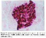
    Insulin immunoreactive cells were situated in the central regions with high frequency, and a few of these cells were also demonstrated in the mantle zones. Spherical to spindle shaped insulin- immunoreactive cells were located in the central pancreatic islet region with numerous frequency and rarely round to oval shaped cells of variable size were also observed. In addition, a few frequency cells were also observed in the mantle zone intermingled with other immunoreactive cells, especially glucagon and somatostatin immunoreactive cells. However, no insulin- immunoreactive cells were found in the peripheral regions and exocrine portions (Fig 1).


    Click Here to Zoom
    Figure 1: Insulin-immunoreactive cells in the pancreatic islets of Martes foina. Insulin-positive cells appear as intensely labelled, dark cells. 40 x 5.

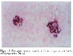
    Glucagon immunoreactive cells were mainly restricted to the mantle zones. In addition, a few of these cells were also demonstrated in the central zones (Fig 2). Howewer; rare examples were found in the peripheral regions and, no glucagon immunoreactive cells were found in the exocrine portions.


    Click Here to Zoom
    Figure 2: Glucagon-immunoreactive cells in the pancreatic islets of Martes foina. 20 x 5.

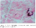
    Somatostatin immunoreactive cells were detected in the mantle zones and peripheral regions with moderate and rare frequencies, respectively. However, also a few somatostatin immunoreactive cells were observed in the central regions where numerous insulin immunoreactive cells were found (Fig 3). Somatostatin immunoreactive cells were not demonstrated in the exocrine portions.


    Click Here to Zoom
    Figure 3: Cells containing somatostatin were scattered throughout the islets. 40 x 5.

  • Top
  • Summary
  • Introduction
  • Methods
  • Results
  • Disscussion
  • References
  • Discussion
    This study revealed that the principal pancreatic islets of the Martes foina contained insulin, glukagon and somatostatin immunoreactive-cells. Additionally, somewhat different distributional patterns of these three types of immunoreactive cells were observed in the Martes foina.

    Insulin is synthesized in the B cells of the pancreatic islets and regulates the serum glucose levels 20. In the mammals, the regional distribution and relative frequency of insulin immunoreactive cells in the pancreas were reported in the hamster 10, C57BL/6 mouse 11, gerbil 13, wood Mouse 14, voles 21, opossum 22, and various laboratory animals 15. From these reports, it is well recognized that insulin immunoreactive cells are situated in the central regions of mammalian pancreatic islets and that other cells, such as glucagon and somatostatin immunoreactive cells, surrounded them. However, somewhat contradicting the finding of other researchers, Redy et al. 23 reported that these immunoreactive cells are observed in the majority of islets where they occur peripherally as groups of cells and within the pancreatic islets of several marsupial species. In the present study, most of the insulin immunoreactive cells were restricted to central regions of islets in the Martes foina, which is similar to previous reports on rodents 9,10,14,15,21.

    Glucagon is synthesized in the A cells of the pancreas and regulates blood glucose levels 20. Morphologically similar cells are also present in the digestive tract of the dog 15. In the present study, glucagon immunoreactive cells were mainly restricted to the mantle zones but a few of these cells were also demonstrated in the central zones in the pancreas of the marten. This results were found to be similar to that of mammalian pancreatic islets 9,10,15,21,22. Although it is seldom in rodents, cell clusters consisted of glucagon immunoreactive cells located in the connective tissue regions of pancreatic duct portions that are generally detected in higher mammals 24.

    Somatostatin which consisted of 14 amino acid, was isolated from hypothalamus of sheep for the first time and it could be divided into straight form and cyclic form 25. This substance inhibits the secretion of the gastrin, cholecystokinin, secretin, glucagon, insulin, motilin and gastric acid 26 and absorption of amino acid, glucose and fatty acids in the gastrointestinal tract 27. In the pancreas of Martes foina, somatostatin immunoreactive cells were detected in the mantle zones and peripheral regions with moderate and rare frequencies, respectively. However, somatostatin immunoreactive cells were not demonstrated in the exocrine portions. This results are paralelled that somatostatin immunoreactive cells have been found in the outermost regions of mammalian pancreatic islets 9,10,14,15,21,22. However, in the present study, most of these immunoreactive cells were found in the mantle zones and mixed with glucagon immunoreactive cells. These presented topographically different distributional patterns in mammalian species 9,10,14,15,21,22.

    In conclusion, the regional distribution of endocrine cells in the pancreatic islets of Martes foina was found to be similar to that of other mammals, especially rodents, exept for the topographically different distribution of somatostatin compared to that of other rodents.

  • Top
  • Summary
  • Introduction
  • Methods
  • Results
  • Discussion
  • References
  • References

    1) Kuru M. Omurgalı Hayvanlar. Basımevi, Erzurum, Atatürk Univ. 1987; 551-564.

    2) Demirsoy A. Rodentia. Yaşamın Temel Kuralları. Meteksan Anonim Şirketi, Ankara 1992; 695-729.

    3) Jansson L, and Sandler S. The influence of cyclosporin A on the vascular permeability of the pancreatic islets and on diabetes induced by multiple low dose of streptozotocin in the Mouse. Virchows Archiv A Pathol Anat Histopathol., 1988; 412 : 225-230.

    4) Orci L. Macro and micro domains in the endocrine pancreas. Diabetes, 1982; 31(8pt1) : 538- 564.

    5) Sternberger L.A. The Unlabelled Antibody Peroxidase- Antiperoxidase (PAP) Method. In: Brazeau P, Immunocytochemistry (L. A. Stenberg, ed). 1979;Pp. 104-169. John Wiley and Sons. New York.

    6) Sternberger L.A. Immunocytochemistry. 3rd ed. Wiley, New York, 1986.

    7) Perez-Tomas R, Ballesta J,. Pastor L.M,. Madrıd J.F., Polak J.M,: Comparative immunohistochemical study of the Gastroenteropancreatic Endocrine System of Three Reptiles. Gen Comp Endocrinol., 1989; 76 : 171-191.

    8) D'este L., Bufa R,. Pelagı M, Sıccardı AG, and. Renda T, Immunohistochemical localization of chromogranin A and B in the endocrine cells of the alimentary tract of the gren frog, Rana esculanta. Cell Tissue Res., 1994; 277: 341-349.

    9) Gomez Dumm C.L, Console G.M, Lunna G.C.,. Dardenne M., Goya RG. Quantitative immunohistochemical changes in the endocrine pancreas of nonobase diabetic (NOD) mice. Pancreas, 1995; 11: 396-401.

    10) Camıhort G, Del Zotto H,. Gomez-Dumm., Gaglıardıno J.J. Quantitative ultrastructural changes induced by sucrose administration in the pancreatic B cells of normal hamsters. Biocell, 2000; 24 : 31-37.

    11) Donev S, Petkov P, Marquıe G,. Duhault J., Jablenska R. Immunohistochemical investigations of the endocrine pancreas in normoglycemic sand rat (Psammomys obesus). Acta Diabetol. Lat., 1989; 26 : 309-313.

    12) Reedy S.N, Bıbby N.J., Ellıott R.B, Cellular distribution of insulin, glucagon, pancreatic polypeptide and somatostatin in the fetal and adult pancreas of guinea pig: a comparative immunohistochemical study. Eur. J. Cell Biol., 1985; 38(2) : 301-305.

    13) Ku S.K, Lee H.S, Park K.D., Lee J.H. An immunohistochemical study on the pancreatic islets cells of the Mongolian gerbils, Meriones unguiculatus. J. Vet. Sci., 2001; 2 : 9-14.

    14) Yukawa M, Takeuchı T, Watanabe T., Kıtamura S. Proportions of various endocrine cells in the pancreatic islets of wood mice (Apodemus specious). Anat. Histol. Embryol., 1999; 28(1) : 13-16.

    15) Wıeczorek G, Pospıschıl A., Perentes E.A. Comparative immunohistochemical study of pancreatic islets in laboratory animals (rats, dogs, minipigs, nonhuman primates). Exp. Toxicol. Pathol., 1998; 50 (3) : 151-172.

    16) Kong H.S, Lee J.H, Park K.D, Ku S.K., Lee H.S. Immunohistochemical study of the endocrine cells in the pancreas of the carp, Cyprinus carpio (Cyprinidae). J. Vet. Sci., 2002; 3: 303-14.

    17) Ku S.K, Lee H.S., Lee J.H, An immunohistochemical study on the pancreatic endocrine cells of the C57BL/6 mouse. J. Vet. Sci., 2002; 3: 327-33.

    18) Lee J.H, Ku S.K, Lee H.S., Kıtagawa H. An immunohistochemical study of endocrine cells in the pancreas of the Red-bellied frog (Bombina orientalis). Eur. J., Histochem., 2003; 47: 165-72.

    19) Shu S, Ju G., Fan L. The Glucose Oxidase-DAB-Nickel Method in Peroxidase Histochemistry of the Nevous System. Neurosc.i Lett., 1988; 85: 169-171.

    20) Hsu W.H., Crump M.H, The endocrine pancreas. In: Mcdonald, L.E. and Pineda MH (ed), Veterinary endocrinology and reproduction, pp. Lea & Febiger, Philadelphia, 1989; 186-201.

    21) Sasakı M, Araı T, Usuı T., Okı Y. Immunohistochemical, ultrastructural, and hormonal studies on the endocrine pancreas of voles (Microtus arvalis) with monosodium aspartate-induced diabetes. Vet. Pathol., 1991; 28(6): 497-505.

    22) Krause W.J, Cuts J.H., Yamada J. Immunohistochemical study of the developing endocrine pancreas of the opossum (Didelphis virginiana). Acta Anat (Basel) 1989; 135(1) : 84-96.

    23) Reedy S.N, Bıbby N.J, Fısher S.L., Ellıott R.B. Immunolocalization of insulin, glucagon, pancreatic polypeptide and somatostatin in the pancreatic islets of the possum, Trichosorus vulpecula. Gen. Comp. Endocrinol., 1986; 64(1) : 157-162.

    24) Ku S.K, Lee H.S., Lee J.H. Immunohistochemical of glucagon- immunoreactive cells in the developing pancreas of the korean native goat (Capra hircus). Korean J. Biol. Sci., 1999; 3: 187-191.

    25) Brazeau P, Vale W, Burgurs R. et al Hypothalmic polypeptide that inhibits the secretion of immunoreactive pituitary growth hormone. Science, 1973; 179: 77-79.

    26) Kıtamura N, Yamada J, Calıngasan N. Y., Yamashıta T. Immunocytochemical distribution of endocrine cells in the gastrointestinal tract of the horse. Equine Vet. J. 1984; 16(2) : 103-107.

    27) Polak J.M, Adrıan T.E,. Bryant M.G, Bloom S.R., Heıtz Pearse P.H. A.G.E. Pancreatic polypeptide in the insulomas, gastrinomas and glucagonomas. Lancet, 1976; 55: 328-330.

  • Top
  • Summary
  • Introduction
  • Methods
  • Results
  • Discussion
  • References
  • [ Başa Dön ] [ Özet ] [ PDF ] [ Benzer Makaleler ] [ Yazara E-Posta ] [ Editöre E-Posta ]
    [ Ana Sayfa | Editörler | Danışma Kurulu | Dergi Hakkında | İçindekiler | Arşiv | Yayın Arama | Yazarlara Bilgi | E-Posta ]