[ Ana Sayfa | Editörler | Danışma Kurulu | Dergi Hakkında | İçindekiler | Arşiv | Yayın Arama | Yazarlara Bilgi | E-Posta ]
Fırat Üniversitesi Sağlık Bilimleri Tıp Dergisi
2009, Cilt 23, Sayı 2, Sayfa(lar) 095-101
[ Özet ] [ PDF ] [ Benzer Makaleler ] [ Yazara E-Posta ] [ Editöre E-Posta ]
Pigmente Deri Lezyonlarının Tanısı ve Ayırıcı Tanısında Dermatoskopinin Önemi
Başak KANDİ1, Betül DEMİR1, Demet ÇİÇEK1, Reşat ÖZERCAN2
1Fırat Üniversitesi, Tıp Fakültesi, Dermatoloji Anabilim Dalı, Elazığ, TÜRKİYE
2Fırat Üniversitesi, Tıp Fakültesi, Patoloji Anabilim Dalı, Elazığ, TÜRKİYE
Anahtar Kelimeler: Dermatoskopi, pigmente deri lezyonları
Özet
Dermatoskopi, pigmente deri lezyonların tanısında ve benign-malign ayırımının yapılmasında yaygın olarak kullanılmakta olan noninvazif bir yöntemdir.

Bu çalışmada pigmente deri lezyonları klinik muayene, dermatoskopik ve histopatolojik inceleme yöntemleri ile değerlendirildi. Değerlendirme sonucunda lezyonların tanısı, melanositik-nonmelanositik ve benign-malign ayırımında klinik ve dermatoskopik muayenelerin sensitivite ve spesifitesinin araştırılması amaçlanmıştır.

Çalışmamız 2005-2006 yılları arasında FıratÜniversitesi Tıp Fakültesi Dermatoloji polikliniğine başvuran 45 kadın ve 22 erkek toplam 67 hastada bulunan 94 adet pigmente deri lezyonunda yapıldı. Hastaların mevcut pigmente lezyonları klinik, dermatoskopik ve histopatolojik olarak incelendi. Lezyonların klinik, dermatoskopik ve histopatolojik tanıları karşılaştırıldı.

Lezyonların klinik ön tanıları ile dermatoskopik ön tanıları karşılaştırıldığında 73'ünün (%77.7), klinik ön tanıları ile histopatolojik tanıları karşılaştırıldığında 63'ünün (%67), dermatoskopik ön tanıları ile histopatolojik tanıları karşılaştırıldığında ise 82'sinin (%87) birbiri ile uyumlu olduğu gözlendi. Melanositik-nonmelanositik ayırımında klinik muayenenin istatistiksel olarak %96.6 sensitif ve %61.5 spesifik, dermatoskopinin ise %97.7 sensitif ve %88.8 spesifik olduğu, benign-malign ayırımında klinik muayenenin istatistiksel olarak %92.4 sensitif ve %72.7 spesifik, dermatoskopinin ise %97.7 sensitif ve %80 spesifik ve olduğu saptandı.

Sonuç olarak dermatoskopi pigmente deri lezyonların tanısında ve benign-malign ayırımının yapılmasında klinik muayene yöntemine göre daha sensitif ve daha spesifik bir yöntemdir.

  • Başa Dön
  • Özet
  • Giriş
  • Materyal ve Metot
  • Bulgular
  • Tartışma
  • Kaynaklar
  • Giriş
    Morphological imaging of the skin is a significant part of dermatological examination. An experienced clinician can recognize and interpret many important three-dimensional morphological structures like size, form, symmetry, color, elevation and ulceration upon inspection with naked eye. However, some dubious images need a closer inspection. Close inspection is also of utmost importance in the examination of pigmented skin lesions1.

    Dermoscopy is a non-invasive method used in the diagnosis of pigmented skin lesions2-4. It enables the clinician to recognize pigmented lesions and skin tumors which have a complex morphology and cannot be decided with naked eye, and to identify lesions which do not allow benign-malignant distinction2,5,6. It produces more accurate results than other methods employed in the diagnosis of pigmented skin lesions7. When compared to clinical examination alone, it increases the diagnostic performance of dermatologists5,8,9.

    Dermoscopy is 10 to 27% more sensitive than clinical criteria of ABCD (asymmetry, border regularity, color distribution, diameter) particularly in the early diagnosis of cutaneous melanoma7. This is of great value in the early recognition and reducing the mortality of melanoma8,10-17. Dermoscopy has become a practical method suggestive of the melanocytic or nonmelanocytic character of any pigmented skin lesion18.

    Previous papers have mostly studied the sensitivity and specificity of dermoscopy relative to clinical examination. In the present study, we compared clinical and dermatoscopic pre-diagnoses with histopathologic diagnoses of 94 pigmented skin lesions and investigated the sensitivity and specificity of clinical and dermatoscopic examinations in melanocytic-nonmelanocytic and benign- malignant distinction.

  • Başa Dön
  • Özet
  • Giriş
  • Materyal ve Metot
  • Bulgular
  • Tartışma
  • Kaynaklar
  • Materyal ve Metot
    This study included skin biopsy material taken from 94 pigmented skin lesions collected from a total of 67 patients, 45 female and 22 male, who presented to the Dermatology outpatient clinic of Firat University Medical School between the years 2005 and 2006. Age, sex, occupation, city of residence, presentation complaints, family history with regard to melanoma, localization of lesions, duration for which the lesion was present, presence of other pigmented skin lesions, if any, were recorded for all patients. Skin type identification was based on Fitzpatrick’s skin phototype classification and the patients were classified as Type 1-5, according to their skin colors and history of sun burn19. The patients’ current pigmented lesions were examined first by naked eye and then with dermatoscopic images and dermatoscopic photographs were taken.

    All pigmented lesions were assigned lesion numbers, their macroscopic characteristics were evaluated on the basis of clinical ABCDE criteria, which are asymmetry, border, color, diameter and elevation, and the findings were recorded. Pre-diagnoses were made depending on these findings and the lesions were classified as melanocytic-nonmelanocytic and benign-malignant according to these pre-diagnoses.

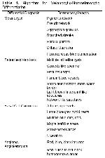
    Before dermatoscopic examination, macroscopic photographs of the pigmented lesions were taken using the Grimed MMicrosoft WDM Image Capture (Win 32) camera and Scalar USB microscope M2 macrolens of the dermatoscope. After that, immersion oil was dropped on the pigmented lesion, and dermatoscopic examination was conducted after placing the glass base of the 30X magnification lens of the computerized digital dermatoscope. The images obtained as such were uploaded to the computer. In the first place, the lesions first differentiated as melanocytic or nonmelanocytic on the basis of the algorithm approved by Consensus Net Meeting Board (Table 1) 2. In the second phase, lesions which were considered nonmelanocytic were differentiated as benign and malignant according to dermatoscopic specific images, and those which were considered melanocytic were differentiated by dermatoscopic imaging. Menzies15 method was used to differences the lesions of face, pattern analyse used to differences the lesions of acral area lesions and ABCD rule used to differences the lesions of the other area lesions. Thus, the lesions were grouped as benign and malignant, irrespective of the melanocytic-nonmelanocytic differentiation.


    Büyütmek İçin Tıklayın
    Table 1: Algorithm for Melanocytic-Nonmelanocytic Differentiation.

    Taking account of traumatization, which can change dermatoscopic ABCD (Asimetri, Border, Color, Differential structure) score, each lesion was assigned a total dermoscopy score, calculated by multiplying the individual scores by weighted coefficients as indicated in the ABCD rule, and was recorded (A: 1.3, B: 0.1, C: 0.5, D: 0.5). Melanocytic lesions were classified as benign or malignant according to the dermoscopy score, and dermatoscopic pre-diagnoses based on dermatoscopic findings were given and recorded.

    All inspected lesions were removed by excisional biopsy with the consent of the patients. Before the surgical procedure, the lesions were cleaned with antiseptic solution, and local anesthesia was induced using epinephrine and 2% lidocaine injection. The lesions were removed by fusiform excision method, using lancet no 15, and the excision site was closed with appropriate suture material. The specimens obtained were sent to the Pathology Laboratory within transport media containing 10% formalin.

    Preparations were stained with hematoxylin eosin for histopathologic examination at the pathology laboratory. The lesions that were suspected malignant melanoma were stained with melan A, S-100 and HMB 45. All the cross sections were evaluated by the same pathologist for standardization, and the histopathologic preparations were photographed. Diagnoses was made by the histopathologic findings were classified as benign and malignant. Objective, clinical and dermatoscopic pre-diagnoses were compared with histopathologic diagnoses.

    Statistical analyses were performed using SPSS 12.0 package software.

  • Başa Dön
  • Özet
  • Giriş
  • Materyal ve Metot
  • Bulgular
  • Tartışma
  • Kaynaklar
  • Bulgular
    Ninety four pigmented skin lesions were obtained from the 67 cases who presented at the Dermatology outpatient clinic of Fırat University Medical School. Of the cases, 45 (67.2%) were female and 22 (32.8%) were male. Mean age of male patients was 36.13±18.16 and mean age of female patients was 38.59±16.01 (0-80 years-range). Demographical characteristics of the cases, as well as the distribution of pigmented skin lesions by body areas are presented in Table 2.


    Büyütmek İçin Tıklayın
    Table 2: Demographical Characteristics of Cases and Distribution of Pigmented Skin Lesions.

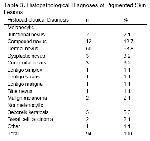
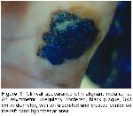
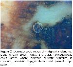
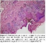
    Revealed histopathologic examination that of the lesions, 86 were melanocytic, 8 were nonmelanocytic, 86 were benign and 8 were malignant (2 malignant melanoma, 1 lentigo maligna, 3 dysplastic nevus, 2 pigmented basal cell carcinoma). Histopathologic diagnoses of the lesions are presented in Table 3 (Figures 1, 2, 3).


    Büyütmek İçin Tıklayın
    Table 3: Histopathological Diagnoses of Pigmented Skin Lesions.


    Büyütmek İçin Tıklayın
    Figure 1: Clinical appearance of malignant melanoma. An asymmetric, irregularly bordered, black plaque, 3x3 cm in diameter, with an ulcerated and crusted center on the left hand hypothenar area.


    Büyütmek İçin Tıklayın
    Figure 2: Dermatoscopic image of malignant melanoma. Light to dark brown, white and black heterogeneous color, areas where pigment network structure is impaired, scar-like depigmentation and paralel ridge pattern.


    Büyütmek İçin Tıklayın
    Figure 3: Histopathologic image of malignant melanoma. Large globules formed by pigmented and atypical melanocytes on epidermis and superficial dermis; atypical melanocyte increase on basal layer HEx200.

    When clinical and dermatoscopic pre-diagnoses of the lesions were compared statistically, 73 (77.7%) were found consistent and 21 (22.3%) were found inconsistent with prediagnosis. When clinical pre-diagnoses and histopathologic diagnoses were compared, 63 (67%) were consistent and 31 (33%) were inconsistent; and when dermatoscopic pre-diagnoses were compared with histopathologic diagnoses, 82 (87%) were found consistent and 12 (13%) were found inconsistent.

    Considering the histopathologic diagnoses, it was established that clinical examination, the sensitivity was 96.6 % and the spesifity was 61.5 % while dermoscopy was 97.7% sensitive and 88.8% specific in differentiating between melanocytic and nonmelanocytic pigmented skin lesions. Consistency of clinical, dermatoscopic and histopathologic examinations in the differentiation between melanocytic and nonmelanocytic lesions is shown in Tables 4, 5 and 6.


    Büyütmek İçin Tıklayın
    Table 4: Clinics and Dermatoscopic Diagnosis in Melanocytic-Nonmelanocytic Differentiation.


    Büyütmek İçin Tıklayın
    Table 5: Clinics and Histopathological diagnosis in Melanocytic-Nonmelanocytic Differentiation.


    Büyütmek İçin Tıklayın
    Table 6: Dermatoscopy and Histopathologicaly diagnosis in Melanocytic-Nonmelanocytic Differentiation.

    Considering the histopathologic diagnoses, it was established that clinical examination, the sensitivity was 92.4 % and the spesifity was 72.7 % while dermoscopy was 97.7% sensitive and 88.8% specific in differentiating between benign-malignant differentiations of the pigmented skin lesions. Consistency of clinical, dermatoscopic and histopathologic examinations in the differentiation between in the benign-malignant differentiation is shown in Tables 7, 8 and 9.


    Büyütmek İçin Tıklayın
    Table 7: Clinics and Dermatoscopic Diagnosis in Benign-Malignant Differentiation.


    Büyütmek İçin Tıklayın
    Table 8: Clinics and Histopathologicaly Diagnosis in Benign-Malignant Differentiation.


    Büyütmek İçin Tıklayın
    Table 9: Dermatoscopy and Histopathological Diagnosis in Benign-Malignant Differentiation.

  • Başa Dön
  • Özet
  • Giriş
  • Materyal ve Metot
  • Bulgular
  • Tartışma
  • Kaynaklar
  • Tartışma
    Dermoscopy is a non-invasive technique used in the diagnosis of pigmented lesions and producing more accurate results compared to other methods2-4,7. Clinical data may not always be adequate in the evaluation of pigmented skin lesions. Dermoscopy is used as a technique to help the physician in the diagnosis of melanocytic lesions like benign melanocytic lesions, dysplastic nevus, and malignant melanoma, in the benign-malignant differentiation, as well as the differential diagnosis of other pigmented nonmelanocytic lesions. It is particularly useful in the correct evaluation of morphological structures which cannot be decided on with naked eye and uncertain pigmented skin lesions, as well as in the pre-operative diagnosis of pigmented skin lesions planned for excision3,4,6,20-22.

    Dermatoscopic diagnoses are given in line with diagnostic algorithms, which are the ABCD rule developed by Nachbar et al.14, Menzies et al.15 method, the seven point checklist designed by Argenziano et al.16, and the pattern analysis developed by Pehamberger et al.13. Dermoscopy uses a two-step algorithm, where the first step involves melanocytic-nonmelanocytic differentiation of the pigmented lesion and the second step consists of the benign-malignant differentiation of the lesion by dermatoscopic criteria24,25. Use of these dermatoscopic methods by experienced physicians increases diagnostic accuracy by 5% to 30%, sensitivity of clinical examination by 19% and specificity 6.2%; thereby the number of patients sent to biopsy decreases by 42%24-27.

    Recently, dermoscopy has become a practical and suggestive method in the differentiation between melanocytic and nonmelanocytic pigmented skin lesions18. Of the nonmelanocytic skin lesions, pigmented basal cell carcinoma in particular can be clinically confused with benign pigmented lesions. Specific dermatoscopic images of pigmented basal cell carcinoma can enable differentiation of any of its types from other tumors28. In a two-year study Demirtaşoğlu et al.29 found that dermoscopy raised the rate of accuracy from 60% to 90% in the diagnosis of pigmented basal cell carcinoma and reported that dermoscopy is a valuable diagnostic tool in the diagnosis of pigmented basal cell carcinoma. White et al.30 who reported a case of pigmented basal cell carcinoma that developed from a burn scar noted that basal cell carcinoma could be clinically confused with malignant melanoma, particularly in patients with dark skin, and stated that dermatoscopic examination helped diagnosis. In our study, dermatoscopic findings of two cases who were clinically considered to be melanocytic were consistent with seborrheic keratosis, and their seborrheic keratosis pre-diagnoses were confirmed with histopathology. A case who was clinically suspected malignant melanoma was found by dermatoscopic examination to be consistent with pigmented basal cell carcinoma, which diagnosis was confirmed by histopathologic investigation. On the contrary, in a case who was clinically given the pre-diagnosis of seborrheic keratosis dermatoscopic findings suggested a melanocytic lesion and the histopathologic diagnosis showed dermal nevus. We established that in differentiating between melanocytic and nonmelanocytic pigmented skin lesions, clinical examination was statistically 96.6% sensitive and 61.5% specific, while dermoscopy was 97.7% sensitive and 88.8% specific.

    Dermoscopy assumes importance in differentiating between benign and malignant pigmented skin lesions of uncertainty31. Since tumor thickness is the major prognostic factor in melanoma, early diagnosis of melanoma is very important and extremely valuable in reducing the mortality associated with melanoma. In this context dermoscopy assists the physician to recognize malignant melanoma at its onset8,10-17. Blum et al.32 reported that physicians could accurately diagnose cutaneous melanoma clinically at a rate of 65% to 80% and dermoscopy increased sensitivity by 10% to 27% in accurate diagnosis, while Westerhoff et al.33 noted a 39% increase in the latter. Wollina et al.34 emphasized that early diagnosis was the main step in the improvement of prognosis in malignant melanoma, and reported that, having a high sensitivity and specificity in the diagnosis of early melanoma, dermoscopy was an easy-to-use method that dermatologists can employ in the accurate diagnosis of pigmented skin lesions. In a study carried out on 206 skin lesions, Bono et al.35 found that clinical examination was 43% sensitive and 91% specific, whereas dermoscopy was 83% sensitive and 69% specific in the diagnosis of cutaneous melanoma, and reported that dermoscopy was evidently useful in diagnosing very small cutaneous melanoma. Barzegari et al.21 pointed out that computerized dermatoscopic analysis was 93% sensitive and 95% specific in melanoma diagnosis. In our study, of the 2 lesions to which we gave malignant melanoma pre-diagnoses clinically, one was an ulcerated plaque in the palmar area of the hand and the other was a heterogeneous black-gray macula, 2-3 cm in diameter, located between toes. Dermatoscopic findings of both lesions supported our pre-diagnoses, which were then histopathologically confirmed to be malignant melanoma. Clinical and dermatoscopic pre-diagnoses of the lesions were not difficult to give. In our study, lentigo maligna, dysplastic nevus, malignant melanoma and pigmented basal cell carcinoma were accepted as malignant, and it was found that clinical examination was statistically 72.7% specific and 92.4% sensitive, whereas dermoscopy was 80% specific and 97.7% sensitive in the benign-malignant differentiation.

    Unwarranted excisions performed on low-risk pigmented lesions located on the face, genital and acral areas in particular can cause significant morbidity. In cases like these, dermoscopy helps the physician, and enables avoidance of unnecessary excisions in benign tumors36,37,38. Furthermore, dermoscopy makes follow-up of lesions which have a high risk of converting to malignant melanoma, like melanocytic nevus, by physicians safer.

    Besides being an easy-to-use, non-invasive and inexpensive diagnostic method in the diagnosis of pigmented skin lesions, dermoscopy also increases the diagnostic accuracy of clinical examination2-4,17,39-43. In a study including 107 cases with clinically suspected melanocytic skin lesions, Ferrara et al.17 reported that dermatoscopic images and histopathology of lesions were consistent. Burroni et al.39 in their study of 174 cases with pigmented skin lesions established that 71.8% of dermatoscopic pre-diagnoses were consistent with their respective histopathologies. Piccolo et al.40 reported the sensitivity and specificity of digital dermoscopy in the diagnosis of pigmented skin lesions to be 92% and 99%, respectively, for an experienced physician. However, in their study including 9004 pigmented skin lesions, Kittler et al.41 reported that dermoscopy increased the diagnostic accuracy of clinical examination by 35% and that dermoscopy was 89% sensitive and 79% specific in diagnosis. In our study when histopathologic diagnoses of 94 pigmented skin lesions were compared, we found that 63 were consistent with the clinical examination and 82 were consistent with dermoscopy.

    In conclusion, dermoscopy is an easy method that is used in the diagnosis and benign-malignant differentiation of pigmented skin lesions. Dermoscopy not only is agreeable for the patients in terms of early diagnosis, excision and prognosis of malignant lesions, but also offers both the physician and the patients a chance of easy follow-up by preventing unnecessary surgical interventions to low-risk melanocytic lesions.

  • Başa Dön
  • Özet
  • Giriş
  • Materyal ve Metot
  • Bulgular
  • Tartışma
  • Kaynaklar
  • Kaynaklar

    1) Binder M, Braun RP. Principles of dermoscopy. In: Marghoob AA, Braun RP, Kopf AW, Eds. Atlas of Dermoscopy. 1st ed. Newyork: Taylor&Francis; 2004. p. 7-10.

    2) Argenziano G, Soyer HP, Chimenti S, et al. Dermoscopy of pigmented skin lesions: results of a consensus meeting via the internet. J Am Acad Dermatol 2003; 48: 679-693.

    3) Binder N, Schwarz N, Winkler A, et al. Epiluminescence microscopy. A useful tool for the diagnosis of pigmented lesions for formally trained dermatologists. Arch Dermatol 1995; 131: 186-191.

    4) Braun RP, Kaya G, Masouye I, Krischer J, Saurat JH. Histopathologic correlation in dermoscopy: a micropunch tecnique. Arch Dermatol 2003; 139: 349-351.

    5) Jaramillo-Ayerbe F, Vallejo-Contreras J. Frequency and clinical and dermatoskopic features of volar and ungual pigmented melanocytic lesions: a study in schoolchildren of Manizales. Pediatr Dermatol 2004; 21: 218-222.

    6) Mayer J. Systematic review of the diagnostic accuracy of dermatoscopy in detecting malignant melanoma. Med J Aust 1997; 167: 206-210.

    7) Soyer HP, Kenet RO, Wolf IH, Kenet BJ, Cerroni L. Clinicopathological correlation of pigmented skin lesions using dermoscopy. Eur J Dermatol 2000; 10: 22-28.

    8) Soyer HP, Smolle J, Leitinger G, Rieger E, Kerl H. Diagnostic reliability of dermoscopic criteria for detecting malignant melanoma. J Dermatol 1995; 190: 25-30.

    9) Carli P, De Giorgi V, Naldi L, Dosi G. Reability and inter-observer agreement of dermoscopic diagnosis of melanoma and melanocytic nevi. Eur J Cancer Prev 1998; 7: 397-402.

    10) Bafounta ML, Beauchet A, Aegerter P, Saiag P. Is dermoscopy (epiluminescence microscopy) useful for the diagnosis of melanoma? Arch Dermatol 2001; 137: 1343-1350.

    11) Koh HK, Geller AC, Miller DR, Lew RA. The early detection and screening for melanoma. International status. Cancer 1995; 75: 674-683.

    12) Steiner A, Pehamberger H, Binder M, Wolff K. Pigmented Spitz nevi: improvement of the diagnostic accuracy by epiluminescence microscopy. J Am Acad Dermatol 1992; 27: 697-701.

    13) Pehamberger H, Binder M, Steiner A, Wolff K. In vivo epiluminescence microscopy: improvement of early diagnosis of melanoma. J Invest Dermatol 1993; 100: 356-362.

    14) Nachbar F, Stolz W, Merkle T, et al. ABCD rule of dermoscopy. J Am Acad Dermatol 1994; 30: 551-559.

    15) Menzies S, Ingvar C, Mc Carthy WH. A sensitivity and specifitiy analysis of the surface microscopy features of invasive melanoma. Melanoma Res 1996; 6: 55-62.

    16) Argenziano G, Fabbrocini G, Carli P, et al. Epiluminescence microscopy for the diagnosis of doubtful melanocytic skin lesions. Comparison of the ABCD rule of dermoscopy and a new 7-point checklist based on pattern analiysis. Arch Dermatol 1998; 134: 1563-1570.

    17) Ferrara G, Argenziano G, Soyer HP, et al. Dermoscopic and Histopatologic Diagnosis of Equivucal Melanocytic Skin Lesions An Interdisciplinary Study on 107 Cases. Cancer 2002; 1094-1100.

    18) Ferrara G, Argenziano G, Soyer HP, et al. Dermoscopic-Patologic Corelation: an atlas ıf 15 cases. Dermatol Clin 2002; 20: 228-235.

    19) Pathak MA, Nghiem P, Fitzpatrick TB. Acute and Chronic Effects of the sun. In: Freedberg IM, Eisen AZ, Wolff K, et al., Eds. Fitzpatrick’s Dermatology in General Medicine. 5th ed. New York: Mc Graw Hill; 1999. p. 1598-1607.

    20) Braun RP, Rabinovitz HS, Oliviero M, Kopf AW, Saurat JH. Dermoscopy of pigmented skin lesions. J Am Acad Dermatol 2005; 52: 109-121.

    21) Barzegari M, Ghaninezhad H, Mansoori P, et al. Computer-aided dermatoscopy for diagnosis of melanoma. BMC Dermatol 2005; 5: 8.

    22) Stolz W, Semmelmayer U, Johow K, Burgdorf WH. Principles of dermatoscopy of pigmented skin lesions. Semin Cutan Med Surg 2003; 1: 9-20.

    23) Menzies SW, Ingvar C, Crotty KA, McCarthy WH. Frequency and morphologic characteristics of invasive melanomas lacking specific surface microscopic features. Arch Dermatol 1996; 132: 1178-1182.

    24) Scope A, Benvenuto-Andrade C, Agero AL, Marghoob AA. Nonmelanocytic lesions defying the two-step dermoscopy algorithm. Dermatol Surg 2006; 32: 1398-1406.

    25) Menzies SW. Cutaneous melanoma: making a clinical diagnosis, present and future. Dermatologic Ther 2006; 19: 32-39.

    26) Tripp JM, Kopf AW, Margoop AA, Bart RS. Management of dysplastic nevi: a survey of fellows of American Academy of Dermatology. J Am Acad Dermatol 2002; 46: 674-682.

    27) Nehal KS, Oliveria SA, Margoob AA, et al. Use of and beliefs about dermoscopy in the management of patients with pigmented lesions: a survey of dermatology residency programmes in the United States. Melanoma Res 2002; 12: 601-605.

    28) Agero AL, Busam KJ, Benvenuto-Andrade C, et al. Reflectance confocal microscopy of pigmented basal cell carcinoma. J Am Acad Dermatol 2006; 54: 638-643.

    29) Demirtaşoğlu M, İlknur T, Lebe B, et al. Evaluation of dermoscopic and histopathologic features and their correlations in pigmented basal cell carcinomas. J Eur Acad Dermatol Venereol 2006; 20: 916–920.

    30) White EA, Rabinovitz HS, Grene RS, Oliviero M, Kopf A. Pigmented basal cell carcinoma simulating melanoma in a burn scar. Cutis 2003; 71: 404-406.

    31) Elder DE. Precursors to Melanoma and Their Mimics: nevi of special sites. Mod Pathol 2006; 2: 4-20.

    32) Blum A, Rassner G, Garbe C. Modified ABC-point list of dermoscopy: A simplified and highly accurate dermoscopic algorithm for the diagnosis of cutaneous melanocytic lesions. J Am Acad Dermatol 2003; 48: 672-678.

    33) Westerhoff K, McCarthy WH, Menzies SW. Increase in the sensitivity for melanoma diagnosis by primary care physicians using skin surface microscopy. Br J Dermatol 2000; 143: 1016-1020.

    34) Wollina U, Burroni M, Torricelli R, et al. Digital dermoscopy in clinical practise: a three-centre analysis. Skin Res Technol 2007; 13: 133-142.

    35) Bono A, Tolomio E, Trincone S, et al. Micro-melanoma detection: a clinical study on 206 consecutive cases of pigmented skin lesions with a diameter ≤ 3 mm. Br J Dermatol 2006; 155: 570-573.

    36) Zalaudek I, Zanchini R, Petrillo G, et al. Dermoscopy of an Acral Congenital Melanocytic Nevus. Pediatr Dermatol 2005; 22: 188-191.

    37) Blum A, Meztler G, Hoffmann-Wellenhof R, et al. Correlation between dermoscopy and histopatology in pigmented and non-pigmented skin tumours. Hautarzt 2003; 54: 279-291.

    38) Sahin S, Gokoz O. Histopathologic Correlation in Dermoscopy. Turkiye Klinikleri J Int Med Sci. 2007; 3: 74-80.

    39) Burroni M, Sbano P, Cevenini G, et al. Dysplastic naevus vs. in situ melanoma: digital dermoscopy analysis. Br J Dermatol 2005; 152: 679-684.

    40) Pıccolo D, Ferrari A, Peris K, et al. Clinical and Laboratory Investigations Dermoscopic diagnosis by a trained clinician vs. a clinician with minimal dermoscopy training vs. computer-aided diagnosis of 341 pigmented skin lesions: a comparative study. Br J Dermatol 2002; 147: 481–486.

    41) Kittler H, Pehamberger K, Wolff K, Binder M. Diagnostic accuracy of dermoscopy. Lancet Oncol 2002; 3: 159-165.42.

    42) Steiner A, Pehamberger H, Wolff K. In vivo epiluminescence microscopy of pigmented skin lesions 2: diagnosis of small pigmented skin lesions and early detection of malignant melanoma. J Am Acad Dermatol 1987; 17: 584-591.

    43) Braun RP, Rabinovitz H, Oliviero M, et al. Dermatoscopy of pigmented lesions. Ann Dermatol Venereol 2002; 129: 187-202.

  • Başa Dön
  • Özet
  • Giriş
  • Materyal ve Metot
  • Bulgular
  • Tartışma
  • Kaynaklar
  • [ Başa Dön ] [ Özet ] [ PDF ] [ Benzer Makaleler ] [ Yazara E-Posta ] [ Editöre E-Posta ]
    [ Ana Sayfa | Editörler | Danışma Kurulu | Dergi Hakkında | İçindekiler | Arşiv | Yayın Arama | Yazarlara Bilgi | E-Posta ]