[ Ana Sayfa | Editörler | Danışma Kurulu | Dergi Hakkında | İçindekiler | Arşiv | Yayın Arama | Yazarlara Bilgi | E-Posta ]
Fırat Üniversitesi Sağlık Bilimleri Veteriner Dergisi
2010, Cilt 24, Sayı 2, Sayfa(lar) 077-080
[ Özet ] [ PDF ] [ Benzer Makaleler ] [ Yazara E-Posta ] [ Editöre E-Posta ]
Erişkin Porsuk (Meles meles) Testis'i Üzerinde Işık ve Elektron Mikroskopik İncelemeler
Meryem KARAN, Sadık YILMAZ, Ömer ATALAR, Gürsel DİNÇ
Fırat Üniversitesi, Veteriner Fakültesi, Anatomi Anabilim Dalı, Elazığ, TÜRKİYE
Anahtar Kelimeler: Testis, porsuk, Meles meles, ışık mikroskop, elektron mikroskop
Özet
Çalışmada, ışık ve elektron mikroskobu (TEM) kullanarak Doğu Anadolu bölgesinde yakalanan porsuk (Meles meles) testis'leri incelendi. Bu amaçla 5 erişkin porsuk kullanıldı.

Testis parenşiminin, seminifer tubulün sıralı hücreleri ve onların kanalları ile interstisyal hücrelerden oluştuğu gözlendi. Sertoli hücrelerinin tubulün bazal laminası üzerinde olduğu saptandı. Bunların az sayıda olduğu ve tubul boyunca aralıklı olarak dizildikleri tespit edildi. Sitoplazmaları farklı şekillerde çok sayıda mitokondriye sahipti. Leydig hücreleri büyük ve asidofilik hücrelerdi. Spermatogonia, yoğun kromatinli büyük, yuvarlak veya oval bir çekirdekle karakterize idi. Primer spermatositler, seminifer tubul içinde görülen en büyük germ hücreleriydi. Spermatid'in apikal başı sertoli hücre sitoplazmasına girmişti.

  • Başa Dön
  • Özet
  • Giriş
  • Materyal ve Metot
  • Bulgular
  • Tartışma
  • Kaynaklar
  • Giriş
    The badger (Meles meles,), which is the subject of this study, belongs to the Mustelidae family, order carnivora. With its striking black and white striped head, the badger is one of our most instantly recognisable mammals 1, 2.

    The light and electron microscopic structure of the normal testis has been described in several species: human 3-5, rat 6, 7, rabbit 8, 9, dog 10, 11. However, there was no study on the histologic structure of testis in badger.

    Therefore, this study was focused on the investigation of the histology of testis in adult badgers to make a contribution to knowledge in this field.

    The purpose of this study was to examine, with the light and TEM, the structure of testis in adult badger, in order to compare the results with those previous reports in other rodentia.

  • Başa Dön
  • Özet
  • Giriş
  • Materyal ve Metot
  • Bulgular
  • Tartışma
  • Kaynaklar
  • Materyal ve Metot
    Five adult badgers (Meles meles, Linnaeus, 1758) were trapped in Eastern Anatolia. Tubular diameter were measured by using ocular micrometry for morphometric analysis.

    Light Microscopy: Under penthotal-induced (6 ml/kg) anaesthesia, the testes were removed. Samples of testes were fixed in Bouin's solution. Tissue samples were routinely processed through a graded series of alcohols, cleared in xylol and embedded in parafin. 7 µm thick sections were obtained and stained with haematoxylin and eosin and PAS.

    Electron Microscopy: Pieces of testes were fixed in cacodylat-buffered 4.5% glutaraldehyde. They were cut into smaller pieces (approximately 1 mm3), post-fixed in 1% osmium tetroxide. The fixed tissues were dehydrated by passage through a graded series of ethanol, and embedded in Araldite.

    Serial semithin sections were cut at approximately 1 μm thickness, stained with toluidine blue, and observed under light microscopy. Thin sections were cut for electron microscopy with glass knives with ultratome. Sections were stained with either lead citrate or double stained with uranyl acetate followed by lead citrate, and examined under a Zeiss EM-9A electron microscope.

  • Başa Dön
  • Özet
  • Giriş
  • Materyal ve Metot
  • Bulgular
  • Tartışma
  • Kaynaklar
  • Bulgular
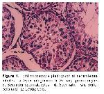
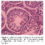
    Light microscopy: The two principal parenchymal components of the testis were the seminiferous tubules and endocrine cells of the interstitium. The basement membrane was surrounded the tubules. Each seminiferous tubule was approximately 437 μm in diameter. A few of them were convoluted and the others were usually oval or round in shape. Intertubular areas of testis were narrow. As a result of this, interstitial tissue was scarce. It was seen capillaries in the connective tissue. The tubules consisted of a few sertoli cells and spermatogenic cells. Leydig cells within interstitial regions were visible. Myoid cells was formed a single layer in the peritubular tissue. The nuclei of myoid cells was flat and long (Figure 1, 2).


    Büyütmek İçin Tıklayın
    Figure 1: Light microscope photograph of seminiferous tubules. a: Spermatogonium b: Srimary spermatocytes c: Secunder spermatocytes d: Spermatid sh: Sertoli cells and L: Leydig cells.

    Sertoli cells were tall and triangular, with cytoplasm. Cellular margins were difficult to distinguish by light microscopy. Sertoli cell nuclei were readily identified and were usually found towards the basement membrane of the seminiferous tubule. The sertoli cell nucleus was ovoid in shape with a prominent nucleolus. Sertoli cells had an extensive cytoplasm (Figure 1, 2).


    Büyütmek İçin Tıklayın
    Figure 2: Light microscope photograph of seminiferous tubules. a: Spermatogonium b: Primary spermatocytes c: Spermatid sh: Sertoli cell m: Myoid cell k: Capillary.

    The spermatogonia were located on or near the tubule basal lamina. It had relatively little cytoplasm. Spermatogonia were characterised by a large round or oval nucleus with condensed chromatin. Primary spermatocytes were readily recognised by their extensive cytoplasm and large nuclei. They were the largest germ cells seen within the seminiferous tubule. The spermatid was a spherical cell, and had a medium-size pale, round, or oval nucleus. The spermatozoon was an extremely elongated structure (Figure 1,2).

    In the surrounding interstitial tissue, scattered large leydig cells were observed. Leydig cells were generally found singly and sometimes they were in groups in the supporting tissue. They were generally around capillaries. Leydig cell nucleus was large, round or oval with dispersed chromatin, and it was usually exantrically situated. The nuclei had a clear nuclear membrane. Leydig cells had an eosinophilic cytoplasm (Figure 1).

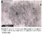
    Transmission electron microscopy: Cytoplasm of the sertoli cell was clear. There were a number of mitochondria in their cytoplasm. They have elongated, oval and round shaped. Elongated mitochondria were striking. This represented active of sertoli cells (Figure 3).


    Büyütmek İçin Tıklayın
    Figure 3: Electron microscope photograph of cytoplasm of sertoli cell. It was seen a lot of long and oval shaped mitochondria

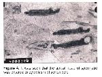
    The apical head of spermatid was entered to cytoplasm of sertoli cell and the apical head of slightly elongated spermatids was occasionally surrounded by the laminated sER (Figure 4).


    Büyütmek İçin Tıklayın
    Figure 4: It was seen that the apical head of spermatid was entered to cytoplasm of sertoli cell.

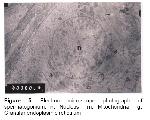
    Spermatogonia were characterised by a large round or oval nucleus with condensed chromatin. Granular endoplasmic reticulum was seen within the cytoplasm of spermatogonia. A number of mitochondria were also recognized. They had various shapes and sizes (Figure 5).


    Büyütmek İçin Tıklayın
    Figure 5: Electron microscope photograph of spermatogonium. n: Nucleus m: Mitochondria g: Granular endoplasmic reticulum

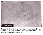
    Primary spermatocytes had a large and round nuclei. There were nuclear pores on the nuclear membrane. A well developed golgi apparatus and mitochondria were found within their cytoplasm (Figure 6).


    Büyütmek İçin Tıklayın
    Figure 6: The structure of primary spermatocyte. n: Nucleus m: Mitochondria G: Golgi apparatus e: Smooth endoplasmic reticulum

    The nucleus of spermatid was small and oval in shape. Mitochondria were seen in the cytoplasm of spermatid.

  • Başa Dön
  • Özet
  • Giriş
  • Materyal ve Metot
  • Bulgular
  • Tartışma
  • Kaynaklar
  • Tartışma
    It has been reported that the interstitial distance between seminiferi contorti was quite large in the adult rabbits 9 and in the adult squirrels 12. In the present study, interstitial distance was narrow. The diameter of tubulus seminiferus contortus was notified as 186 μm 13 and 181.5 μm 11 for the adult dogs. This was found as 437 μm for badger in this study.

    There were different findings about the shape of sertoli cells as pyramid 14, columnar 3 and irregular 11. In this study, the shape of sertoli cells was determined as triangular in badgers.

    The present study showed that there was a lot of oval and long mitochondria in the cytoplasm of sertoli cells in the adult badgers. Similar results have been reported for human 3, 15 and water buffalo 16.

    Leydig cells were found singly or colony of various sized cells with large and vesicular single nucleus in human (

    ). Leydig cells were defined as pyramidal shaped and colonized with various size around blood vessel in the adult rabbits 9. In rats, leydig cells were defined as irregular polygonal shaped with eosinophylic cytoplasm and large nucleus without chromatin 6, 7. In this study, leydig cells were determined as generally single or rarely grouped with eosinophylic cytoplasm and large nucleus in the adult badgers.

    It was reported that nuclei of leydig cells situated exantricallly in rats 6. We also found an eccentric nucleus in leydig cells of the adult badger.

    It was reported that pig, horse and opossum have more leydig cells that occupied nearly all intertubular space 3. In this study, it was observed that leydig cells were present in intertubular areas as single or small groups in badgers.

    Ross et al 15 and Leeson et al 14 reported that myoid cells composed a single layer in rodentia. However, peritubular tissue was formed by 3-5 myoid cells layer in human. Prakash et al 17 presented that myoid cells were mostly two to three layered in bonnet monkey. In the present study, it was observed that myoid cells composed only a single layer in badgers.

    Jurado et al 18 found that apical head of spermatid was inserted in the cytoplasm of sertoli cell. We also determined similar result in badger.

    In conclusion, light and electron microscopy examinations results of this study showed that the structure of badger's testis has many similarities with that of the other mammalian.

  • Başa Dön
  • Özet
  • Giriş
  • Materyal ve Metot
  • Bulgular
  • Tartışma
  • Kaynaklar
  • Kaynaklar

    1) Kuru M. Omurgalı Hayvanlar. Erzurum: Atatürk Üniv Basımevi 1987.

    2) Demirsoy A. Yaşamın Temel Kuralları. Ankara: Meteksan Anonim Şirketi 1992.

    3) Fawcett WD, Bloom WA. A Text Book of Histology. 11th Edition, Igaku-Shoin/Saunders: W.B. Saunders Company, 1986.

    4) Livni N, Palti Z, Segal S, Laufer A. Fine structure of sertoli and leydig cells in azoospermic human testis. Arch Pathol Lab Med 1977; 101: 442-445.

    5) Thomas D, Trainer MD. Histology of the normal testis. The Am J Surg Pathol 1987; 11(10): 797-809

    6) Gözil R, Erdoğan D, Kadıoğlu D, Aydoğan S. Testis'de steroid salgı (testesteron) oluşturan leydig hücrelerinin ışık mikroskop düzeyinde çeşitli histokimyasal yöntemlerle değişik sıçan yaş gruplarında incelenmesi. Gazi Üniv Tıp Fak Derg 1988; 4(1): 71-81.

    7) Knorr DW, Vanha-Perttula T, Lipsett MB. Structure and function of rat testis through pubescence. Endocrinology 1970; 86: 1298.

    8) Aydın A, Yılmaz S. Tavşanlarda testislerin postnatal dönemde morfolojik gelişimi. F Ü Sağlık Bil Derg 14 (1): 163-170, 2000.

    9) Öztürk F, Cengiz N, Vardı N, Otlu A. Erişkin tavşan testisleri üzerinde histolojik ve bazı histoşimik araştırmalar. Fırat Tıp Derg 1997; 1(3): 141-144.

    10) Evans H, Christensen G. Miller's Anatomy of the Dog. Philidelphia: Saunders Company 1979.

    11) Yılmaz S. Köpeklerde testislerin morfolojik gelişimi. Doğa Tr J Vet Anim Sci 1992: 16: 493-503.

    12) Karan M, Timurkaan S, Aydın A. Erişkin sincaplarda (Sciurus vulgaris) testislerin ışık mikroskopik yapısı. F Ü Sağlık Bil Derg 2006; 20(3): 185-187.

    13) Woodal PF, Johnstone IP. Dimensions and allometry of testes, epididymides and spermatozoa in the domestic dog (Canis familiaris). J Reprod Fertil 1988; 82: 603-609.

    14) Leeson TS, Leeson CR, Paparo AA. Text/Atlas of Histology. Philadelphia: W.B. Saunders Company 1988.

    15) Ross MH, Romrell LJ, Koye GI. Histology. A text and atlas. 3rd Edition, Baltimore: Williams & Wilkins 1995; 636-651.

    16) Kurohmaru M, Yamashiro S, Azmi TI, Basrur PK. An ultrastructural study of the sertoli cell in the water buffalo (Bubalus bubalis). Anat Histol Embryol 1992; 21(1): 82-90.

    17) Prakash S, Prithiviraj E, Suresh S. Developmental changes of seminiferous tubule in prenatal, postnatal and adult testis of bonnet monkey (Macaca radiata). Anat Histol Embryol 2008; 37: 19-23.

    18) Jurado SB, Kurohmaru M, Hayashi Y. An ultrastructural study of the sertoli cell in the Shiba goat. J Vet Sci 1994; 56(1): 33-38.

  • Başa Dön
  • Özet
  • Giriş
  • Materyal ve Metot
  • Bulgular
  • Tartışma
  • Kaynaklar
  • [ Başa Dön ] [ Özet ] [ PDF ] [ Benzer Makaleler ] [ Yazara E-Posta ] [ Editöre E-Posta ]
    [ Ana Sayfa | Editörler | Danışma Kurulu | Dergi Hakkında | İçindekiler | Arşiv | Yayın Arama | Yazarlara Bilgi | E-Posta ]