[ Ana Sayfa | Editörler | Danışma Kurulu | Dergi Hakkında | İçindekiler | Arşiv | Yayın Arama | Yazarlara Bilgi | E-Posta ]
Fırat Üniversitesi Sağlık Bilimleri Veteriner Dergisi
2013, Cilt 27, Sayı 2, Sayfa(lar) 099-103
[ Özet ] [ PDF ] [ Benzer Makaleler ] [ Yazara E-Posta ] [ Editöre E-Posta ]
Elazığ Yöresinde Pnömonili Keçi Akciğerlerinde Küçük Ruminant Vebası (PPR) Viral Antijeninin İmmunohistokimyasal Olarak Belirlenmesi
Songül ÇERİBAŞI1, Mustafa ÖZKARACA2, Ali Osman ÇERİBAŞI1, Harun ÖZER1
1Fırat Üniversitesi, Veteriner Fakültesi, Patoloji Anabilim Dalı, Elazığ, TÜRKİYE
2Elazığ Veteriner Kontrol Enstitüsü, Patoloji Laboratuvarı, Elazığ, TÜRKİYE
Anahtar Kelimeler: Peste des Petits Ruminants virus, keçi, pnömoni, immunohistokimya
Özet
Bu çalışmada Elazığ yöresindeki pnömonili keçi akciğerlerinde immunohistokimyasal yöntemle küçük ruminant vebası (PPR) viral antijenlerinin varlığı ve yaygınlığının tespiti amaçlanmıştır. Bu amaçla, Elazığ'daki bir mezbahada kesilen, 889 erkek kıl keçisinin akciğerleri muayene edildi. Çalışmada makroskobik olarak 152 olguda (17.10%) pnömoni tespit edildi, pnömonilerde apikal ve kardiyak loplarda lezyon şiddeti değerlendirildiğinde 97'sinde hafif, 39'unda orta, 17'sinde şiddetli konsolidasyon bulunduğu saptandı. Pnömonili 152 akciğer mikroskobik olarak kataral-purulent (41, %26.97), intersitisyel (72, %47.37), fibrinli (34, %22.37), verminöz (2, %1.32), pulmoner adenomatoz (3, %1.97) olarak sınıflandırıldı. PPR viral antijenlere; histopatolojik olarak hafif şiddette konsolidasyona sahip kataral-purulent bronkopnömoni (n=3), orta şiddette konsolidasyona sahip intersitisyel (n=2) ve fibrinli bronkopnömoni (n=2) belirlenen, 7 olguda (%4.61) rastlandı. Viral antijenlere ilişkin immunboyama bronş ve bronşiyol epitelinde daha yoğun olmak üzere lümendeki eksudat içerisinde de tespit edildi. Sonuç olarak etkenin keçi pnömonilerinde önemli etiyolojik ajanlardan biri olabileceği kanısına varılmıştır. Ek olarak, sunulan çalışmada histopatolojik lezyonların nonspesifik olduğu PPR kaynaklı pnömoniler dikkate alındığında immunohistokimyasal yöntemin PPR enfeksiyonlarının teşhisinde ileri laboratuvar metodlarına alternatif bir yöntem olarak kullanılabileceği kanaatine varılmıştır.
  • Başa Dön
  • Özet
  • Giriş
  • Materyal ve Metot
  • Bulgular
  • Tartışma
  • Kaynaklar
  • Giriş
    Peste des Petits Ruminants (PPR), which can progress as an acute or chronic endemic viral disease in sheep and goats, resembles rinderpest both clinically and pathologically, with a high morbidity and mortality1-4. The cause of the disease is an RNA virus from the morbilli virus genus of the Paramyxoviridae family5.

    The main pathological findings of PPR are observed in the gastrointestinal and the respiratory systems6. As in rinderpest, it is usually characterized with severe erosive necrotic stomatitis and enterocolitis4. Contrary to rinderpest, the most consistent findings in PPR infections are lung lesions characterized by bronchitis, bronchiolitis or bronchointerstitial pneumonia7,8. Eosinophilic intracytoplasmic and intranuclear inclusion bodies and syncytial cells in some alveoli lumen are observed in this infection2,8-10. The diagnosis of PPR infection can be established by immunohistochemistry, cell culture, ELISA, and PCR methods11-15. It has been reported that the viral antigens in the lungs are frequently observed in the bronchi and the bronchial epithelium, syncytial cells and alveolar macrophages, together with exudates in the bronchi and bronchial lumen immunohistochemically (IHC)10,16.

    Immunohistochemical studies in goats have been carried out in natural and experimental PPR infections9,16,17. However, there is a limited number of studies performed on PPR pneumonia with natural origin in goats8,10.

    In this study, it was aimed to identify by immunohistochemistry the presence and the extent of distribution of PPR viral antigens in the goat lungs in the Elazig region.

  • Başa Dön
  • Özet
  • Giriş
  • Materyal ve Metot
  • Bulgular
  • Tartışma
  • Kaynaklar
  • Materyal ve Metot
    1. Sample collection: In the present study, the lungs of 889 with apparently healthy male hair goats (> 1 year old), which were submitted to slaughter between January and June 2011 in Elazig were examined. The macroscopic findings were observed in the apical and cardiac lobes of only 152 lungs. On the macroscopic examination, the severity of pneumonia in the apical and cardiac lobes was scored according to the extent of the consolidation. The lesions in the apical and cardiac lobes were scored as mild “less than 10% of the volume affected”, moderate “consolidation of 10-20% of the volume affected” and severe “more than 20% of the volume affected”. After fixation of the tissue samples with macroscopic consolidation in 10% buffered formalin for 48 h, they were embedded in paraffin wax before sectioning. The tissue sections were then stained with hematoxylin and eosin (HE), and were examined under the light microscope.

    2. Immunohistochemistry: The IHC method was applied in only 147 lung sections with pneumonia, which microscopically had catarrhal purulent bronchopneumonia and interstitial or fibrinous bronchopneumonia. The remaining five lung sections were not examined microscopically due to the presence of verminous and pulmonary adenomatosis. IHC staining was performed according to the avidin-biotin-peroxidase complex (ABC) procedure. Polyclonal rabbit antirinderpest serum (1/1000, The Pirbright Institute, ENGLAND) was used as the primary antibody. The IHC kits used for the other stages (Ultravision Detection System, Anti-polyvalent, HRP/DAB, Thermo Scientific, Cat No: TP-015-HD) were provided by a firm and the staining was carried out according to the manufacturer’s instructions. In the immunohistochemical staining, deparaffinized and rehydrated tissue sections were placed in the citrate buffer solution (10 mM citric acid, pH: 6.0) and were kept in a microwave for 20 min for antigen retrieval stage. The sections were incubated in 3% H2O2 solution for 10 min in order to inhibit endogenous peroxidase activity and washed three times by the phosphate buffered saline (PBS, pH: 7.4). Then the sections were treated with the blocking solution for 10 min. After draining the blocking serum, the sections were incubated with the primary antibody, diluted to 1: 200 rate in PBS at 4°C overnight in a humidified chamber. Following three sequential washes in PBS, the sections were treated with biotinylated anti-goat polyvalent secondary antibody for 10 min. The sections washed three times in PBS were then treated with the peroxidase-conjugated streptavidin solution for 10 min. Following another PBS bath, the sections were incubated with 3,3-diaminobenzidine (DAB). Following the colour changes, the sections were washed with tap water and then counterstained with Mayer’s hematoxylin (M-H). A healthy goat lung was used as a negative control. For positive control, non-immune mouse serum was used instead of primary serum for the lung sections with pneumonia.

  • Başa Dön
  • Özet
  • Giriş
  • Materyal ve Metot
  • Bulgular
  • Tartışma
  • Kaynaklar
  • Bulgular
    1. Macroscopic Findings: One hundred fifty-two (17.1%) of 889 goat lungs were macroscopically pneumonic. Although the severity and distribution of the lesions varied, all pneumonia lesions were seen to be macroscopically characterized by patchy or diffuse, purple-red or gray, focal or irregular lobular atelectatic foci. Among the 152 cases, 97 (63.82%) exhibited mild lesions, 39 (25.66%) had moderate lesions and 16 (10.53%) had severe lesions according to the extent of consolidation.

    2. Histopathological Findings: One hundred fifty-two lungs with pneumonia were classified as catarrhal-purulent (n=41, 26.97%), interstitial (n=72, 47.37%), fibrinous (n=34, 22.37%), verminous (n=2, 1.32%), and pulmonary adenomatosis (n=3, 1.97%).

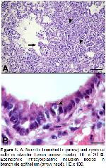
    In the interstitial and fibrinous pneumonia which were determined PPR viral antigen-positive by IHC staining, necrotic bronchitis and bronchiolitis were observed together with exudates in the alveoli, bronchi and bronchiole lumen, which was also rich in macrophages, neutrophils and mononuclear cells. In addition, syncytial cells in the alveolar lumen (Figure 1A) and eosinophilic intracytoplasmic inclusion bodies in the bronchi and bronchiole epithelium were observed in four cases which had interstitial and fibrinous pneumonia (Figure 1B). Some syncytial cells had intranuclear inclusion bodies. Most of the interalveolar septae in the PPR-positive interstitial pneumonia were thickened at irregular degrees due to infiltration of lymphocyte and a small number of macrophages and, had increased connective tissue. There was also lymphoid hyperplasia around the bronchi and the bronchioles. The alveoli lumens of the fibrinous pneumonia with PPR viral antigens were filled with oat-shaped leukocytes, which are characteristic finding for Pasteurella spp. infections.


    Büyütmek İçin Tıklayın
    Figure 1: A. Necrotic bronchiolitis (arrow) and syncytial cells in alveolar lumen (arrow heads), HE x 20 B. Eosinophilic intracytoplasmic inclusion bodies in bronchiole epithelium (arrow head), HE x 100.

    In the catarrhal-purulent pneumonic lesions with PPR viral antigens, the alveolar oedema was predominantly appeared while no syncytial cell formations in the alveolar lumens and no inclusion bodies in the bronchi and the bronchiole epithelium were found. There was vacuolar degeneration in the bronchi and the bronchiole epithelium with diffuse neutrophil leukocytes in the lumen. Few alveolar macrophages were detected in the alveolar lumen.

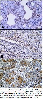
    3. Immunohistochemical Findings: The PPR viral antigens were detected in 7 (4.76%) of 147 pneumonic lungs. Of these three had catarrhal-purulent bronchopneumonia with mild consolidation, two had interstitial and the remaining had fibrinous bronchopneumonia with moderate consolidation. Strong immunolabelling of viral antigen occurred in cytoplasm and occasionally the nuclei of epithelial cells in respiratory mucosa. Cytoplasmic labelling appeared diffuse or granular. Specific staining related with the viral antigens was found more intensely in the bronchi and the bronchiole epithelium (Figure 2A) and, also cellular exudates in the lumen (Figure 2B). Similar staining was present in the bronchial glandular epithelium. Furthermore, the syncytial cells in the bronchiole and alveolar lumen showed severe positivity. While the alveolar epithelium displayed staining on the luminal surface, type II pneumocytes, alveolar macrophages and cellular exudates in the alveolar lumen showed immunepositivity (Figure 2C). The healthy goat lung (normal control) and the pneumonic lung sections with nonimmune mouse serum (serum control) did not show positive immune staining.


    Büyütmek İçin Tıklayın
    Figure 2: A. Specific staining related with PPR viral antigens in bronchiole epithelium, MH, ABC-P x 10. B. PPR viral antigens in cellular exudates, MH, ABC-P x 40. C. Severe PPR immunopositivity in syncytial cells and cellular exudates in alveolar lumen, MH, ABC-P x 40.

  • Başa Dön
  • Özet
  • Giriş
  • Materyal ve Metot
  • Bulgular
  • Tartışma
  • Kaynaklar
  • Tartışma
    PPR has spread into many African and Middle East countries1,7,19,20 depending on the density of animal movements, after it has first been defined in goats in 1942 at Côte d'Ivoire18. The disease was first defined in Turkey in 1993 with pathological and immunohistochemical findings in sheep11. Later, it was reported in sheep and goats in different regions of Turkey with characteristic and introductory findings10,14,16,17,21-25.

    PPR infection in sheep and goats is observed in three forms being hyperacute, acute and subclinical26. Bronchointerstitial pneumonia usually occurs in natural and experimental PPR infections6,8. It has been reported that bronchopneumonia is macroscopically characterized with consolidation and atelectasis and the anterior and the cardiac lobes of bronchopneumonic lungs are particularly dark red in colour and hard in consistency8. In the present study, PPR viral antigens were also observed in three cases which had catarrhalpurulent pneumonia with mild consolidation. Although no clinical examinations were performed in this study, the three cases were considered as subclinical PPR infection due to the presence of mild consolidation and mild histopathological lesions in the lungs.

    Microscopic lung lesions in PPR infections are characterized with bronchitis, bronchiolitis or bronchointerstitial pneumonia, viral inclusion bodies and proliferation of epithelial cells in the respiratory tract7,8,10,11. The syncytial cells in the alveoli and the bronchiole lumen and the intracytoplasmic inclusion bodies in the trachea, bronchi and the bronchiole epithelium are considered as pathognomonic for PPR2,8. In the present study, there were necrotic bronchitis, broncholitis, interstitial pneumonia, syncytial cells and inclusion bodies in some samples. However, in the three cases in which PPR viral antigens were detected by the IHC method, there were no necrotic changes, inclusion bodies or syncytial cells in the respiratory tract epithelium. Similarly, it has been reported that eosinophyllic intracytoplasmic inclusion bodies and syncytial cells are seen in some bronchopneumonias16. Yener et al. reported that the absence of the viral inclusion bodies in the respiratory tract epithelium of goats with PPR-related pneumonia may be due to the status of the animals in the acute or recovery period of the disease10. In an experimental study, it was stated that syncytial cells may be identified through the last stage of the infection27. On the other hand, the inclusion bodies and syncytial cell formations observed in lungs with PPR virus infections have also been reported in parainfluenza (PI) and respiratory syncytial virusoriginated infections4,6,28. Furthermore, it has been stated that these lesions are suppressed when the secondary bacterial infections are involved6; therefore, it has been reported that the PCR and IHC methods may be used for the definitive diagnosis of PPR virus pneumonia23,27. Similarly, it has been emphasized that identification of PPR viral antigens is necessary for the differential diagnosis24.

    It has been previously reported that the PPR virus may cause impairment in the mucociliary barrier or the macrophage defence system as in PI type 3 virus infections in cattle6. However, the results of molecular studies have disclosed that Pasteurella spp. is secondarily involved in the pathogenesis of the lung lesions following invasion of the PPR virus to the pneumocytes29. Similarly, PPR induced-pneumonia may develop into a bacterial pneumonia that results in death and the primary viral aetiology may be overlooked2,7-9,11. In the present case, the presence of oatformed leukocytes in fibrinous pneumonia and diffuse neutrophil leukocytes in catarrhal pneumonia were recorded as the histological changes that show the development of the secondary bacterial infections in lungs, with the pulmonary defence system being affected by the PPR virus.

    Because of its strong cross reaction in previous immunohistochemical studies, polyclonal rabbit antirinderpest serum was fixed with formaldehyde as the primary antibody and it was successfully used for identification of the PPR viral antigens in paraffin embedded tissues8,10,11,16,17,24. In this study, among 152 natural pneumonia cases, seven (4.61%) had PPR viral antigens by the IHC method using polyclonal rabbit anti-rinderpest serum. In recent regional studies in Turkey, the rate of PPR-positivity has been found to be 40% in goats with pneumonia10 and 11.42% in sheep with pneumonia24. On the other hand, in another study, the prevalence of PPR infection was determined between 0.87 and 82.6% and it was higher in sheep (29.2%) than in goats (20%)14. It has been stated that animal movements may be effective in the differences in prevalence, as well as many factors such as climate, infection time, virulence, the virus amount, age and species of the animal, care and nutritional conditions4,14.

    In conclusion, the rate of PPR viral antigens in goat with pneumonia in the Elazig region was found to be 4.61% by IHC. It has been concluded that PPR viruses may be regarded as one of the aetiological agents in goat pneumonia. Furthermore, when non-specific histopathological lesions in the pneumonia with PPR origin, as evidenced in the presented study, are considered, the immunohistochemistry can be used as an alternative method to advanced laboratory methods for diagnosis of PPR infections in goats.

  • Başa Dön
  • Özet
  • Giriş
  • Materyal ve Metot
  • Bulgular
  • Tartışma
  • Kaynaklar
  • Kaynaklar

    1) Elzein EMEA, Hassanien MM, Al-Afaleq AI, Elhadi MAA, Housawi FMT. Isolation of peste des petits ruminants from goats in Saudi Arabia. Vet Rec 1990; 127: 309-310.

    2) Obi TU, Ojo MO, Durojaiye OA, et al. Peste des petits ruminants (PPR) in goats in nigeria: clinical, microbiological and pathological features. Zbl Vet Med 1983; 30: 751-761.

    3) Rowland AC, Bourdjn P. The histological Relationship between peste des petits ruminant and kata in West Africa. Rev Elev Med Vet Pays Trop 1970; 23: 301-307.

    4) Scott GR. Peste des petits ruminants (goat plaque). In: Dinter Z, Morein B. (Editors). Virus Infections of Ruminants. 1st Edition, Amsterdam, Oxford, New York, Tokyo: Elsevier 1990: 355-361.

    5) Gibbs EPJ, Taylor WP, Lavman MJP, Bryant J. Classification of peste des petits ruminants virus as the fourth member of genus Morbillivirus. Intervirology 1979; 11: 268-274.

    6) Caswell JL, Williams K. The respiratory system. In: Maxie MG, Kennedy J. (Editor). Palmer’s Pathology of Domestic Animals. 5th Edition, Edinburgh: Saunders Elsevier 2007: 594-622.

    7) Aruni AW, Lalitha PS, Mohan AC, Chitravelu P, Anbumani SP. Histopathological study of a natural outbreak of peste des petits ruminants in goats of Tamilnadu. Small Rumin Res 1998; 28: 233-240.

    8) Brown CC, Mariner JC, Olander HJ. An immunhistochemical study of the pneumonia caused by peste des petits ruminants virus. Vet Pathol 1991; 28: 166-170.

    9) Bundza A, Afshar A, Dukes WD, Myers DJ, Dulac GC, Becker SAWE. Experimental peste des petits ruminants (goat plaque) in goat and sheep. Can J Vet Res 1988; 52: 46–52.

    10) Yener Z, Sağlam YS, Temur A, Keleş H. Immunohistochemical detection of peste des petits ruminants viral antigens in tissues from cases naturally occuring pneumonia in goats. Small Rumin Res 2004; 51: 273-277.

    11) Alçığır G, Vural SA, Toplu N. Türkiye‘de kuzularda peste des petits ruminants virus enfeksiyonunun patomorfolojik ve immunhistolojik ilk tanımı. Ankara Üniv Vet Fak Derg 1996; 43: 181-189.

    12) Couacy-Hymann E, Roger F, Hurard C, et al. Rapid and sensitive detection of peste des petits ruminants virus by a polymerase chain reaction assay. J Virol Methods 1992; 100: 17-25.

    13) Diallo A, Libeau G, Couacy-Hymann E, Barbron M. Recent developments in the diagnosis of rindirpest and peste des petits ruminants. Vet Microbiol 1995; 44: 307-317.

    14) Ozkul A, Akça Y, Alkan F, et al. Prevalence, distribution, and host range of peste des petits ruminants virus, Turkey. Emerg Infect Dis 2002; 8: 708-712.

    15) Saliki JT, House JA, Mebus CA, Dubovi EJ. Comparison of monoclonal antibody-based sandwich enzyme-linked immunosorbent assay and virus isolation for detection of peste des petits ruminants virus in goat tissues and secretions. J Clin Microbiol 1994; 1349-1353.

    16) Toplu N. Characteristic and non-characteristic pathological findings in peste de petits ruminants (PPR) of sheep in the Ege district of Turkey. J Comp Pathol 2004; 131: 135-141.

    17) Epikmen TE. Doğal Küçük Ruminant Vebası (Peste des Petits Ruminants)’nın Tanısında Patolojik ve İmmunohistokimyasal İncelemeler. Yüksek Lisans Tezi, Aydın: Adnan Menderes Üniversitesi, Sağlık Bilimleri Enstitüsü, 2009.

    18) Gargedennec L, Lalanne A. La peste des petits ruminants. Bulletin des Services Zootechniques et des Epizooties de L’Afrique Occidental Francaise 1942; 5: 16-21.

    19) El-Sergany MA, Reda IM, Sakr EEA, Abourwash AA. Pathology of peste des petits ruminants (PPR) in sheep in Egypt. Egyptian J Comparative Pathol 1992; 5: 25-29.

    20) Wamwayi HM, Rossiter PB, Kariukl DP, et al. Peste des petits ruminants in East Africa. Vet Rec 1995; 136: 199-200.

    21) Aytekin I, Mamak N, Ulucan A, Kalınbacak A. Clinical, haemotological, biochemical and pathological findings in lamb with Peste des Petits Ruminants. Kafkas Univ Vet Fak Derg 2011; 17: 349-355.

    22) Gul Y, Dabak M, İssi M, Başbuğ O. Elazığ’da 1999 yılında koyun ve keçilerde gözlenen peste des petits ruminants (PPR) olguları. Fırat Üniv Sağ Bil Derg 2001; 15: 31-38.

    23) Kul O, Kabakçı N, Atmaca HT, Özkul A. Natural peste des petits ruminants virus infection: novel pathologic findings resembling other morbillivirus infections. Vet Pathol 2007; 44: 479-486.

    24) Sağlam YS, Temur A. Immunohistochemical Detection of Peste des Petits Ruminants (PPR) Viral Antigen from the Cases of Naturally Occurring Pneumonia in Sheep. Kafkas Univ Vet Fak Derg 2009; 15: 423-428.

    25) Yesilbag K, Yılmaz Z, Golcu E, Ozkul A. Peste des petits ruminants outbreak in western Turkey. Vet Rec 2005; 27:260.

    26) Lefevre PC, Diallo A. Peste des petits ruminants. Rev Sci Tech Off Int Epiz 1990; 9: 951-965.

    27) Gulyaz V, Ozkul A. Pathogenicity of a local peste des petits ruminants virus isolate in sheep in Turkey. Trop Anim Health Prod 2005; 37: 541-547.

    28) Ceribasi S, Özkaraca M, Özer H, Ceribasi AO. Prevalence of parainfluenza type 3 virus antigens in pneumonic caprine lungs by direct immunofluorescence and immunoperoxidase techniques. Small Rumin Res 2012; 108: 127-132.

    29) Kumar P, Tripathi BN, Sharma AK, et al. Pathological and immunohistochemical study of experimental peste des petits ruminants virus infection in goats. J Vet Med B 2004; 51: 153-159.

  • Başa Dön
  • Özet
  • Giriş
  • Materyal ve Metot
  • Bulgular
  • Tartışma
  • Kaynaklar
  • [ Başa Dön ] [ Özet ] [ PDF ] [ Benzer Makaleler ] [ Yazara E-Posta ] [ Editöre E-Posta ]
    [ Ana Sayfa | Editörler | Danışma Kurulu | Dergi Hakkında | İçindekiler | Arşiv | Yayın Arama | Yazarlara Bilgi | E-Posta ]