
Pnömonili Sığır Akciğer Örneklerinde Mycoplasma bovis’in Real Time PCR ile Araştırılması
Cilt 36, Sayı 3
Ekim 2022 · Sayfalar 218-223
Pnömonili Sığır Akciğer Örneklerinde Mycoplasma bovis’in Real Time PCR ile Araştırılması
Investigation of Mycoplasma bovis in Bovine Lung Samples with Pneumonia by Real Time PCR
Sifa KARAHAN1, Ismail Hakki EKIN2
2Van Yuzuncu Yil University, Faculty of Veterinary Medicine, Department of Microbiology, Van, TÜRKİYE
Bu çalışmada, pnömonili sığır akciğerlerinde Mycoplasma (M.) bovis’in varlığı, gen spesifik primerlerin kullanıldığı real time polimeraz zincir reaksiyonu (PCR) yöntemi ile araştırıldı. Makroskobik olarak pnömoni tanısı koyulan 100 adet sığır akciğer örneğinin 52’sinde real time PCR ile M. bovis saptandı. Örneklerin 58’inde konvansiyonel bakteriyolojik yöntemler ile çeşitli bakteriyel etkenlerin varlığı tespit edildi. Çalışmada M. bovis’den sonra en yüksek oranda (%25) Escherichia coli tespit edilirken, Pasteurella multocida, Klebsiella pneumoniae, Sphingomonas paucimobilis, Mannheimia haemolytica, Staphylococcus epidermidis ve Staphylococcus spp. örneklerin sırasıyla %13, %7, %7, %4, %1 ve %1’inden izole edildi. Akciğer örneklerinin 42’sinden ise konvansiyonel bakteriyolojik yöntemler ile bakteriyel etken izole edilmedi. Çalışmadan elde edilen bulgular ile M. bovis’in sığırlarda solunum sistemi hastalıklarına neden olan önemli bir etken olduğu belirlenmekle birlikte hastalığın teşhis ve tedavisinde diğer bakteriyel etkenlerin varlığının da göz önünde bulundurulması gerektiği kanaatine varıldı.
The aim of this study was to investigate Mycoplasma (M.) bovis in bovine lungs with pneumonia by real time polymerase chain reaction (PCR). In this study, 100 lung tissue samples taken from cattle with pneumonia that were macroscopically diagnosed and brought to Adana Veterinary Control Institute were used. Real time PCR was used for the detection of M. bovis in the samples and conventional bacteriological culture method was used for the detection of other bacterial agents. M. bovis was detected in 52 of 100 bovine lung samples by real time PCR. In 58 of these samples, various bacteriological agents were isolated and identified but bacterial agent was not isolated from 42 of lung samples by conventional bacteriological methods. Escherichia coli was detected in the highest rate (25%) after M. bovis while Pasteurella multocida, Klebsiella pneumoniae, Sphingomonas paucimobilis, Mannheimia haemolytica, Staphylococcus epidermidis and Staphylococcus spp. were isolated from 13%, 7%, 7%, 4%, 1% and 1% of the samples, respectively. The findings of the study indicated that M. bovis was an important causative agent of respiratory diseases in cattle, and that the presence of other bacterial agents should be considered in the diagnosis and treatment of the disease.
Giriş
Respiratory system diseases, which are frequently encountered in cattle in Türkiye as well as all over the world, are an important health problem in terms of animal health [1-3]. Various predisposing factors such as infectious agents, care and feeding are important in the formation of respiratory system diseases. While viral agents such as Bovine Respiratory Syncytial Virus, Parainfluenza Virus Type 3, Infectious Bovine Rhinotracheitis, Bovine Viral Diarhea and Bovine Herpes Virus are common in the disease, bacterial agents include Pasteurella multocida, Mannheimia haemolytica, Histophilus somni and Mycoplasma spp. draws attention. Among bacterial agents, especially mycoplasmas cause serious economic losses. Mycoplasma bovis (M. bovis) is the most frequently isolated species in bovine pneumonia caused by mycoplasmas [4].Diagnosis of the respiratory disease is often difficult because significant clinical symptoms cannot be observed in the early period. In the late period, it causes heavy economic losses due to loss of body condition after infection, regression in growth, pneumonia and secondary infections [5].
Isolation of M. bovis from the lungs of cattle with pneumonia is often difficult and requires good experience. Cultural isolation may show a wide species distribution and therefore sometimes M. bovis cannot be detected. On the other hand, the use of PCR method for molecular detection of M. bovis is critical for early diagnosis and treatment. Various PCR techniques are used in the molecular diagnosis of the agent. Therefore, PCR-based molecular methods can be more advantageous in such analyzes and false negative rates are further reduced [6-11].
Kleinschmidt et al. [11] reported that M. bovis was detected by PCR method at a rate of 25-33% in cattle with respiratory system disease in countries such as England, Spain, Denmark, France, Switzerland, Germany and Israel.
Due to the isolation and identification problems of mycoplasma species by cultural methods, a rapid diagnosis cannot be made in the early stages of the disease. Despite this fact, it is seen that there are few molecular epidemiological studies on this subject in Türkiye [12-18].
Sayın et al. [19] determined that, the presence of respiratory system infections due to mycoplasma were in 17 (80.9%) of 21 different dairy farms located in 7 geographical regions in Türkiye. In their study, M. bovis was isolated and identified by cultural methods while 149 (87.6%) of 172 clinical samples found to be positive for Mycoplasma spp. by PCR.
Karahan et al. [20] were investigated M. bovis in different clinical samples that were taken from 148 animals located in three different farms in the Eastern Anatolia Region. As a result, all 3 lung samples, 23.5% of 51 nasal swabs and 21.1% of 90 milk samples were found to be positive by cultural and PCR analyses.
It has been reported that [13] they observed macroscopic pneumonia lesions in 100 (3.89%) of 2565 lung samples taken from bovine in the slaughterhouse. As a result of the PCR analysis, they stated that M. bovis, M. dispar and M. bovinrhinis were identified in 19 of the samples
The prevalence of M. bovis was determined as 7.5% in calf pneumonia in the Trakya and Marmara Region by bacteriologic culture analysis [21]. In another study conducted for the diagnosis of M. bovis in bovine lung samples in Erzurum province by PCR method, 36% positivity was detected, while this rate was reported to be 4% in Kars province [13].
In some studies, it was stated that obtaining the collected samples, especially from groups with clinical signs of respiratory system diseases and with herd problems, was effective in the high rate of positivity for M. bovis [22, 23].
It is possible to isolate the agent from the lesioned tissues of the lung, lymph nodes and pleural fluid by cultural methods [8]. In some studies, for the diagnosis of M. bovis by PCR, it was reported that positivity was higher especially in cases of pus-necrotic bronchopneumonia [13, 14, 24, 25].
In this study, it was aimed to investigate the presence and prevalence of M. bovis by real time PCR method in lung necropsy samples with pneumonia taken from cattle in South region of Türkiye.
Gereç ve Yöntem
Ethical Statement: Ethical approval for this study was obtained from Ministry of Agriculture and Forestry, Adana Veterinary Control Institute Animal Experiment Local Ethics Committee (13.11.2017 / 11-3847).Animal Samples: In this study, 100 lung tissue samples taken from cattle with pneumonia and brought to Adana Veterinary Control Institute were used in 2017 and 2018. Two samples were taken from each lung material which brought to the laboratory. One was stored at -80⁰C until examined for detection of M. bovis by real time PCR. Another sample was examined by culture method to investigate the presence of other pathogens.
Cultural Analysis: For the isolation of respiratory tract pathogens other than M. bovis, the samples were inoculated on to Blood Agar Base (Merck-110886, Germany) supplemented with 5% sheep blood and Mac Conkey Agar (Merck-105465, Germany) and incubated for 24-48 hours at 37⁰C. Growing colonies were firstly subjected to Gram staining, hemolysis, and growth test on Mac Conkey Agar. Finally, preliminary identified colonies were identified using the ID panel by the VITEK® 2 Compact (BIOMÉRIEUX-France) instrument.
DNA Isolation: In order to homogenize the lung tissue samples to be examined, 25 mg were taken and placed in MagNA Lyser Green Beads tubes, and 800 μL of 0.9% saline water was added. Then, it was homogenized for 60 seconds at 6000 rpm in a MagNA Lyser (Roche Diagnostics - USA) device and then kept in the Aluminum Cooling Block (Scienfocus lab - USA) for 3-5 minutes. The samples were centrifuged at 8000 rpm for 5 minutes and supernatant was taken into Eppendorf tubes to be used in the tests.
DNA isolations of the samples were performed in the QIAcube (Qiagen, Germany) fully automatic nucleic acid isolation device. QIAamp DNA Mini Kit (Qiagen, UK) was used for DNA isolation and it was performed automatically by the device according to the protocol recommended by the company. The obtained DNA samples were stored in a deep freezer at -20⁰C [17,27].
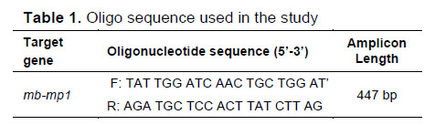
Real Time PCR: For the amplification of M. bovis specific membrane protein gene region by real time PCR, M. bovis specific primers were used according to Foddai et al. [27] previously reported (Table 1). Preferred primers amplify a 447 bp gene region.
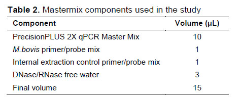
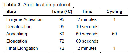
Commercial Standard real time PCR Detection Kit for M. bovis (Primer Design, GENESIG® UK) was used (Table 2) to detect M. bovis in DNA samples by real time PCR. The protocol was applied as recommended (Table 3).
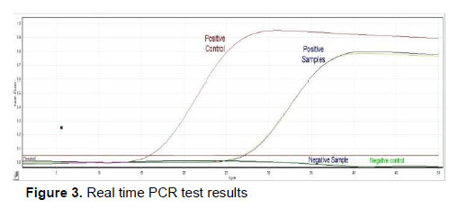
The results in the real time PCR device were evaluated by comparing them with the positive (logarithmic curve) and negative control (solid line). The positive/negative result of a sample was determined by the ct value. If the cycle threshold (ct) value is ˂30, it is considered positive, if the ct value is between 30-35 and there is a logarithmic curve, it is considered weakly positive, and if the ct value is ˃35 and the amplification curve has a linear structure, it is accepted as negative.
Bulgular
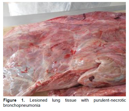
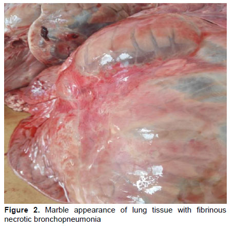
Macroscopic Examinations: The lung samples were macroscopically examined in detail by opening the lumen of the bronchi and bronchioles. On the outer and cross-sectional surfaces of the lobes, diffuse areas of necrosis were observed mostly in the appearance of a typical mottled marble landscape, as well as gray-cream colored partly hard consistency and nodular character. It was determined that the lesioned lung tissues had a harder consistency than the normal lung tissue, and necrosis particularly affected the lung regions around the bronchi and bronchioles. On the cross-sectional surfaces of some lesioned areas, the presence of a necrotic-purulent exaudate surrounded by a connective tissue capsule and the presence of coagulation necrosis in some samples were observed (Figure 1). In some samples, it was observed that the lungs were affected at the lobar level, and these regions were viscous and voluminous (Figure 2).
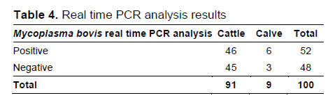
Real Time PCR Test Results: In the examination performed with real time PCR, 52 of 100 samples were found positive for M. bovis (Table 4, Figure 3). Of the positive samples, 46 were obtained from adult cattle, 6 from calves.
Fortyeight of the samples were found negative for M. bovis by real time PCR. Of these samples, 45 were adult cattle and 3 were calves (Table 4).
Bacteriological Culture Analysis Results: Bacterial agents were isolated from 50 of 100 Lung samples examined in the study. Nine of culture positive lung samples were contained more than one bacterium and a single agent was isolated from 41 of them.
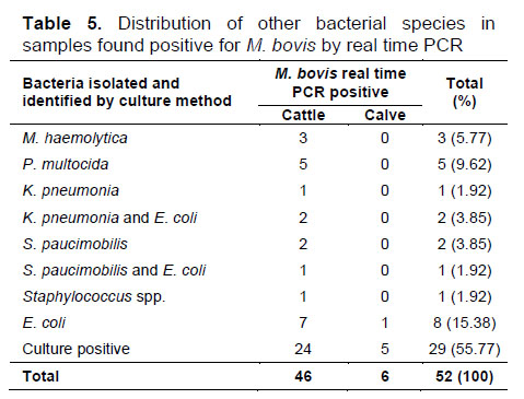
No other bacterial agents could be isolated by bacteriological culture in 29 of 52 samples that were found to be M. bovis positive by real time PCR method. In 23 of the samples, different types and numbers of bacteria were grown, while 2 different bacteria were detected in 3 of them. In the other 20, a single bacterial species was isolated and identified (Table 5,
M. bovis positive samples by real time PCR were also analyzed by bacteriological culture method. According to the results obtained, Escherichia coli, Pasteurella multocida, Mannheimia haemolytica, Klebsiella pneumonia, Sphingomonas paucimobilis and Staphylococcus spp. were isolated in different rate and identified (Table 5).
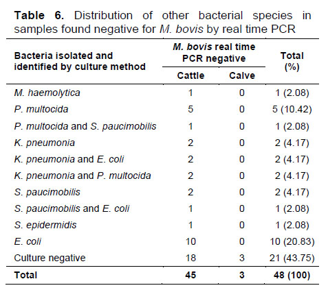
Bacterial agent was isolated by culture method from 27 of 48 lung samples that were negative for M. bovis by real time PCR method, but no bacterial agent could be isolated from 21. While a single agent was isolated in 21 of the culture-positive samples, more than one agent was isolated in 6 of them.
E. coli from 10, P. multocida from 6, K. pneumonia from 6, S. paucimobilis from 3, M. haemolytica from 1 and S. epidermidis from 1 of M. bovis negative samples by real time PCR were identified by culture (Table 6).
In studies conducted in various countries, differences in M bovis isolation rates from pneumonia cases were observed. It has been reported that M. bovis is isolated in 11.8% in Hungary [29], 13-23% in Ireland [30, 31], 25% in Italy [32], 30% in France [7] and 91% in Canada [33].
In various studies conducted in different regions of Türkiye, it is reported that the isolation rate of M. bovis varies between 4% and 81.3% [21, 28, 34-36].
In this study, a total of 100 bovine lung samples were examined by molecular and cultural methods. Out of 52 (52%) examined samples were found positive for M. bovis by real time PCR. In studies on the prevalence of M. bovis in various countries (11.8-91%) and Türkiye (4-81.3%), it has been reported that it can be isolated at different rates. The results obtained in this study are similar to the isolation rates obtained in both Türkiye and other countries. In particular, isolation by molecular or bacteriological culture method may cause proportional differences since the sensitivity of molecular methods is higher than the bacteriological culture method.
Since the host defense system is weakened in M. bovis infections, a predisposing situation may occur in terms of secondary pathogens. In studies of mixed infections, Arcangioli et al. [37] stated that M. bovis was detected in the early stage of pneumonia and M. bovis may be the primary or predisposing factor in the formation of bovine respiratory disease outbreaks.
Haines et al. [26] reported that they detected M. bovis antigen by immunohistochemical method in the 35 lungs and the 22 joints of 49 cattle with chronic pneumonia that did not respond to treatments. They reported that out of M. bovis antigen positive cases, 39% were BVDV, 10% H. somni, 20% M. haemolytica, and 12% vere found to be both BVDV and M. haemolytica antigen positive.
Bacterial agents isolated together with M. bovis include M. haemolytica, P. multocida, Trueperella pyogenes and less frequently H. somni, and viral agents such as bovine respiratory syncytial virus, BHV-1 and BPIV-3. agents are reported [38]. Brice et al. [30] also stated that they isolated 20.5% M. haemolytica, 9.06% T. pyogenes and 8.36% P. multocida in 287 animals with positive M. bovis. Şahin [34] isolated various mycoplasma species (6 M. bovis, 4 M. bovirhinis and 2 M. arginine) from 12 of 109 cattle lungs with pneumonia. Seven of them were P. multocida, M. haemolytica and Staphylococcus spp. reported positive. Byrne et al. [31] reported that in 66% of the M. bovis positive cases they detected in Ireland, they also detected viral agents such as BHV-1 and BPIV-3, as well as other bacteria such as P. multocida and M. haemolytica.
Various bacterial agents were detected by culture method in 50 of 100 lung samples examined in this study. While 9 of the culture positive samples contained more than one agent, only one agent was isolated from 41 of them. Different species and numbers of bacteria were detected in 23 of 52 samples found positive for M. bovis by real time PCR. While 2 different bacteria were detected in 3 of them, a single bacterium was isolated and identified in the other 20. In this study, bacterial culture positivity was detected in 23 of the samples found positive for M. bovis by real time PCR. Out of 23 M. bovis positive samples, E. coli from 8, P. multocida from 5, M. haemolytica from 3, K. pneumonia from 3, S. paucimobilis from 3, and Staphylococcus spp. from 1 sample were isolated and identified. Among these samples, E. coli was also isolated and identified in 2 K. pneumonia and 1 S. paucimobilis positive samples. Various bacterial agents were isolated by culture method from 27 of 48 lung samples that were negative for M. bovis by real time PCR. A single agent was isolated in 21 of the samples that were positive for bacteriological culture. More than one agent was isolated in 6 of them.
In studies on the subject, it was observed that other pathogens isolated from M. bovis positive animals showed similarities with the isolated strains in this study. As can be seen, suppression of the immune system in pneumonia cases and other predisposing factors that develop in the tissues provide an environment for the inclusion of secondary factors. The existence of multifactorial pneumonia cases observed in other studies was once again demonstrated in this study.
The results obtained in this study show that M. bovis is a common and important (52%) pneumonia agent in southern Türkiye. In the study, different types and numbers of bacteria were isolated in 44.2% of the samples that were positive for M. bovis by real time PCR, while 2 different bacteria were isolated and identified in 3 of them, and a single bacterium was isolated and identified in the other 20. In such studies, it is very difficult to determine the primary factor. On the other hand, the inability to detect different agents in M. bovis positive samples increases the possibility of M. bovis being the primary agent.
It was concluded that the high rate of M. bovis positivity in the study was due to the real time PCR method, which is shown as a reference method among diagnostic methods in the literature and used in this study.
As a result, pneumonia cases are frequently seen in cattle in Türkiye as well as all over the world and cause significant economic losses in many regions. For this reason, pneumonia cases are an important problem that should be carefully considered. In the prevention of disease, business management errors, negative care conditions and stress factors should not be ignored in terms of herd management.
Tartışma
Pneumonia are one of the important problems in cattle breeding. Mycoplasmas are detected at a significant rate among the agents of pneumonia, and the most common among them is M. bovis. Pneumonia cases caused by mycoplasmas have been reported for different years [14, 18, 28].References
- YEMLERLE ALINAN YÜKSEK DÜZEYDE İYOTUN SIĞIRLARDA BAZI KLİNİK HEMATOLOJİK VE BİYOKİMYASAL PARAMETRELER ÜZERİNE ETKİSİ
- SIĞIR THEİLERİOSİSİ
- ŞAP AŞISI UYGULANAN BESİ SIĞIRLARINDA ANTİOKSİDAN VİTAMİNLERİN KLİNİK VE BAZI HEMATOLOJİK PARAMETRELER İLE ANTİOKSİDAN ENZİM VE LİPİT PEROKSİDASYON DÜZEYLERİNE ETKİLERİ
- SIĞIRDA FARİNGEAL PENETRAN BİR YORGAN İĞNESİ OLGUSU
- KIŞ MEVSİMİNDE KARS YÖRESİNDE ZAVOT SIĞIRLARININ GENEL, YAŞ VE CİNSİYETE GÖRE VENÖZ KAN GAZI DEĞERLERİ