
Kedi ve Köpeklerde Göz Hastalıklarının Değerlendirilmesi
Cilt 39, Sayı 2
Temmuz 2025 · Sayfalar 122-129
Kedi ve Köpeklerde Göz Hastalıklarının Değerlendirilmesi
Evaluation of Eye Diseases in Cats and Dogs
Sena ÇAKI, Ali Said DURMUŞ
Kedi ve köpeklerde yaygın olarak görülen göz hastalıkları genellikle enfeksiyon etkenlerine ve travmalara bağlı olarak şekillenebilmektedir. Yapılan bu çalışmada kedi ve köpeklerde yaygın olarak gözlenen göz hastalıklarının değerlendirilmesi amaçlanmıştır. Çalışmanın materyalini göz hastalıklarına sahip olan farklı yaş, ırk ve cinsiyetteki 107 adet kedi ile 92 adet köpek oluşturmuştur. Hasta hayvanların ırkı, yaşı, cinsiyeti, anamnez bilgileri, şikayetlerin ne zaman başladığı ve hastalığın oluş şekli belirlenerek kayıt altına alınmıştır. Göz muayeneleri gerçekleştirilen olguların saptanan anormal oluşumlarının fotoğrafları alınmıştır. Hasta hayvanlarda uygun tedavi yöntemleri gerçekleştirilerek takiplerine iyileşme sağlanıncaya kadar devam edilmiştir. Yapılan bu çalışmada kedi ve köpeklerde en çok karşılaşılan göz hastalığının konjunktivitis (%40.20) olduğu belirlenmiştir. Ayrıca symblepharon (%15.57), blefaritis (%12.56), keratokonjunktivitis sicca (%8.54), kornea ülseri (%7.54), strabismus (%4.52), üçüncü göz kapağı bezinin prolapsusu (cherry eye) (%3.02), trişiazis (%2.01), keratitis (%1.51), entropion (%1.01), thelazia (%1.01), meibomitis (%0.50), kist dermoid (%0.50) ve yabancı cisimlere (%1.51) bağlı göz hastalıkları da tespit edilmiştir. Bu çalışmanın bulguları, iyi alınmış bir anamnez, detaylı bir muayene, doğru bir teşhis ve tedavi ile hassas bir organ olan gözde gelişen hastalıkların başarıyla tedavi edilebileceğini ortaya koymuştur.
Eye diseases, which are common in cats and dogs, can generally occur due to infectious agents and trauma. The aim of this study was to evaluate eye diseases commonly observed in cats and dogs. The material of the study consisted of 107 cats and 92 dogs of different ages, breeds and genders with eye diseases. The breed, age, gender, anamnesis information, when the complaints started and how the disease occurred were determined and recorded. Photographs of abnormal formations detected in the cases whose eye examinations were performed were taken. Appropriate treatment methods were applied to sick animals and their follow-up continued until recovery was achieved. In this study, conjunctivitis (40.20%) was found to be the most common eye disease in cats and dogs. In addition, symblepharon (15.57%), blepharitis (12.56%), keratoconjunctivitis sicca (8.54%), corneal ulcer (7.54%), strabismus (4.52%), third eyelid gland prolapse (cherry eye) (3.02%), trichiasis (2.01%), keratitis (1.51%), entropion (1.01%), thelazia (1.01%), meibomitis (0.50%), cyst dermoid (0.50%) and eye diseases due to foreign bodies (1.51%) were also detected. The findings of this study revealed that with a well-taken anamnesis, a detailed examination, a correct diagnosis and treatment, diseases that develop in the eye, which is a sensitive organ, can be successfully treated.
Giriş
Eyelid diseases such as blepharitis, entropion, ectropion, distichiasis, trichiasis, eyelid coloboma and eyelid tumours are observed in cats and dogs. Entropion may be seen in some breeds including Chow Chow, Sharpei, English Cocker Spaniel, American Cocker Spaniel, English Bulldog, Toy Poodle, Miniature Poodle, Saint Bernard, Great Danes and Bull Mastiff[1]. Despite its low prevalence in cats, it occurs with a predisposition in Persian breeds [2]. It has been reported that entropion may lead to conjunctivitis, keratitis, corneal ulceration and corneal sequestrum if left untreated[3].Allergy, eyelid lesions, foreign bodies, bacteria and viruses play a role in the formation of conjunctivitis, which is a commonly observed eye disease in cats[4,5]. Conjunctivitis may be observed in one eye or both eyes[6]. It has been reported that conjunctivitis is observed more commonly in dog breeds such as Pug, Pekingese, Schnauzer, Cocker Spaniel, Bulldog and Shih-Tzu [7].
Eye diseases such as conjunctivitis follicularis, cyst dermoid, cataract, lens dislocation and uveitis are also observed in dogs[4].
Although third eyelid gland prolapse (cherry eye) is rare in cats, it is reported to be observed more frequently in dogs, especially in dogs under one year of age. Treatment is performed operatively [4,8].
Keratitis, eosinophilic keratitis, chronic superficial keratitis, corneal ulcer, corneal necrosis, keratoconjunctivitis sicca and corneal oedema are frequently observed corneal diseases in cats and dogs [9-12].
In this study, it was aimed to determine the incidence of eye diseases in cats and dogs, to evaluate the causes, treatment and prevention of diseases.
Gereç ve Yöntem
Research and Publication Ethics: The Local Ethics Committee for Animal Experiments approved this study at Fırat University (Approval number: 20.04.2022, 2022/07).The material of the study consisted of 107 cats of different ages, breeds and genders and 92 dogs of different ages, breeds, and genders with eye diseases brought to Fırat University Faculty of Veterinary Medicine Animal Hospital Surgery Clinic and some veterinary clinics in Gaziantep province. Informed consent forms were obtained from the owners and eye examinations of the sick animals brought with the complaint of eye disease were performed. The owners were asked questions that would help the diagnosis such as the disease status observed in the animals, when the case started and how it continued, whether such a situation had been encountered before and how this situation affected their normal life.
A direct ophthalmoscope and light source were used during eye examination. Local anaesthetics (lidocaine) were administered in the form of drops or subconjunctival injections when necessary.
Patients were examined in detail after their movements, gait, head position, eye size, posture, symmetry, photophobia, and the presence of any discharge were examined. After this procedure, palpation was applied for ocular and facial examinations. Responses to stimuli were observed. Eye movements, presence of blink reflex, response to visual stimulation using hand movements, pupillary constriction or dilation under different lights were evaluated to assess neuro-ophthalmic conditions.
Pressure was applied to the bulbus oculi to detect the symptoms. The presence of foreign body in the bulbus oculi and intraocular pressure were checked. Pupillary reflex was checked with a flashlight. Eyelids were checked for movement, colour and swelling. Conjunctiva was examined for the presence of different formations such as discolouration, adhesions or masses. The sclera was examined for colour, irregularity, swelling and the presence of masses. The presence of vascularization, pigmentation, ulceration, foreign body, abscess and necrosis on the corneal surface were checked and evaluated. The third eyelid was examined for position, presence or absence of foreign body, discolouration and mass.
The findings obtained from the anamnesis until the recovery after treatment were collected, evaluated and reported. Descriptive statistics of the data obtained were given as frequency and/or percentage. Additionally, eye diseases between cats and dogs were analyzed by Chi-square test with the help of SPSS 22 package program. The statistical significance level was accepted as p<0.05.
Bulgular
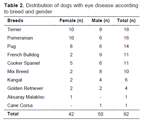
The breed and gender distribution of cats with eye disease given in Table 1 and the breed and gender distribution of dogs given in Table 2.
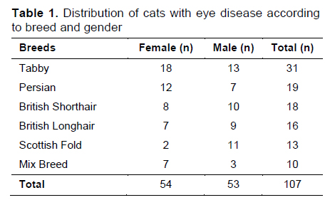
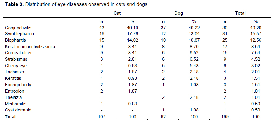
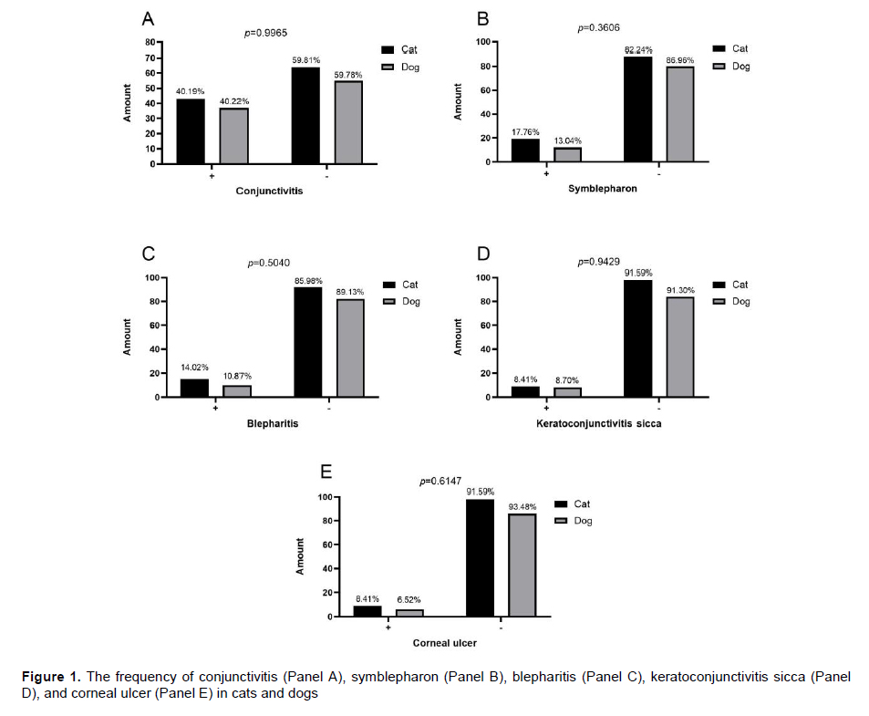
The distribution of eye diseases observed in cats and dogs given in Table 3. In the comparison, no statistically significant difference was found between the frequencies of the five most common eye diseases in cats and dogs (p>0.05) (Figure 1).
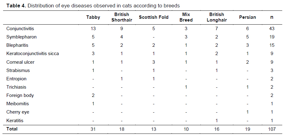
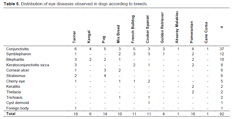
The distribution of eye diseases according to breeds in cats given in Table 4 and according to breeds in dogs given in Table 5.
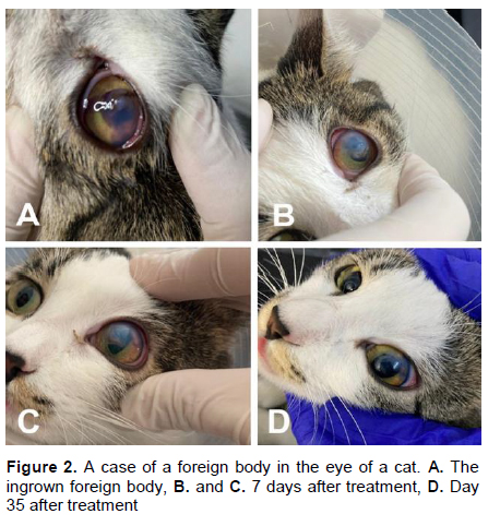
In the cases, corneal penetration, mucopurulent discharge, pain and closure of the eye were observed in which a foreign body was detected (Figure 2).
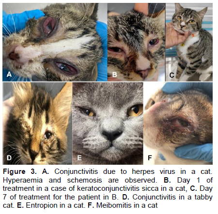
In cases of conjunctivitis with systemic viral infection, general disturbance, increased body temperature, heart and respiratory rate were observed. Ocular chemosis was detected (Figure 3A). Seromucous discharge, pigmentation and vascularization were noted in cases with keratoconjunctivitis sicca (Figure 3B, Figure 3C). Results of Schirmer tear tests were under normal limits. In patients with neonatal conjunctivitis, adhesions on the upper and lower eyelids and swelling of the eye were observed. It was determined that the eye discharge had a mucopurulent consistency (Figure 3D). Serous tear discharge, wetness in the upper eyelid, photophobia, conjunctivitis, blepharospasm and purulent discharge were observed in cases with entropion (Figure 3E). In meibomitis, mucopurulent discharge was noted (Figure 3F). In cases of trichiasis, serous tear discharge with pigmentation in the traumatised area was determined.
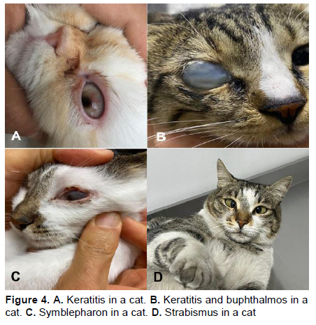
In cases of keratitis, an opaque cornea without gloss was observed. Corneal ulceration was also recorded (Figure 4A, Figure 4B). Herpes virus infections were recorded in cats with symblepharon. Adhesions between conjunctiva and sclera were determined (Figure 4C). It was determined that the majority of the 19 cats and 12 dogs with symblepharon were stray animals and all of them lacked the sense of vision. In most of the cases, the cornea and bulbar conjunctiva were severely adhered to each other. Although these adhesions were opened in the operated animals, recurrence occured. No other abnormal condition was observed in the case of congenital strabismus (Figure 4D).
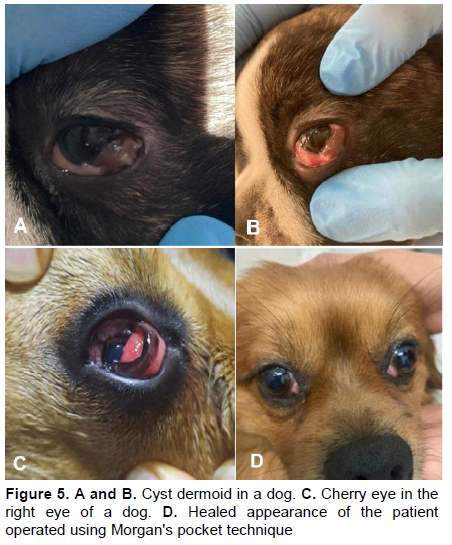
In corneal ulcers, pain, tissue loss in the ulcerated area, serous, seromucous and purulent tear discharge were observed. Serous tear discharge, vascularization and pruritus were found in cases of thelazia. Serous, seromucous and purulent tear discharge and pain were observed in cases with dermoid cyst (Figure 5A, Figure 5B). In the patients with cherry eye, redness, dryness and vascularization were observed (Figure 5C, Figure 5D). The patients with cherry eye were treated with Morgan's pocket technique.
Tartışma
As the number of companion animals such as cats and dogs is increasing day by day, veterinarians are more frequently encountering eye problems observed in these animals. Breed predispositions, infection and traumatic causes are also known to increase the incidence of eye diseases in cats[13].Sarıerler and Kılıç[14] reported 172 eye diseases (153 dogs, 19 cats) in 1048 dogs and 180 cats in a study they conducted. In this study, 107 of 199 patients with eye diseases were cats and 92 were dogs. However, no statistically significant difference was found between the incidence of the five most common eye diseases in cats and dogs.
Tamilmahan et al.[15], in a study conducted in 425 dogs, found that the number of male dogs was 260 and the number of female dogs was 165. Kumar et al.[16] reported that 65% (n=39) of 60 dogs with eye disease were male and 35% (n=21) were female. In this study, it was determined that 50 of 92 dogs were male and 42 were female. In the present study, eye diseases were found to be 54.35% in males and 45.65% in females. These findings are similar to the results of the study conducted by Kumar et al.[16]. It is thought that the higher incidence of eye diseases in male dogs compared to females may be due to the fact that males are more exposed to traumatic eye injuries due to their more aggressive behavior.
Şengöz Şirin et al.[17], reported that the cat breed with the highest incidence of eye disease was mix breeds with a rate of 29.8% and the dog breed with the highest incidence of eye disease was Golden Retriever breed with a rate of 82.3%. In another study[16], it was reported that eye diseases were mostly observed in Pug breed dogs with a rate of 28.3%. In contrast to the findings of Şengöz Şirin et al.[17], in this study, a lower rate (4.35%) of eye diseases was found in Golden Retriever breed dogs. Similar to the findings of Kumar et al.[16], 92 dogs were found to have eye diseases in this study and the first three breeds were Terrier (18 dogs), Pomeranian (16 dogs) and Pug (14 dogs).
Uzunlu et al.[18] reported the incidence of conjunctivitis as 42.31% in cats and 24.44% in dogs in a study conducted in 78 cats and 90 dogs. Çatalkaya et al.[19] reported that conjunctivitis was the most common eye disease with a rate of 21.15% in a study of 291 cats with eye diseases. In this study, 199 patients (92 dogs and 107 cats) were diagnosed with eye diseases, the incidence of conjunctivitis was 40.22% in dogs and 40.19% in cats. This result shows that the incidence of conjunctivitis is higher than other eye diseases among the eye diseases observed in cats and dogs, similar to what has been reported in previous studies.
Uzunlu et al.[18] reported keratitis cases as 11.11% in dogs and 15.58% in cats. Çatalkaya et al.[19] reported that keratitis cases in cats were the most common eye disease after conjunctivitis with a rate of 19.59%. Sanchez et al.[20] reported keratitis in 39 of 229 dogs (17.03%). In another study [16], keratitis was detected in 13 (21.7%) of 60 dogs with eye diseases. In this study, unlike the other studies, keratitis was encountered in 2 of 92 dogs (2.18%) and 1 of 107 cats (0.93%). The reason for the differences between the studies is thought to be due to periodic and seasonal changes.
Clinical findings in keratoconjunctivitis sicca include dryness, foreign body and stinging sensation, itching, pain, photophobia, redness and blurred vision[10,21]. Keratoconjunctivitis sicca has been reported to occur in brachycephalic dogs due to aging, genetic predisposition, hormonal changes and systemic diseases. Cavalier King Charles spaniel, English bulldog, Lhasa Apso, Shih Tzu, West Highland White terrier, Pug, Bloodhound, American cocker spaniel, English cocker spaniel, English springer spaniel, Pekingese, Boston terrier, Miniature Schnauzer and Samoyed breeds are reported to be prone to this disease[22,23]. In this study, the observation of keratoconjunctivitis sicca in Terrier, French Bulldog, Cooker Spaniel and Pomerian breeds is consistent with the literature.
Cherry eye is a common ocular problem in dogs. In cats, on the contrary, it is rarely observed. Bulldog, Pekingese, Cocker Spaniel, Neapolitan Mastiff, Beagle and Basset Hound were reported to be the breeds predisposed to cherry eye[24]. In another study [25], it was reported that 1 of 8 cases of cherry eye found in cats and 7 cases found in dogs. In this study, cherry eye was detected in 1 cat and 5 dogs. The findings showed that the incidence of cherry eye was higher in dogs than in cats. In addition, the observation of cherry eye in Bulldog and Cocker Spaniel breeds was consistent with the literature. However, it is noteworthy that cherry eye was also found in a Terrier dog among other breeds in this study.
It has been reported that cherry eye are successfully corrected using Morgan’s pocket technique, which does not alter tear production or the morphology of the third eyelid gland ducts. It has been reported that this technique is easy to apply and has a higher success rate and fewer complications than other treatment methods such as conservative and excision of cherry eye[26,27]. In this study, cherry eye was successfully treated with Morgan's pocket technique.
Saraiva and Delgado[28] reported that ocular dermoids were observed only in dogs in a study conducted in 103 dogs and 20 cats. In this study, the observation of ocular dermoids in only one dog among 107 cats and 92 dogs with eye diseases is consistent with the literature.
Symblepharon is adhesion of the palpebral conjunctiva to the bulbar conjunctiva or cornea. It has been reported that surgical treatment may be a solution, however, recurrence may be observed in cases where adhesions are prominent[29,30]. In this study, it was noteworthy that most of the 19 cats and 12 dogs with symblepharon were stray animals and all of them lacked the sense of sight. The recurrence of these adhesions in the operated animals despite the opening of the adhesions is consistent with the literature.
Many dog breeds[4] and Persian cats[2] are reported to be prone to entropion. Ergin et al.[31] reported that entropion was observed more frequently in brachycephalic cats. In this study of 107 cats and 92 dogs with eye disease, entropion was detected in only 2 brachycephalic breeds, British Shorthair and Scottish Fold. Although there were 19 Persian cats with eye diseases in the study, it was found remarkable that entropion was not observed in these cats.
In conclusion, in this study, it was observed that the eye is sensitive to eye diseases due to its location and texture. It is concluded that each case should be approached in detail and carefully. The detailed anamnesis, clinical examinations and techniques used in this study have shown that the diseases are not only limited to the eye, but can sometimes occur as a result of a systemic infection. For this reason, it is concluded that a detailed, multidimensional examination of patients with eye disease complaints will help to make the correct diagnosis and the success of the treatment.
References
- Weingart C, Kohn B, Siekierski M, Merle R, Linek M. Blepharitis in dogs: A clinical evaluation in 102 dogs. Vet Dermatol 2019; 30: 222-e69.
- Laus JL, Vicenti FAM, Talieri IC, Jorge AT, Bolzan AA. Primary entropion in Persian cats. Ciência Rural 1999; 29: 737-740.
- White JS, Grundon RA, Hardman C, O’Reilly A, Stanley RG. Surgical management and outcome of lower eyelid entropion in 124 cats. Vet Ophthalmol 2012; 15: 231-235.
- AlSaad KM. A common problems of dog eyes (a review). IOSR Journal of Agriculture and Veterinary Science 2021; 22: 19-26.
- Hartmann A, Hawley J, Werckenthin C, Lappin M, Hartmann KJ. Detection of bacterial and viral organisms from the conjunctiva of cats with conjunctivitis and upper respiratory tract disease. J Feline Med Surg 2010; 12: 775-782.
- Kumar K, Kumari K, Praveen PK, Ganguly S. Clinical management of conjunctivitis in dog: A case study. Indian Journal of Animal Health 2016; 55: 167-168.
- Nadias GK, Novac CS, Matei IA, et al. Prevalence of antimicrobial resistant bacteria from conjunctival flora in an eye infection prone breed (Saint Bernard). Molecules 2021; 26: 1-9.
- Deveci MZY, İşler CT, Yurtal Z, Altuğ ME, Kirgiz Ö. Evaluation of Morgan’s pocket technique in the treatment of nictitans gland prolapse in dogs. Turk J Vet Anim Sci 2020; 44: 521-527.
- Farghali HA, AbdElKader NA, AbuBakr HO, et al. Corneal ulcer in dogs and cats: novel clinical application of regenerative therapy using subconjunctival injection of autologous platelet-rich plasma. Front Vet Sci 2021; 8: 641265.
- Kulualp K, Kılıç S. Kuru göz sendromu. FÜ Sağ Bil Vet Derg 2012, 26: 115-124.
- Laguna F, Leiva M, Costa D, Lacerda R, Peña Gimenez T. Corneal grafting for the treatment of feline corneal sequestrum: A retrospective study of 18 eyes (13 cats). Vet Ophthalmol 2015; 18: 291-296.
- Singh RB, Singh RB, Das S, et al. Paradox of complex diversity: Challenges in the diagnosis and management of bacterial keratitis. Prog Retin Eye Res 2022; 88: 101028.
- Parlak K, Akyol ET, Zamirbekova N, et al. Eye injuries in cats with head trauma. Kocatepe Veterinary Journal 2021; 14: 482-491.
- Sarıerler M, Kılıç N. Adnan Menderes Üniversitesi (ADÜ) Veteriner Fakültesi Cerrahi Kliniğine getirilen hastalara toplu bir bakış (1999-2003). Uludağ Üniversitesi Veteriner Fakültesi Dergisi 2003; 22: 75-79.
- Tamilmahan P, Zama MMS, Pathak R, Muneeswaran NS, Karthik K. A retrospective study of ocular occurrence in domestic animals: 799 cases. Vet World 2013; 6: 274-276.
- Kumar T, Punia M, Agnihotri D, Sindhu N, Jain VK. Incidence of ophthalmic affections in dogs-A short study. Int J Cur Microbiol App Sci 2018; 7: 1560-1565.
- Şengöz Şirin Ö, Çetin MN, Neyse B. Evaluation of eye diseases in cats and dogs: A retrospective study: 200 cases (2021-2022). MAE Vet Fak Derg 2023; 8: 44-49.
- Uzunlu EO, Aras S, Zamirbekova N, Akyol ET, Arıcan M. Kedi ve köpeklerde görülen yaygın göz hastalıkları: retrospektif çalışma (2018-2019). Bozok Vet Sci 2020; 1: 17-22.
- Çatalkaya E, Ersöz-Kanay B, Yayla S, Saylak N, Bayat A. Evaluation of eye diseases encountered in cats: A retrospective study (2020-2023). Dicle Üniv Vet Fak Derg 2023; 16: 108-111.
- Sanchez RF, Innocent G, Mould J, Billson FM. Canine keratoconjunctivitis sicca: Disease trends in a review of 229 cases. J Small Anim Pract 2007; 48: 211-217.
- Johnston L. Keratoconjunctivitis sicca (dry eye). SA Pharmaceutical Journal 2013; 80: 27-31.
- Avci N, Gumus F, Boztok Ozgermen B, Ozen D. Keratoconjunctivitis sicca in intact Aksaray Malakli breed dogs: Evaluation of 50 cases. Vet Ophthalmol 2024; 00: 1-10.
- Dodi PL. Immune-mediated keratoconjunctivitis sicca in dogs: current perspectives on management. Vet Med (Auckl) 2015; 6: 341-347.
- Arıcan M, Erol H, Parlak K, Kamış Ü, Yavru N. Comparison of the effects of intraocular pressure with phacoemulsification and extracapsular cataract extraction methods in dogs with cataract. Eurasian J Vet Sci 2014; 30: 188-194.
- Han MC, Sağlıyan A, Polat E, İstek Ö. Bazı evcil hayvanlarda karşılaşılan göz hastalıklarının değerlendirilmesi: Retrospektif bir çalışma: 278 olgu: (2002-2013). Harran Üniv Vet Fak Derg 2019, 8: 104-107.
- Kumar G, Reddy AK. Surgical management of cherry eye in a cocker spaniel dog. The Pharma Innovation 2020; 9: 267-268.
- ELAZIĞ’DA KÖPEKLERDE TESPİT EDİLEN TRAVMATİK MYİASİSLER
- STUMP PYOMETRALI BİR KÖPEKTE OVARİAN REMNANT SENDROMU
- KÖPEKLERDE DENEYSEL TİBİA DEFEKTLERİNİN ONARIMINDA SİRKÜLER EKSTERNAL FİKSATÖRÜ KULLANILARAK YAPILAN KEMİK KAYDIRMA TEKNİĞİ UYGULAMASI
- KÖPEKLERDE UCU YİVLİ VE UCU YİVSİZ İNTRAMEDULLAR PİNLERİN AKSİYEL EKSTRAKSİYON GÜÇLERİNİN DENEYSEL OLARAK ARAŞTIRILMASI: HİSTOLOJİK BULGULAR
- BİR KÖPEKTE RASTLANAN TUBEROSİTAS TIBİAE’NİN AVULSİON KIRIĞI OLGUSU