[ Ana Sayfa | Editörler | Danışma Kurulu | Dergi Hakkında | İçindekiler | Arşiv | Yayın Arama | Yazarlara Bilgi | E-Posta ]
Fırat Üniversitesi Sağlık Bilimleri Veteriner Dergisi
2016, Cilt 30, Sayı 3, Sayfa(lar) 187-194
[ Özet ] [ PDF ] [ Benzer Makaleler ] [ Yazara E-Posta ] [ Editöre E-Posta ]
Prepubertal Ratlarda Di-(2-Etilhekzil) Fitalat'ın Oluşturduğu Testiküler Toksisite Üzerine Ginkgo Biloba Ekstrakt 761'in Koruyucu Etkisi
Tuba PARLAK AK1, Mine YAMAN2
1Munzur Üniversitesi, Sağlık Yüksekokulu, Beslenme ve Diyetetik Bölümü, Tunceli, TÜRKİYE
2Fırat Üniversitesi, Veteriner Fakültesi, Histoloji Embriyoloji Anabilim Dalı, Elazığ, TÜRKİYE
Anahtar Kelimeler: Apoptozis, di-(2-etilhekzil) fitalat, ginkgo biloba ekstrakt 761, testiküler toksisite, TUNEL
Özet
Bu çalışmada, ratlara farklı sürelerde aynı dozda di-(2-etilhekzil) fitalat (DEHP) verilerek oluşturulan testiküler hasar üzerine ginkgo biloba ekstrakt 761 (EGb 761)'in koruyucu etkisi incelendi. Yetmiş iki adet 28 günlük Wistar Albino cinsi rat; kontrol, DEHP, DEHP + EGb 761 ve EGb 761 gruplarına ayrıldı. Çalışmanın 7., 14., ve 28. günlerinde hayvanlar dekapite edildi. DEHP uygulamasının kontrol grubu ile karşılaştırıldığında; malondialdehit (MDA), glutatyon peroksidaz (GSH-Px) ve katalaz (CAT) seviyelerinde, testiküler dokuda dejenerasyon, nekroz, atrofi, disorganizasyon, Leydig hücre hiperplazisi ve bazal membran kalınlaşması ile birlikte apoptotik indekslerde belirgin artışlara sebep olmasına karşın, testislerin ağırlıklarında, seminifer tubüllerin çaplarında, germinal hücre tabakası kalınlıklarında, serum testosteron ve glutatyon (GSH) seviyelerinde belirgin azalışlara neden olduğu belirlendi. DEHP uygulanan ratlara EGb 761 verilmesi ile bozulmuş oksidan/antioksidan dengesinde, azalmış testosteron konsantrasyonunda, indüklenmiş testiküler apoptoziste belirgin gelişme ve testiküler dokunun histopatolojik görünümünde kademeli olarak düzelme sağlandığı gözlendi. DEHP' ın uygulanan süre ve dozda testis dokusu için son derece toksik olduğu ve ciddi yapısal hasara sebep olduğu; antioksidan etkiye sahip olan EGb 761'in ise büyük ölçüde bu hasarın önüne geçtiği tespit edildi.
  • Başa Dön
  • Özet
  • Giriş
  • Materyal ve Metot
  • Bulgular
  • Tartışma
  • Sonuç
  • Kaynaklar
  • Giriş
    Di-(2-ethylhexyl) phthalate (DEHP) is a well-known reproductive toxicant. Experimental studies indicate that DEHP caused atrophy in testes and seminiferous tubules, spermatogenic disorder, and a decrease in testis weight and fertility1-3. It is reported that the oral administration of DEHP increased free radical production by induced oxidative stress in testis and these radicals damaged seminiferous tubules2. It has been presented in experimental studies related to the testicular toxicity of DEHP, that antioxidants such as vitamin and selenium have been reported to have reparative effects against this toxicity4,5. It is suggested that there may be their positive effects against this toxicity, especially the variety of herbal products commonly found in the environment and being its versatile effect.

    A standardized extract of Ginkgo biloba leaves is a well-defined product and contains 24% flavone glycosides and 6% terpene lactones6. The studies with the testicular germ cell apoptosis indicate that there is a protective effect of Ginkgo biloba extract 761 (EGb 761) against apoptosis and testicular damage7,8. It is reported that EGb 761 modulates expression of apoptotic related genes, reduces generation of free radicals and increases activity of antioxidant enzymes different animal tissues9-11.

    In recent years, experimental studies have been conducted showing the protective effect of EGb 761 against toxic agents. However, there was not any study related to the therapeutic effect of the herbal products against DEHP formed testicular toxicity. It was aimed with the present study to investigate the protective effect of EGb 761 as known a powerful antioxidant and the anti-apoptotic properties on DEHP formed sub-acute toxicity in prepubertal rat testis.

  • Başa Dön
  • Özet
  • Giriş
  • Materyal ve Metot
  • Bulgular
  • Tartışma
  • Sonuç
  • Kaynaklar
  • Materyal ve Metot
    Animals and experimental design: In this study, seventy-two healthy adult male Wistar albino rats, weighing 60-80 g and averaging 28-days-old were used. Rats were kept at 25 °C in a 12h light-dark cycle and allowed free access to a pellet diet and drinking water. The protocol for the animal use was approved by the Institutional Review Board of the National Institute of Health and Local Committee on Animal Research (FÜDAM-07.07.2010/11-64).

    DEHP (Acros, New Jersey, USA) was dissolved in corn oil (5 mL/kg) and administered to the animals by gavage at a dose of 2 g/kg/every other day. The dose used in this study were based on previous reports showing the adverse effects of DEHP on male reproductive organ in young rats12. EGb 761 (Abdi Ibrahim, Istanbul, Turkey) was administered 50 mg/kg13 by the same method one hour after DEHP treatment. All treatments were maintained for 28 days. A total of 72 male Wistar albino rats were randomly divided into four experimental groups of 18 animals each. These groups were arranged as follows: Control; treated with 5 mL/kg corn oil during 28 days. DEHP; treated with 5 mL/kg corn oil + DEHP 2 g/kg during the first 14 days; treated with 5 ml/kg corn oil during the last 14 days. DEHP+EGb 761; treated with DEHP 2 g/kg + 50 mg/kg EGb 761 during the first 14 days; treated with 50 mg/kg EGb 761 during the last 14 days. EGb 761; treated with 50 mg/kg EGb 761 during 28 days.

    Sample collection and homogenate preparation: The animals were sacrificed under slight ether anaesthesia exactly 24 h after the last administration at the end of 7, 14 and 28th days (into 3 sacrificed groups of six animals each), testes were removed. They were divided into two pieces; the first testis section was used for the histopathological examination, the second testis was stored at -20 °C until biochemical analyses.

    Biochemical analysis: Lipid peroxidation level was measured based on the concentration of thiobarbituric acid reactive substances, and the amount of produced malondialdehyde (MDA) was used as an index of LPO14. Catalase activity was spectrophotometrically determined by measuring the decomposition of hydrogen peroxide (H2O2) at 240 nm according to the method of Aebi15. Reduced glutathione (rGSH) content of the testis homogenate was measured at 412 nm using the method of Sedlak and Lindsay16. Glutathione peroxidase (GSH-Px) activity was determined according to the method of Lawrence and Burk17. The protein content in the testis was measured by method of Lowry et al.18. The serum testosterone level was measured by ELISA method using Elisa testosterone kit (Dia Metra, Italy) according to the manufacturer’s instructions and expressed as ng/mL.

    Histological examination: Testis tissues were fixed by Bouin’s solution, embedded in paraffin, and were stained with haematoxylin-eosin and PAS- haematoxylin. A total of 25 seminiferous tubules (ST) were randomly examined per section and, their diameters and germinal cell layer thickness (GCLT; from the basal membrane towards the lumen of the tubule) were measured using an ocular micrometer in a light microscope and, and the mean size of ST and GCLT were calculated. Johnsen’s testicular scoring19 was used to categorise the spermatogenesis in control and treatment groups. Twenty-five ST from each section were evaluated, and a score between 1 (very poor) and 10 (perfect) was given to each tubule according to Johnsen’s criteria.

    Determination of apoptotic cells in testis: The apoptotic germ cells were evaluated by using terminal transferase dUTP nick-end labelling (TUNEL, In Situ Cell Death Detection Kit, POD; Roche, Mannheim, Germany) according to the manufacturer’s instructions. Briefly, tissue sections were deparaffinized and digested with 20 mg/mL proteinase K for 10 min, followed by treatment with 3% H2O2 for 5 min to inhibit endogenous peroxidases. After washing with PBS, sections were then incubated with the TUNEL reaction mixture containing TdTenzyme and digoxigenin–11–dUTP in humidified atmosphere at 37 °C for 1 h and then stop/wash buffer was applied for 30 min at 37 °C. Labeling was revealed using 3-amino-9-etilcarbazole substrate. Counterstaining was performed using Mayer’s hematoxylin and sections were dehydrated and mounted with glycerol. For negative controls, sections were incubated with solution without terminal transferase. Apoptotic index was calculated as percentage of TUNEL-positive cells by the following equation: apoptotic cells %= (number of TUNEL-positive cells/total number of cells) × 10020.

    Statistical analyses: Data are presented as mean ± SEM. The degree of significance was set at P<0.05. Nonparametric Kruskal Wallis Varyans Analyses were used to determine the differences between the groups with respect to all parameters. Also, Mann Whitney-U test was used to determine the groups that causing this differences. In addition, the differences between the groups in terms of ST diameter, GCLT parameters, Johnsen testicular score, and TUNEL positive apoptotic cell index were determined with ANOVA (One Way of Analysis Variance) using Duncan test. The SPSS for Windows version 15.0 program (SPSS, Chicago, IL, USA) was used for statistical analyses.

  • Başa Dön
  • Özet
  • Giriş
  • Materyal ve Metot
  • Bulgular
  • Tartışma
  • Sonuç
  • Kaynaklar
  • Bulgular
    The average of the body and testis weights of the control and experimental groups have been given in Table 1. The body and testis weights decreased in DEHP groups during experiment, but it showed an increase in according to this group in the DEHP + EGb 761 groups (P<0.05). Significant (P<0.05) decreases in diameters of ST, GCLT, and Johnsen’s testicular score were determined in DEHP group as compared to the control group. However, EGb 761 administration to DEHP-treated animals significantly (P<0.05) improved the DEHP-induced damages in these parameters (Table 2).


    Büyütmek İçin Tıklayın
    Table 1: Mean±SEM values of body (BW) and testis weights (TW) in different treatment groups. (DEHP= Di-(2-ethylhexyl) phthalate, EGb 761= Ginkgo biloba extract 761).


    Büyütmek İçin Tıklayın
    Table 2: Mean±SEM values of diameters ST, GCLT, Johnsen testicular score, and TUNEL positive apoptotic cell indices in different treatment groups. (DEHP= Di-(2-ethylhexyl) phthalate, EGb 761= Ginkgo biloba extract 761).

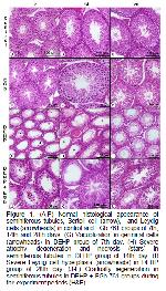
    The histopathological changes in the testes of all experimental groups were given in Table 3. It was observed that histological appearences of testes of control and EGb 761 groups were normal in the 7th day (Figure 1A and D). The histopathological changes such as necrosis, degeneration, desquamation, vacuolization, disorganization and reduction in germinal cells, atrophy in tubules were observed in DEHP group (Figure 1G). These types of histopathological damage were more milder in DEHP+EGb 761 group (Figure 1J).


    Büyütmek İçin Tıklayın
    Figure 1: (A-F) Normal histological appearance of seminiferous tubules, Sertoli cell (arrow), and Leydig cells (arrowheads) in control and EGb 761 groups of 7th, 14th and 28th days. (G) Vacuolization in germinal cells (arrowheads) in DEHP group of 7th day. (H) Severe atrophy, degeneration and necrosis (stars) in seminiferous tubules in DEHP group of 14th day. (I) Severe Leydig cell hyperplasia (arrowheads) in DEHP group of 28th day. (J-L) Gradually regeneration in seminiferous tubules in DEHP + EGb 761 groups during the experiment periods (H&E).


    Büyütmek İçin Tıklayın
    Table 3: The existence of some pathological lesions in testes of different treatment groups. (DEHP= Di-(2-ethylhexyl) phthalate, EGb 761= Ginkgo biloba extract 761).

    It was observed that ST was smooth of morphology and started of spermatogenesis in control and EGb 761 groups in the 14th day. (Figure 1B and E). In DEHP group pointed out which deepened the tubular atrophy due to necrosis in ST and continued the degeneration (Figure 1H). In addition to multinucleated giant cell formations in some tubules, Leydig cell hyperplasia, interstitial edema and capillary congestion was observed significantly. In DEHP + EGb 761 group it was seen that the intensive atrophic and necrotic changes replaced the mild degenerative changes. Furthermore, the presence of regeneration and spermatogenesis in many of the ST was detected in this group. And compared with DEHP treated group, interstitial regions showed a near normal appearence, but mild Leydig cell hyperplasia was found. (Figure 1K).

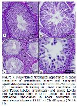
    In the 28th day, the histological structures of control and EGb 761 groups were normal and were formed a complete spermatogenesis in ST (Figure 1C and F, 3A and B). In DEHP group continued the necrotic and degenerative changes together with more intensive Leydig cell hyperplasia (Figure 1I), and thickening of ST basal membrane were seen as the most evidently in this period (Figure 3C). In DEHP+EGb 761 group, there were only mild degenerative changes in ST (Figure 1L). It was determined that the basal membrane and Leydig cells were normal in appearence compared with those in DEHP group (Figure 3D).

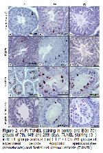
    Figure 2 illustrates apoptosis, demonstrated by TUNEL-staining in the testes of control and treated groups (Figure 2). The apoptotic cell index of DEHP group was significantly (P<0.05) higher than that of the control and other groups. However, a significant (P<0.05) decrease was observed in apoptotic cell indices of DEHP+EGb 761 group compared with that of the DEHP group only (Table 2).


    Büyütmek İçin Tıklayın
    Figure 2: (A-F) TUNEL-staining in control and EGb 761 groups of 7th, 14th and 28th days. TUNEL-staining (G-I) in DEHP groups and (J-L) in DEHP + EGb 761 groups of experiment periods. Apoptotic spermatocytes (arrowheads) and Sertoli cell (arrow) indicate (TUNEL).


    Büyütmek İçin Tıklayın
    Figure 3: (A-B) Normal histological appearance in basal membrane of seminiferous tubules and elongated spermatids (arrowheads) in control and EGb 761 groups. (C) Prominent thickening in basal membrane of seminiferous tubules (arrowheads) and severe Leydig cell hyperplasia (star) in DEHP group. (D) Normal histological appearance in basal membrane of seminiferous tubules in DEHP + EGb 761 group (PAS & H).

    Biochemical findings and testosterone levels of all groups were given in Table 4. It was determined that MDA, GSH-Px and CAT levels significantly increased in DEHP and DEHP+EGb 761 groups than control and EGb 761 groups. It was determined that rGSH and serum testosterone levels decreased in DEHP group during experimental period (P<0.05). Additionally, It was seen marked increase in the serum testosterone levels in DEHP+EGb 761 group compared to DEHP group in the 28th days (P<0.05).


    Büyütmek İçin Tıklayın
    Table 4: Mean±SEM values of malondialdehyde (MDA), reduced glutathione (rGSH), glutathione-peroxidase (GSH-Px) and catalase (CAT) activities and serum testosterone levels in different treatment groups. (DEHP= Di-(2-ethylhexyl) phthalate, EGb 761= Ginkgo biloba extract 761).

  • Başa Dön
  • Özet
  • Giriş
  • Materyal ve Metot
  • Bulgular
  • Tartışma
  • Sonuç
  • Kaynaklar
  • Tartışma
    DEHP is well known for its testicular toxicity in laboratory animals12,21,22. This toxicity caused a decrease in the relative testis weights with the body weight in experimental studies23,24 also a decrease in GCLT and in the average ST diameter25. In this study, DEHP administration significantly caused decrease in GCLT and in the average ST diameter with the weights of body and testes. These findings are supported by previous studies23-25.

    The lipid peroxidation (LPO) induced with toxic substances indicates the presence of oxidative stress in tissue with high concentrations of MDA, low concentrations of GSH9,26. The previous studies related to DEHP toxicity indicated that it was free radical trigger which caused increase in GSH-Px and CAT enzymes together with decrease in SOD and GSH levels2,27. In this study, biochemically the determination of increase in the concentration of MDA, decrease in GSH level, increase in the enzymes levels such as GSH-Px and CAT indicate that our findings were similar to the findings in the literature.

    Many studies stated that DEHP toxication negatively affected testosterone production of Leydig cells22,28,29. Miura et al.30 indicated that the concentration of testosterone significantly decreased in association with Leydig cell dysfunction in DEHP treated animals. In addition, it was reported that DEHP toxicity caused to a decrease in the antioxidant enzyme activities, and an increase in the ROS and lipid peroxides22. Leydig cells are sensitive to the extracellular sources of ROS leading to ultrastructural changes in their structures31. In accordance with literature data given above, it is thought that the increase in the lipid peroxide levels and the inhibition of the testicular steroidogenesis caused Leydig cell dysfunction. Akingbemi et al.21 reported that Leydig cell hyperplasia induced as a result of DEHP exposure and this was associated with serum testosteron and LH levels. During experiment of our study, it was observed that there was a significant decrease in testosterone levels of DEHP treated group compared to the others. But, it was seen an increase in 28th day compared to previous experiment periods. It has been thought that these changes in testosterone levels which may be depend on Leydig cells dysfunction and hyperplasia.

    A repeated administration of DEHP was reported to cause testicular DNA damage5,12,23. The apoptotic cell indices of DEHP groups were found to be significantly higher than those of the control groups in our study.

    Ablake et al.32 reported that DEHP firstly caused the spermatogenic deterioration and focal degeneration. Then, It was determined spermatogenic arrest found in Sertoli cells together with just a few spermatogoniums, but there was regeneration in the some tubules at the end of the experimental process. Additionally, while Park et al.12 suggested that DEHP toxicity caused edema in interstitial region and giant cells with the multinucleate with germ cell origin, the other researchers argued that DEHP induced hyperplasia in Leydig cells21,33,34. Similar to the literature findings in our experimental study; it was observed that the testicular damage and interstitial edema began in 7th day of the treated, this damage was still more deepened and multinucleate giant cell formation seen on 14th day. It was also observed that degenerative effects and Leydig cell hyperplasia to be more intense on 28th day. But there were a few tubule regeneration. Testicular damage observed in this work may be elucidated with the direct or indirect effect of DEHP, which later induces lipid peroxidation that is a chemical mechanism capable of corrupting the structure and function of testis.

    EGb 761 has eliminated the testicular damage with variety mechanisms such as stabilized the membranes and prevention LPO35. Toxicity studies showed that EGb 761 protected testis by suppressing ROS production and combating antioxidant enzyme deactivation against oxidative stress. In other words, it was determined that EGb 761 treatment neutralized the abnormalities in both MDA and the GSH levels, also transformed to normal the antioxidant enzyme activities such as SOD, GSH-Px and CAT8,36. In this study significant decreases in MDA level, GSH-Px and CAT activities and marked increase in rGSH level of EGb 761 treated rats was observed. These findings demonstrate that EGb 761 has a potent antioxidative effect.

    It was reported that EGb 761 was a herbal agent which has a suppressive role against the sexual function dysfunction37 and increased the sexual performance by increasing their testosterone levels in the young rats38. In addition, it was stated that the testosterone level decreasing the result of Leydig cell damage in diabetic rats gradually increased with EGb 76139. This study showed that DEHP induced the decreased testosterone levels increased in EGb 761 treatment.

    Kanter40 demonstrated that the decrease in ST diameter and germinal cell degeneration composed with ischemia / reperfusion injury approached to the control group with EGb 761. EGb 761 administration to DEHP treated rats significantly increased in ST diameter and in GCLT in parallel with the literatures in our study. This result may be associated with beneficial effects of EGb 761 being the spermatic cell proliferation in tubules.

    The previous studies indicated that EGb 761 has antiapoptic properties13,40. Yeh et al.8 stated that EGb 761 pressured doxorubicin induced germ cell apoptosis and had antiapoptotic effect on testis. Also, it was determined that the ischemia / reperfusion and the testicular torsion increased the germ cell apoptosis and decreased these negative effects with EGb 7617,13,40. In this study, it was determined that DEHP induced testicular apoptosis gradually improved depending on antiapoptotic properties of EGb 761.

  • Başa Dön
  • Özet
  • Giriş
  • Materyal ve Metot
  • Bulgular
  • Tartışma
  • Sonuç
  • Kaynaklar
  • Sonuç
    This study suggested that DEHP caused highly significant testicular damage considering the duration of application and applied dose while EGb 761 had antioxidant and antiapoptotic effects and effectively prevented the occurence of these damages. Long term administration of DEHP is anticipated to cause more significant damage. With the studies to be performed in the future, the possible mechanisms of the DEHP induced toxic damage can be delineated and the potential effects of EGb 761 on such mechanisms could be identified.
  • Başa Dön
  • Özet
  • Giriş
  • Materyal ve Metot
  • Bulgular
  • Tartışma
  • Sonuç
  • Kaynaklar
  • Kaynaklar

    1) Andriana BB, Tay TW, Tachiwana T, et al. Effects of mono (2-ethylhexyl) phtlate (MEHP) on testes in rats in vitro. Okajimas Folia Anat Jpn 2004; 80: 127-136.

    2) Kasahara E, Sato EF, Miyoshi M, et al. Role of oxidative stress in germ cell apoptosis induced by di-(2-ethylhexyl) phthalate. Biochem J 2002; 365: 849-856.

    3) Staples CA, Peterson DR, Parkerton TF, Adams WJ. The environmental fate of phthalate esters: A literature review. Chemosphere 1997; 35: 667-749.

    4) Ishihara M, Itoh M, Miyamoto K, et al. Spermatogenic disturbance induced by di-(2-ethylhexyl) phthalate is significantly prevented by treatment with antioxidant vitamins in the rat. Int J Androl 2000; 23: 85-94.

    5) Erkekoğlu P, Zeybek ND, Giray B, Asan E, Hincal F. The effects of di-(2-ethylhexyl) phthalate exposure and selenium nutrition on sertoli cell vimentin structure and germ-cell apoptosis in rat testis. Arch Environ Cont Toxicol 2012; 62: 539-547.

    6) Anonymous. EGb 761: Ginkgo biloba extract. Ginkor Drug Res Develop 2003; 4: 188-193.

    7) Akgül T, Ayyıldız A, Nuhoğlu B, et al. Ginkgo biloba (EGb 761) usage attenuates testicular injury induced by testicular ischemia / reperfusion in rats. Int Urol Nephrol 2008; 40: 685-690.

    8) Yeh YC, Liu TJ, Wang LC, et al. A standardized extract of ginkgo biloba suppresses doxorubicin-induced oxidative stress and p53-mediated mitochondrial apoptosis in rat testes. Br J Pharmacol 2009; 156: 48-61.

    9) Timioğlu O, Kutsal S, Ozkur M, et al. The effect of EGb 761 on the doxorubicin cardiomyopathy. Res Commun Mol Pathol Pharmacol 1999; 106: 181-192.

    10) Logani S, Chen MC, Tran T, Le T, Raffa RB. Actions of ginkgo biloba related to potential utility for the treatment of conditions involving cerebral hypoxia. Life Sci 2000; 67: 1389-1396.

    11) DeFeudis FV, Papadopoulos V, Drieu K. Ginkgo biloba extracts and cancer: a researches area in its ınfancy. Fundam Clin Pharmacol 2003; 17: 405-417.

    12) Park JD, Habeebu Sultan SM, Klaassen CD. Testicular toxicity of di-(2-ethylhexyl) phthalate in young sprague-dawley rats. Toxicology 2002; 171: 105-115.

    13) Akgül T, Karaguzel E, Surer H, et al. Ginkgo biloba (EGb 761) affects apoptosis and nitric-oxide synthases in testicular torsion: An experimental study. Int Urol Nephrol 2009; 41: 531-536.

    14) Placer ZA, Cushman LL, Johnson BC. Estimation of product of lipid peroxidation (malonyl dialdehyde) in biochemical systems. Anal Biochem 1966; 16: 359-364.

    15) Aebi H. Catalase. In: Bergmeyer HU (Editor). Methods in enzymatic analysis. NewYork: Academic Press 1983: 276-286.

    16) Sedlak J, Lindsay RH. Estimation of total, protein- bound and nonprotein sülfhydryl groups in tissue with ellman's reagent. Anal Biochem 1968; 25: 192-205.

    17) Lawrence RA, Burk RF. Glutathione peroxidase activity in selenium-deficient rat liver. Biochem Biophys Res Commun 1976; 71: 952-958.

    18) Lowry OH, Rosebrough NJ, Farr AL, Randall RJ. Protein measurement with folin phenol reagent. J Biol Chem 1951; 193: 265-275.

    19) Johnsen SG. Testicular biopsy score count a method for registration of spermatogenesis in human normal values and results in 335 hypogonadal males. Hormones 1970; 1: 2-25.

    20) Türk G, Çeribaşı S, Sönmez M, et al. Ameliorating effect of pomegranate juice consumption on carbon tetrachloride-induced sperm damages, lipid peroxidation, and testicular apoptosis. Toxicol Ind Health 2013; DOI: 10.1177/0748233713499600.

    21) Akingbemi BT, Ge R, Klinefelter GR, Zirkin BR, Hardy MP. Phthalate-induced leydig cell hyperplasia is associated with multiple endocrine disturbances. Proc Natl Academy Sci of the USA 2004; 101: 775-780.

    22) Kim HS, Saito K, Ishizuka M, Kazusaka A, Fujita S. Short period exposure to di-(2-ethylhexyl) phthalate regulates testosterone metabolism in testis of prepubertal rats. Arch Toxicol 2003; 77: 446-451.

    23) Sobarzo CM, Lusting L, Ponzio R and Denduchis B. Effects of di-(2-ethylhexyl) phthalate on n- cadherin and catenin protein expression in rat testis. Rep Toxicol 2006; 22: 77-86.

    24) Noriega NC, Howdeshell KL, Furr J, et al. Pubertal administration of DEHP delays puberty, suppresses testosterone production and inhibits reproductive tract development in male sprague-dawley and long-evans rats. Toxicol Sci 2009; 111: 163-178.

    25) Cammack JN, White RD, Gordon D, et al. Evaluation of reproductive development following intravenous and oral exposure to DEHP in male neonatal rats. Int J Toxicol 2003; 22: 159-174.

    26) Çeribaşı AO, Sakin F, Türk G, Sönmez M, Ateşşahin A. Impact of ellagic acid on adriamycin-induced testicular histopathological lesions, apoptosis, lipid peroxidation and sperm damages. Exp Toxicol Pathol 2012; 64: 717-724.

    27) Pereira C, Mapuskar K, Rao CV. Effect of diethyl phthalate on rat testicular antioxidant system: A dose-dependent toxicity study. Pestic Biochem Physiol 2008; 90: 52-57.

    28) Akingbemi BT, Youker RT, Sottas CM, et al. Modulation of rat leydig cell steroidogenic function by di-(2-ethylhexyl) phthalate. Biol Reprod 2001; 65: 1252-1259.

    29) Oishi S. Effects of co-administration of di-(2-ethylhexyl) phthalate and testosterone on several parameters in the testis and pharmacokinetics of its mono-de-esterified metabolite. Arch Toxicol 1989; 63: 289-295.

    30) Miura Y, Naitoi M, Ablake M, et al. Short-term effects of di-(2-ethylhexyl) phthalate on testes, liver, kidneys and pancreas in mice. Asian J Androl 2007; 9: 199-205.

    31) Jones HB, Garside DA, Liu R, Roberts JC. The influence of phthalate esters on leydig cell structure and function in vitro and in vivo. Exp Mol Pathol 1993; 58: 179-193.

    32) Ablake M, Itoh M, Terayama H, et al. Di-(2-ethylhexyl) phthalate induces severe aspermatogenesis in mice, however subsequent antioxidant vitamins supplementation accelerates regeneration of the seminiferous epithelium. Int J Androl 2004; 27: 274-281.

    33) Ljungvall K, Karlsson P, Hulten F, et al. Delayed effects on plasma concentration of testosterone and testicular morphology by intramuscular low-dose di-(2-ethylhexyl) phthalate or estradiol benzoate in the prepubertal boar. Theriogenology 2005; 64: 1170-1184.

    34) Foster PM, Mylchreest E, Gaido KW, Sar M. Effects of phthalate esters on the developing reproductive tract of male rats. Hum Rep Update 2001; 7: 231-235.

    35) Chan PC, Xia Q, Fu PP. Ginkgo biloba leave extract: biological, medicinal and toxicological effects. J Environ Sci Health C 2007; 25: 211-244.

    36) Amin A, Mahmoud-Ghoneimio D, Syam MI, Daoud S. Neural network assessment of herbal protection against chemotherapeutic-induced reproductive toxicity. Theor Biol Med Modelling 2012; 9: 1.

    37) Cohen AJ, Bartlik B. Ginkgo biloba for antidepressant-induced sexual dysfunction. J Sex and Marit Therapy 1998; 24: 139-143.

    38) Yeh KY, Pu HF, Kaphle K, et al. Ginkgo biloba extract enhances male copulatory behavior and reduces serum prolactin levels in rats. Horm Behavior 2008; 53: 225-231.

    39) Wu XY, Wang WY, Wang RR, et al. Ginkgo biloba extract enhances testosterone synthesis of leydig cells in type 2 diabetic rats. Natl J Androl 2008; 14(4): 371-376.

    40) Kanter M. Protective effects of ginkgo biloba (EGb 761) on testicular torsion/detorsion-induced ischemia-reperfusion injury in rats. Exp Mol Pathol 2011; 91: 708-713.

  • Başa Dön
  • Özet
  • Giriş
  • Materyal ve Metot
  • Bulgular
  • Tartışma
  • Sonuç
  • Kaynaklar
  • [ Başa Dön ] [ Özet ] [ PDF ] [ Benzer Makaleler ] [ Yazara E-Posta ] [ Editöre E-Posta ]
    [ Ana Sayfa | Editörler | Danışma Kurulu | Dergi Hakkında | İçindekiler | Arşiv | Yayın Arama | Yazarlara Bilgi | E-Posta ]