[ Ana Sayfa | Editörler | Danışma Kurulu | Dergi Hakkında | İçindekiler | Arşiv | Yayın Arama | Yazarlara Bilgi | E-Posta ]
Fırat Üniversitesi Sağlık Bilimleri Veteriner Dergisi
2017, Cilt 31, Sayı 1, Sayfa(lar) 021-025
[ Özet ] [ PDF ] [ Benzer Makaleler ] [ Yazara E-Posta ] [ Editöre E-Posta ]
Tavşanlarda Deneysel Osteoartrit Modelinde Sığır Amniyotik Sıvısı ve Hiyaluronik Asidin Eklem İçi Enjeksiyonlarının Kıkırdak Doku Üzerindeki Etkilerinin Karşılaştırılması: Radyolojik Sonuçlar
Murat TANRISEVER, Sait BULUT
Fırat Üniversitesi, Veteriner Fakültesi, Cerrahi Anabilim Dalı, Elazığ, TÜRKİYE
Anahtar Kelimeler: Sığır amniyotik sıvısı, kıkırdak koruyucu etki, deneysel osteoartrit, hiyaluronik asit, radyoloji
Özet
Bu deneysel çalışmada tavşanların genu eklemlerinde oluşturulan osteoartritis (OA) modelinde sığır amniyotik sıvısının kıkırdak doku üzerinde oluşturacağı etkilerin araştırılması ve hiyaluronik asit ile karşılaştırılması amaçlanmıştır. Çalışmada 21 adet tavşan kullanılmış olup, sağ dizlerinin ön çapraz bağları OA oluşturmak amacıyla kesildi. Tavşanlar 4 hafta sonra rastgele 3 eşit gruba ayrıldı. Birinci grup tavşanlara birer hafta ara ile 3 kez eklem içi 0.5 mL sığır amniyotik sıvısı enjekte edildi (SAS). İkinci grup tavşanlara birer hafta ara ile 3 kez eklem içi 0.5 mL hiyaluronik asit uygulandı (HA). Üçüncü grup tavşanlar ise kontrol grubu (KONT) olarak ayrıldı ve herhangi bir uygulama yapılmadı. Tüm deneklerin operasyona alınmadan önce ve operasyonu takiben 30, 90 ve 120. günlerde sağ genu eklemlerinin posterio-anterior ve medio-lateral pozisyonda radyografileri alındı. 90 ve 120. günlerde SAS grubu ile HA grupları arasında radyografi skorları açısından anlamlı bir fark bulunmazken KONT grubuyla diğer iki grup karşılaştırıldığında aralarında önemli dercede fark bulundu (P<0.01). Sonuç olarak, büyüme faktörlerinden zengin inert bir sıvı olan SAS’ın, tavşan deneysel OA modelinde kıkırdak dokudaki dejeneratif değişikliklere karşı koruyucu etkisinin olması nedeniyle kıkırdak koruyucu tedavi ajanlarına alternatif olabileceği kanısına varıldı.
  • Başa Dön
  • Özet
  • Giriş
  • Materyal ve Metot
  • Bulgular
  • Tartışma
  • Kaynaklar
  • Giriş
    Osteoarthritis (OA) is a disease that affects the synovial joints and is characterised by degradation and loss of articular cartilage with subchondral bone remodelling, osteophyte formation and inflammation of the synovium. Clinical signs include joint pain, stiffness, swelling and loss of mobility, increasing in severity as the disease progresses1,2.

    Today, no definitive treatment method has been found for OA. Studies are underway to reduce progressive cartilage damage, which is the basic pathology of the disease. The cartilage healing effects of hyaluronic acid and its effects on slowing the progression of OA have been known and it is widely used today in the treatment of OA3-6. In addition, human amniotic fluid has been used as an alternative to hyaluronic acid in the prevention of the cartilage damage in OA and in cartilage healing in a few studies7-9.

    The aim of this study was the demonstration of the efficacy of bovine amniotic fluid, which can be an alternative to human amniotic fluid, which is much easier to access and more economical, on cartilage tissue in OA the radiological investigation of its effects compare with the widely used hyaluronic acid.

  • Başa Dön
  • Özet
  • Giriş
  • Materyal ve Metot
  • Bulgular
  • Tartışma
  • Kaynaklar
  • Materyal ve Metot
    This study was conducted with the approval number 46 dated 04.04.2013 by the Fırat University Rectorate Animal Experiments Ethics Board Presidency. The experimental animals were obtained from the Firat University Experimental Research Center (FUERC). The study was conducted at the FUERC and Firat University Animal Hospital Small Animal Clinic operation rooms.

    Five-months old male and female 21 New Zealand rabbits with an average weight of 2700-3000 g were used in the study. The method of excising their anterior cruciate ligaments was employed in order to create experimental OA in the rabbits.

    The rabbits were anaesthetized with a 5 mg/kg xylazine hydrochloride (Rompun®; 23.32mg/mL, Bayer, Istanbul, Turkey) and 35 mg/kg ketamine hydrochloride (Ketalar®; 50 mg/mL, Eczacibasi, Istanbul, Turkey) intramuscularly.

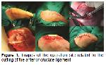
    After anesthesia, the right knees of the rabbits were shaved and they were laid down in the supine position. Before the incision, the knees were sterilized with 10% Povidone iodine. Then, approaching with a latero-anterior longitudinal incision, the patella was deviated to the medial following lateral parapatellar arthrotomy and the anterior cruciate ligament was cut. After the anterior drawer test which was used to evaluate whether the cruciate ligament had been completely cut, the patellar tendon and skin of the rabbits were closed with separate stitches using polyglactin 2/0 suture material (Figure 1).


    Büyütmek İçin Tıklayın
    Figure 1: Images of the operation site related to the cutting of the anterior cruciate ligament

    The experimental animals were allowed normal cage activity after the operation. 4 weeks after the surgical intervention, the rabbits were randomly divided into 3 equal groups. The first group of rabbits (n=7) were given 0.5 mL intra-articular bovine amniotic fluid (BAF) injections 3 times with one week in between. The second group of rabbits (n=7) were given 0.5 mL intra-articular hyaluronic acid (HA) applications 3 times with one week in between. The third group of rabbits (n=7) were used as a control group and nothing was applied to them (CONT).

    The bovine amniotic fluid was taken post-mortem from an approximately 5-6 months pregnant, healthy cows amniotic sac under aseptic conditions from slaughterhouse with using sterile plastic injectors. It was brought to the laboratory without breaking the cold chain and kept at –20°C. It was allowed to dissolve by waiting for 15 minutes at room temperature before use. Sodium hyaluronate in a ready to use glass injector was used as hyaluronic acid (Ostenil®; 10 mg/mL, Bio-Gen, Ankara, Turkey).

    Radiographs of the right genu joint of all subjects were taken in the posterio-anterior and medio-lateral position prior to operation and on the 30th, 90th and 120th days following the operation using the digital X-ray device found at the Fırat University Animal Hospital. The radiographs were evaluated using the Kellgren-Lawrence scoring system (Table 1). Sclerotic bone changes surrounding the joint edge, osteophytic growths extending outward from the joint edge, narrowing of the joint space and destructive changes in the bone ends were examined in this evaluation.


    Büyütmek İçin Tıklayın
    Table 1: Kellgren–Lawrence scoring10,11

    The statistical analysis of the results was done using the software "Statistical package for Social Sciences 22" (SPSS 22). After assessing whether there was a difference between the groups using the Kruskal-Wallis analysis method. Radiography scores were evaluated between the groups as a percentage using the Mann-Whitney U test in order to determine from which group the difference stemmed and whether it was a significant difference or not.

  • Başa Dön
  • Özet
  • Giriş
  • Materyal ve Metot
  • Bulgular
  • Tartışma
  • Kaynaklar
  • Bulgular
    During the study, no allergic reactions were observed in any rabbits and no rabbits died.

    120th day Radiological Findings: Suspicious narrowing of the joint space and suspicion of osteophytes (stage 1) were observed in 1 case, osteophytes and slight narrowing of the joint space (stage 2) were observed in 3 cases and numerous moderate degree osteophytes, narrowing of the joint space, mild scleroses and deformities of the bone ends (stage 3) were observed in 3 cases in the BAF group (Figure 2).


    Büyütmek İçin Tıklayın
    Figure 2: Images of subject No. 5 from the BAF group. A-B: Pre-operative C-D: Post-operative Day 30, E-F: day 90, G-H: Day 120 posterio- anterior and medio-lateral images

    Osteophytes and slight narrowing of the joint space (stage 2) were observed in 4 cases and numerous moderate degree osteophytes, narrowing of the joint space, mild scleroses and deformities of the bone ends (stage 3) were observed in 3 cases belonging to the HA group (Figure 3).


    Büyütmek İçin Tıklayın
    Figure 3: Radiographies of subject No. 6 from the HA group. A-B: Pre-operative C-D: Post-operative Day 30, E-F: Day 90, G-H: Day 120 posterio- anterior and medio-lateral images

    In 3 cases in the control group, numerous moderate degree osteophytes, narrowing of the joint space, mild scleroses and slight deformities of the bone ends (stage 3) were observed and severe large osteophytes, marked narrowing of the joint space, marked sclerosis and definitive deformities of the bone ends (stage 4) were observed in 4 cases in CONT group (Figure 4).


    Büyütmek İçin Tıklayın
    Figure 4: Radiographies of subject No. 1 from the control group. A-B: Pre-operative C-D: Post-operative Day 30, E-F: Day 90, G-H: Day 120 posterio- anterior and medio-lateral images

    A table containing the total radiology scores of all groups on the 30th, 90th and 120th days according to these radiographic images that were taken was created in a detailed manner (Table 2). The statistical data of the study were separately evaluated and calculated under the heading; radiological scores (Table 3).


    Büyütmek İçin Tıklayın
    Table 2: The total radiology scores of the groups


    Büyütmek İçin Tıklayın
    Table 3: Statistical evaluation of radiological scores

  • Başa Dön
  • Özet
  • Giriş
  • Materyal ve Metot
  • Bulgular
  • Tartışma
  • Kaynaklar
  • Tartışma
    In the experimental OA model that they created by means of cutting the anterior cruciate ligament on rabbit knees and with medial meniscectomy, Bilgili and Orhun12 radiologically and histologically investigated the changes that occurred in the joint tissues. In their radiological evaluation they demonstrated that there was superficial chipping, thickening of the subchondral bone tissue and osteophytic growth on the 30th day.

    With this study, superficial chipping in the joint cartilage mentioned in the literature12, sclerosis in the subchondral bone tissue and osteophytic growth was seen on the 30th day in animals in all groups. In addition, while these lesions in the joint did not grow in the BAF and HA groups on the 90th and 120th days of the study, large osteophytes, narrowing of the joint space, subchondral scleroses and deformities of the bone ends were observed in the CONT group.

    Tunay et al.13 have shown that after the cutting of the anterior cruciate ligament in rabbits, the intraarticular application of hyaluronic acid prevented inflammation and disintegration that formed in the cartilage tissue. Tirelioğlu et al.9 used 0.5 mL intraarticular human amniotic fluid 3 times with one week in between in the OA model that they formed after 4 weeks by cutting the anterior cruciate ligament in rabbits and demonstrated that the damage in the femur and tibia cartilages 12 weeks after the last injection was significantly lower compared to the control group that did not receive any injections.

    The bovine amniotic fluid that we used in the study contains insulin-like growth factors (IGFs) that have a stimulating effect on mesenchymal cells and chondrocytes and other growth factors in addition to macromolecules such as HA and hyaluronic acid activating agent (HASA)14-17. It also contains metalloproteinase inhibitors that can prevent cartilage destruction18. Due to the contents of bovine amniotic fluid and human amniotic fluid being similar, the use of bovine amniotic fluid was decided on in this study.

    In conclusion, it has been confirmed that intraarticular hyaluronic acid, which is widely being used in the treatment of OA in both human and veterinary practice has shown its efficacy with this study. In the radiological examinations it has been determined that there is no significant difference between the intraarticular injections of hyaluronic acid and bovine amniotic fluid and that bovine amniotic fluid does not cause any allergic reaction in the knee joints of rabbits. In the light of these findings, it has been concluded that bovine amniotic fluid, which ha been the main target of this study, can be an alternative to cartilage protecting drugs in the treatment of OA. That being said, the therapeutic effect of bovine amniotic fluid, which is easily obtained and more cost efficient, can be put into practice.

  • Başa Dön
  • Özet
  • Giriş
  • Materyal ve Metot
  • Bulgular
  • Tartışma
  • Kaynaklar
  • Kaynaklar

    1) Borrás-Verderaa A, Calcedo-Bernalb V, Ojeda-Levenfeldb J, et al. Efficacy and safety of a single intra-articular injection of 2% hyaluronic acid plus mannitol injection in knee osteoarthritis over a 6-month period. Rev Esp Cir Ortop Traumatol 2013; 56: 274-280.

    2) Pelletier JP, Martel-Pelletier J. Therapeutic targets in osteoarthritis: From today to tomorrow with new imaging technology. Ann Rheum Dis 2003; 62: 79-82.

    3) Adams ME. Lussier AJ. Peyron JG. A risk–benefit assessment of injections of hyaluronan and its derivatives in the treatment of osteoarthritis of the knee. Drug Safety 2000; 23: 115-130.

    4) Dıraçoğlu D. Osteoartritte intra-artiküler hyalüronik asit tedavisi. Türk Fiz Tıp Rehab Derg 2007; 53: 154-159.

    5) Elmorsy S, Funakoshi T, Sasazawa F, et al. Chondroprotective effects of high-molecular-weight cross-linked hyaluronic acid in a rabbit knee osteoarthritis model. Osteoarthritis and Cartilage 2014; 22: 121-127.

    6) Kılıç S, Karadaş E, İstek Ö, et al. Comparison of the effectiveness of hyaluronic acid, prednisolone and doxycycline on the progression of the pond-nuki model of osteoarthritis in dogs. FÜ Sağ Bil Derg 2001; 15: 387-396.

    7) Baki ME. Kıkırdak İyileşmesinde Amniyon Sıvısı ile Yüksek Molekül Ağırlıklı Hiyaluronik Asitin Karşılaştırılması. Uzmanlık Tezi, Trabzon: Karadeniz Teknik Üniversitesi, Sağlık Bilimleri Enstitüsü, 2008.

    8) Özgenel GY, Gülaydan F, Özcan M. Effects of human amniotic fluid on cartilage regeneration from free perichondrial grafts in rabbits. Br J Plast Surg 2004; 57: 423-428

    9) Tirelioğlu O, Bilgen MS, Atıcı T ve ark. Tavşanda deneysel osteoartrit modelinde insan amniotik sıvısının eklem içi uygulamasının kıkırdak doku ve sinovya üzerindeki etkileri. Uludağ Üni. Tıp Fak Derg 2007; 33: 121-125.

    10) Kellgren JH, Lawrence JS. Radiological assessment of osteoarthrosis. Ann Rheum Dis 1957; 16: 494-502.

    11) Köseoğlu HK, Özdemir BÖ. Osteoartritte radyolojik değerlendirme. RAED 2011; 3: 43-48.

    12) Bilgili H, Orhun S. Tavşan diz ekleminde oluşturulan deneysel osteoartritis’de eklem dokularında gelişen değişikliklerin radyolojik ve histolojik olarak araştırılıması. Vet Cer Derg 2012; 8: 8-12.

    13) Tunay S, Bilgili H, Yıldız C, ve ark. Deneysel osteoartritis’in sağaltımında intraartiküler hyaluronik asit kullanımı: Tavşan diz ekleminde radyolojik ve histopatolojik çalışma. Turk J Vet Anim Sci 2002; 26: 939-947.

    14) Dasari G, Prince I, Hearn MTW. Investigations into the rheological characteristics of bovine amniotic fluid. J Biochem Biophys Methods 1995; 30: 217-225.

    15) Decker M, Chiu E, Moiin A, et al. A hyaluronic acid stimulating factor from the bovine fetus and from breast cancer patients. Cancer Res 1989; 49: 3499-3505.

    16) Longaker M, Adzick NS, Hall JL, et al. Studies in fetal wound healing, VII. Fetal wound healing may be modulated by hyaluronic acid stimulating activity in amniotic fluid. J Pediatr Surg 1990; 25: 430-433.

    17) Ravelich SR, Breier BH, Reddy S, et al. Insulin-like growth factor-I and binding proteins 1, 2, and 3 in bovine nuclear transfer pregnancies. Biology of Reproduction 2004; 70: 430-438.

    18) Bunning RAD, Murphy G, Kumar S, et al. Metalloproteinase inhibitors from bovine cartilage and body fluids. Eur J Biochem 1984; 139: 75-80.

  • Başa Dön
  • Özet
  • Giriş
  • Materyal ve Metot
  • Bulgular
  • Tartışma
  • Kaynaklar
  • [ Başa Dön ] [ Özet ] [ PDF ] [ Benzer Makaleler ] [ Yazara E-Posta ] [ Editöre E-Posta ]
    [ Ana Sayfa | Editörler | Danışma Kurulu | Dergi Hakkında | İçindekiler | Arşiv | Yayın Arama | Yazarlara Bilgi | E-Posta ]