[ Ana Sayfa | Editörler | Danışma Kurulu | Dergi Hakkında | İçindekiler | Arşiv | Yayın Arama | Yazarlara Bilgi | E-Posta ]
Fırat Üniversitesi Sağlık Bilimleri Veteriner Dergisi
2017, Cilt 31, Sayı 3, Sayfa(lar) 213-219
[ Özet ] [ PDF ] [ Benzer Makaleler ] [ Yazara E-Posta ] [ Editöre E-Posta ]
Aydın İlinde Bulunan Sığır Çiftliklerinde İzole Edilen Streptococcus uberis Kökenli Mastitislerin ve Virulens Genlerinin Multiplex Polimeraz Zincir Reaksiyonu ile Belirlenmesi
Uğur PARIN1, Şükrü KIRKAN1, Erdem ÇİÇEK2, Hafize Tuğba YÜKSEL1
1Adnan Menderes Üniversitesi, Veteriner Fakültesi, Mikrobiyoloji Anabilim Dalı, Aydın, TÜRKİYE
2Adnan Menderes Üniversitesi, Sağlık Bilimleri Enstitüsü, Mikrobiyoloji Anabilim Dalı, Aydın, TÜRKİYE
Anahtar Kelimeler: Streptococcus uberis, mastitis, virulans genleri, mPCR
Özet
Sütçü ineklerde mastitise sebep olan etkenlerden olan Streptococcus uberis’in izolasyonu ve tür spesifik hedef genler ile identifikasyonu yapılarak, virulans genlerinin varlığı ve dağılımınının multipleks Polimeraz Zincir Reaksiyonuyla ortaya konması amaçlanmıştır. Araştırmada laboratuvara getirilen süt örneklerinden elde edilen izolatlar, koloni görünümü, Gram boyama ve katalaz testi ve biyokimyasal testler ile identifikasyonları sonucu S.uberis suşları ayrıldı ve moleküler çalışmalar için –20 °C’de saklandı. İzole edilen suşlardan DNA ekstraksiyonu, genomik DNA ekstraksiyon kiti (Fermentas®) ile prosedüre uygun olarak yapıldı. Fenotipik olarak S. uberis olduğu belirlenen tüm suşlarda 16S rRNA ve virulans ilişkili genlerin varlığı incelendi. İncelenen 200 süt örneğinin 35 (%17.5)’inden Streptococcus uberis izolasyonu gerçekleştirildi. Polimeraz zincir reaksiyonu ile, identifiye edilmiş S. uberis suşları incelendiğinde toplam 35 adet izolatın tamamında (%100) 1400 bp fragment aralığında16S rRNAgeni tespit edilmiştir. Virulans genleri tek olarak incelendiğinde, 32 (%91) suştan hasB, 32 (%91) suştan gapC, 32 (%91) suştan skc, 30 (%86) suştan cfu, 30 (%86) suştan hasC, 30 (%86) suştan hasA, 29 (%83) suştan sua, 28 (%80) suştan oppF, 25 (%71) pauA, 9 (%26) suştan Ibp geni identifiye edilmiştir. pauB genine ise rastlanmamıştır. Mastitis vakalarından izole edilen S. uberis etkenlerinin virulans genlerinin dağılımı incelendiğinde etkenin patojenitesi açısından kapsül formasyonunu eksprese eden genlerin önemli role sahip olduğu ortaya konulmuştur, ayrıca serin proteaz enzimi sentezinde görevli olan genlerin de dikkate değer oranda dağılım gösterdiği görülmüştür.
  • Başa Dön
  • Özet
  • Giriş
  • Materyal ve Metot
  • Bulgular
  • Tartışma
  • Kaynaklar
  • Giriş
    Mastitis is defined as inflammatory changes in the mammary gland, characterized by pathological changes in the mammary tissue and an increase in the number of somatic cells in the milk and dairy products, which are very important for human and animal health, nutrition and national economy, can only be obtained from healthy animals. As a result of pathological changes caused by the inflammatory state of the mammary gland, a number of physical and chemical changes occur in the milk. These changes limit the availability of milk yield and dairy products, thus mastitis is an important problem that needs to be carefully considered 1-3.

    It is possible to reduce mastitis-induced losses by applying effective control programs at the enterprise level. Implementation of mastitis control programs and ensuring the sustainability of these programs are the most important indicators of economic efficiency, especially in dairy farms with large capacity. However, due to the fact that the milk production capacity of our country is small especially for dairy farming and the productivity-centered mastitis control programs cannot be established yet, major economic losses occur in milk production. Since wholesome milk is obtained from healthy animals, it is a basic aim to maintain animal health and to diagnose mastitis cases early. Economic losses due to mastitis in the dairy industry result from reduced milk quality and milk yield, resulting in increased use of medicines and veterinary services and increased feed costs. At the same time, antibiotics used for the treatment of infections are left in the milk, antibiotic-resistant bacteria due to drug use unconsciously, and limitations of treatment possibilities. The implementation of routine and conscious control programs ensures that clinical mastitis is reduced and that subclinical infections are diagnosed early 1.

    Environmental factors that cause mastitis include unsuitable environmental conditions, inadequate hygiene at stables and shelters, inadequate ventilation conditions, and stiff and dirty substances. In order to increase milk yield, protein-rich feeding increases the likelihood of mastitis. The fact that milkers do not pay attention to cleaning and disinfection also accelerates the spread of infections among animals. When looking at microbial causes; many bacteria, fungi and viral agents cause the formation of mastitis. Mastitis agents are divided into infectious and environmental pathogens, taking into account their characteristics, as well as their relation to breast and mammary ducts. Microbial pathogens multiply in the bovine mammary gland and bulldog animals during milking. The environmental pathogens Streptococcus uberis, Streptococcus dysgalactiae and Escherichia coli cause environmental mastitis infections. The infectious pathogens include Staphylococcus aureus, Streptococcus agalactiae, Mycoplasma species and Corynebacterium bovis. Tuberculosis agents, Proteus, Leptospira, Listeria and Brucella species also cause mastitis 4

    Streptococcal mastitis is initially insidious and mild. Clinically detectable anomalies are not seen immediately in the teat. Infection usually takes formation during the lactation period, and as time goes by, the clinical signs of the disease begin to form due to the accumulation of water in the mammary gland 5. The major strains of S. agalactiae, S. dysgalactiae and S. uberis are isolated in mastitis infections, which are very common in dairy cattle and economic losses occur. Mastitis control programs, improvements in milk, post-partum hygiene practices, and common antibiotic treatments reduced mastitis infections caused by S. agalactiae and S. dysgalactiae, but no significant decrease in infections caused by S. uberis was identified 6.

    The scope of this study was to determine the isolation of factors from mastitic milk and the role of S. uberis etiology in mastitis etiology, and the distribution of virulence related genes in isolated strains by means of molecular methods and to provide information about the typing of isolated strains and distribution of virulence genes.

  • Başa Dön
  • Özet
  • Giriş
  • Materyal ve Metot
  • Bulgular
  • Tartışma
  • Kaynaklar
  • Materyal ve Metot
    A total of 200 milk samples were collected from Holstein cows with acute clinical mastitis found in 17 private dairy cattle farms which have machine milking systems with 20-150 head capacity in Aydın Province between June and November 2013. The animals were all in lactation, had not received antibiotic treatment in the last 1 month, had at least one calf delivery, between 2-8 year of age. Acute clinical mastitis was determined according to physical examination abnormalities (swelling and tenderness, temperature, pain, erythema in mammary lobes, coagulated milk) of the ill cows. Microbiological analyses of the milk samples for identification of Streptococcus uberis were conducted at the Diagnostic and Analysis Laboratory of the Department of Microbiology, Adnan Menderes University, Faculty of Veterinary Medicine. Milk samples were inoculated onto sheep blood agar and incubated for 48 hours at 37 °C for isolation of Streptococcus spp. At the end of the incubation period, S-type colonies 1-3 mm in diameter were selected in the media and stained by Gram staining method. Cocci that were microscopically Gram positive were tested for catalase reaction and catalase negative colonies were detected as Streptococcus spp 3,7. Streptococcal colonies were inoculated onto Edward's medium and incubated at 37 ⁰C for 24 hours. At the end of the incubation, dark brown-black colored colonies were considered positive for S. uberis, light brown colored or colorless colonies were evaluated as negative for S. uberis. Esculin test was conducted for identification at species level 8,9.

    DNA isolation of the isolates identified as S. uberis was made according to the procedure with the genomic DNA extraction kit (Fermentas®). The extracted DNAs were stored in cryo-tubes in –20 ⁰C freezer until PCR runs. The presence of 16S rRNA and virulence related genes in all strains determined to be S. uberis phenotypically was examined by multiplex PCR method. The preparation of the master mix was as followed; 20 ng of DNA sample was used in a total volume of 50 μL using 1 μM of each primer, 0.4 mM dNTP, 1.5 mM MgCl 2, 1X reaction buffer, 1.5 U Taq DNA polymerase 10. In PCR for identification of virulence related genes, primer mixes were prepared by combining each of the primers of 100 mM cfu, hAs, hasB, hasC, gapC, lbp, oppF, pauA / pauB, skc and sua to be 40 μM each. The primers of S. uberis virulence associated genes used in our study are shown in Table 1.


    Büyütmek İçin Tıklayın
    Table 1: Oligonucleotide primer sequences used in the multiplex PCR

    After the preparation of mastermix, the mixture was added to 0.2 mL PCR tubes, numbered up to the sample number and completed up to 48 μL of volume. Then, 2 μL of the extracted DNA was added, added into the relevant tubes, and the mouths were tightly closed. The prepared tubes were then loaded into the thermal cycler. The thermal cycle and time diagram used in the PCR analyzes of the mastermixes prepared for the identification of S. uberis and virulence associated genes was performed as previously described 11.

    6X of the loading dye was added in the volume of 3 μL on the pipette tip and mixed with 10 μL of PCR products. The mixture was added to the appropriate position on the gel. After the preparation of the samples and markers, the electrophoresis gel was run for 40 min at 100 volts.

  • Başa Dön
  • Özet
  • Giriş
  • Materyal ve Metot
  • Bulgular
  • Tartışma
  • Kaynaklar
  • Bulgular
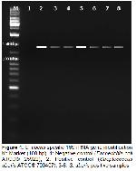
    Biochemical tests, isolation and identification of 35 (17.5%) strains of S. uberis were identified from 200 milk samples examined in our study. Biochemically identified S. uberis strains were examined via Multiplex polymerase chain reaction, and 16S rRNA gene was detected in a total of 35 isolates (100%) with 1400 bp fragment length (Figure 1).


    Büyütmek İçin Tıklayın
    Figure 1: S. uberis spesific 16S rRNA gene identification M: Marker (100 bp), 1: Negative control (Escherichia coli ATCC® 25922), 2: Positive control (Streptococcus uberis ATCC® 700407), 3-8: S. uberis positive samples

    In our study, it is observed that 68, 5% of strains were identified in E, D, B, Q, AH, AN patterns, respectively. 32 (91%) strains expressed hasB, 32 (91%) strains expressed gapC, 32 (91%) strains expressed skc, 30 (86%) strains expressed cfu, 30 (86%) strains expressed hasC, 30 (86%) strains expressed hasA 29 (83%) strains expressed sua, 28 (80%) strains expressed oppF, 25 (71%) pauA, 9 (26%) strains expressed Ibp genes. The pauB gene was not identified.

    When the virulence-related genes of S. uberis isolates were examined, 3 (8.57%) isolates were identified in pattern B (cfu, hasA, hasB, hasC, gapC, pauA/B, skc, sua), 4 (11.4%) isolates in pattern D (cfu, hasA, hasB, hasC, gapC, Ibp, oppF, pauA/B, skc, sua), 8 (22.8%) isolates in pattern E (cfu, hasA, hasB, hasC, gapC, oppF, pauA/B, skc, sua), 2 (5.7%) isolates in pattern G (cfu, hasA, hasB, hasC, gapC, pauA/B, skc, sua), 2 (5.7%) isolates in pattern L (hasA, hasB, hasC, gapC, oppF, pauA/B, skc, sua), 2 (5.7%) isolates in pattern N (hasA, hasB, hasC, gapC, oppF, pauA/B, skc, sua), 1 (2.85%) isolte in pattern P (hasA, hasB, hasC, oppF, pauA/B, skc), 3 (8.57%) isolates in pattern Q (cfu, hasA, hasB, hasC, gapC, oppF, sua), 3 (8.57%) isolates in pattern AH (cfu, gapC, Ibp, oppF, pauA/B, skc), 3 (8.57%) isolates in pattern AN (cfu, hasA, hasB, hasC, gapC, oppF, skc, sua), 2 (5.7%) isolates in pattern AT (cfu, hasB, hasC, Ibp, oppF, skc), 2 (5.7%) isolates in pattern AX (cfu, hasA, hasB, gapC, skc, sua) (Table 2).


    Büyütmek İçin Tıklayın
    Table 2: Distribution of virulence patterns of S. uberis strains (n= 35)

  • Başa Dön
  • Özet
  • Giriş
  • Materyal ve Metot
  • Bulgular
  • Tartışma
  • Kaynaklar
  • Tartışma
    Mastitis is the most important problem of dairy production. In cows, mastitis is an infection that occurs in all farms, including modern farms, and causes significant economic loss. It has been shown that economic losses in cows are more frequent in subclinical mastitis than in clinical mastitis. Mastitis is the leading cause of economic loss in milk production when yield, product losses and treatment costs are evaluated together. Mastitis frequency can be observed up to 40%, especially in modern intensive farms. Despite all hygienic practices, the continuation of the mastitis problem has brought medical preventive measures to the agenda and the development of control programs, including vaccines, has become unavoidable. However, in order to be able to effectively combat an infection, definite etiopathogenesis and its associated epidemiology must be known on the basis of region and herd 10.

    In cattle, mastitis is widespread all over the world and leads to significant economic losses. Pathogens of mastitis usually exhibit a contagious course during transport, and 2 groups are separated as infectious agents from environmental reservoirs. Many streptococcal species are among those isolated as mammary pathogens. S. uberis is an important pathogen in bovine mastitis, and is associated mostly with subclinical and clinical intramammary infections in both culled and untreated cows. These species are often a problem because they are ubiquitous around the dairy farms. Increasing the hygiene procedures against these opportunist pathogens results in the reduction of consequent mastitis, however does not yield an effective result in environmental contamination 12.

    The isolation rates of streptococci from milk samples vary. In a previous research 13, S. dysgalactiae was isolated from 2.3% of the milk samples examined, S. uberis and S. agalactiae was ddetected in the ratio of 0.3% on the farm to have a low isolation rate. Sampimon et al. 14 found the prevalence of S. uberis at 1.1%, S. dysgalactiae at 0.9%, and did not encounter S. agalactiae. Tenhagen et al. 15 have identified S. agalactiae as 0.1%, S. dysgalactiae and S. uberis as 2.3% from cow milk. In a study conducted in UK 16, bacteriological sampling in the milk resulted in the isolation of S. uberis with ratio of 23.5%, whereas S. agalactiae and S. dysgalactiae were not identified.

    Bentley et al. 17 reported that only three of the 206 gram-positive cocci isolated from mastitic milk were S. parauberis. Researchers have indicated that it is difficult to distinguish S. parauberis agents from S. uberis and that there is limited information on epidemiology. Pitkala et al. 18 identified two S. uberis isolates as S. parauberis in their study in Finland and found that the antibiotic resistance of S. parauberis strains resembled that of S. uberis. Devriese et al. 19 reported that catalase-negative and esculin-positive Gram-positive cocci isolated from clinical and subclinical mastitis were mostly identified as S. uberis and that they were not correctly identified.

    In studies conducted in Turkey, Dakman 20 reported that 12.5% of cow mastitis cases were caused by Streptococcus spp. and that 4.5% of these Streptococci were S. agalactiae. Karahan 21 examined 24.3% of mastitic milk samples from Streptococcus spp. 10.4% of isolates were S. agalactiae and 13.9% of other isolates were identified as other Streptococci.

    Şahin et al. 22 found that Streptococci isolation rate from milk was 29.82% and 14.03% of S. agalactiae, 8.77% of S. dysgalactiae and 7.02% of S. uberis were identified.

    The average isolation rate obtained in this study was found to be correlated with the findings obtained from previous studies conducted in Turkey. These findings may arise from the lack of application of mastitis control programs in dairy cows when assessed based on the mean Streptococcal isolation rate. The isolation rates obtained in other studies are at the same level.

    Jayarao and Oliver 23 identified 11 different enzymes in the RFLP-PCR method to isolate mastitic Streptococci at the species level by single enzyme cuts. McDonald et al. 9 performed identification of Streptococci at the species level by using dual enzyme combinations in a single reaction in the RFLP-PCR method.

    Different molecular methods have been used for species-level identification of streptococci. These methods are used especially in the typing of S. uberis 2.

    Duarte et al. 24 identified RFLP-PCR as an efficient method for diagnosing in a single reaction with shorter time than conventional methods. Odierno et al. 25 reported that RFLP-PCR was 100% accurate after RFLP-PCR, but that conventional test results did not fully support RFLP-PCR.

    Jayarao and Oliver 23 compared the results with classical biochemical results in RAPD-PCR evaluation and distinguished near genotypic factors by conventional method.

    Duarte et al. 24 showed that this method could be used in typing microorganisms in the RAPD PCR procedure they applied for group B Streptococci, but the use of additional typing methods in the interpretation of the results increased the reliability.

    Chatellier et al. 26 identified 71 profiles in 114 strains of RAPD-PCR for S. agalactiae and found that these profile differences could be due to the geographical areas where the farms were located.

    In our study, 35 isolates identified as S. uberis by bacteriological culture and biochemical identification method were all identified as S. uberis in PCR result using 16S rRNA gene specific primer. The fact that the primer used has a high specificity and that it is derived from the 16S rRNA gene, which is more stable than other gene regions, indicates that PCR identification can be performed as a reliable and rapid method in routine diagnostic laboratories. The use of molecular methods those are more specific and more sensitive than those of conventional methods with lower sensitivity and would be more beneficial.

    In a study conducted by Hassan et al. 27, positive for cfb gene was detected in S. agalactiae isolates, phenotypically negative for CAMP, by PCR. This suggests that the cfb gene-specific PCR assay can avoid false negative conditions that may arise from the CAMP reaction, which is considered to be one of the key criteria in the biochemical characterization of streptococci.

    Ekin et al. 28 reported a positive phenotype in 73.3% of S. agalactiae isolates obtained from cattle milk in terms of CAMP test in our country. In another study 26.3% of cattle-born Streptococcal isolates were positive for CAMP test 29. Although this test is generally considered to be a determinant test for the identification of Streptococci, it is likely that CAMP testing cannot be used effectively for identification from earlier studies. Reinoso et al. 10 found that the cfu gene distribution in their study was 76.9%.

    The cfu gene responsible for the CAMP factor in S. uberis was identified in 30 (86%) strains in our study. These results were in parallel with findings in our study. Thus, the result that cfu gene identification can be used for identification in S. uberis quickly and reliably.

    The operon in the capsule formation consists of the hasA, hasB and hasC gene loci, which are essential genes for capsule formation 11. HasA, hasB and hasC gene identification rates were 83%, 91% and 86%, respectively, in our study. Reinoso et al. 10 identified the hasA gene as 74.3%, the hasB gene as 66.6% and the hasC as 89.7%. According to the findings obtained, capsule formation in Streptococcal mastitis cases seems to play an important role in the development of infection.

    The oppF gene plays an important role in the milk production of S. uberis agents 30. In our study, oppF gene identification rate was 80%. Reinoso et al. 10 identified the oppF gene by 64.1%. Zadoks et al. 4 reported that this gene could not be isolated from all strains. The findings obtained correlate with the data in our study.

    Serine protease enzymes that convert plasminogenetic plasmids are essential components for the degradation of extracellular matrix proteins and the colonization of bacterial agents in tissues. In addition, the milk proteins that are responsible for the activation of endogenous plasminogenes in the milk are hydrolyzed, thus forming the molecules necessary for S. uberis 11. The skc gene responsible for the synthesis of these enzymes has been identified in 91% of our studies. Reinoso et al. 10 identified 63% of the skc gene in their study.

    The pauA gene 31 that was detected in association with plasminogen activation and intramammary colonization was isolated in 71% of our study. The pauB gene has not been identified in our study. In the study conducted by Smith et al. 30, only one strain of S. uberis was identified as pauB. Expression of pauB genes has not been shown to play a role in the formation of intramammary infections in the direction of the obtained data.

    Multiplex PCR-based molecular assays have been found to be reliable for detecting the genotypic relationships of the virulence genes at the genus level of the S. uberis agent, a common effect of subclinical bovine mastitis cases. In addition, in this study, the close proximity of the homozygous for the identification rates as a result of PCR amplification of the hasA, hasB and hasC gene operon used for the molecular identification of S. uberis has shown that this gene region allows rapid and reliable identification of cattle origin S. uberis agents. It has been found that the molecular methods used in this study may be useful for detecting virulent types among strains of S. uberis which are encountered as an important problem in subclinical mastitis cases and may contribute to putting the pathogen of infection in the basis of field strains. Significant control and strategies to be undertaken in the face of these findings are expected to provide a significant reduction in the incidence of S. uberis-induced mastitis. Thus, effective control programs against S. uberis mastitis, which are very difficult to eradicate or control due to various factors that are effective in the host environment and active agent triplet, will provide important advances both in terms of animal health and public health. It is also believed that the identification of additional genotypic methods and methods for detection of new virulence genes of S. uberis strains as well as speeding up detailed genetic analysis studies will be useful in obtaining more specific results and more detailed information on the epidemiological characteristics of these factors. As a result, when the distribution of virulence genes of S. uberis isolates isolated from mastitis cases is investigated, it has been shown that the genes which exert capsular formation in pathogenicity of the agent play an important role and the genes involved in the synthesis of serine protease enzyme are distributed in a remarkable manner. It is also evaluated that the identification of additional genotypic methods and methods for detection of new virulence genes of S. uberis strains as well as speeding up detailed genetic analysis studies will be useful in obtaining more specific results and more detailed information on the epidemiological characteristics of these factors. The S. uberis factors identified in our study were at the same level as the other studies in our country. The presence of the bacterial agent has been demonstrated in the Aydın region and with the help of regular preventive control strategies it will become possible that the incidence of S. uberis-induced mastitis cases can be lowered and therefore economic losses can be minimized.

  • Başa Dön
  • Özet
  • Giriş
  • Materyal ve Metot
  • Bulgular
  • Tartışma
  • Kaynaklar
  • Kaynaklar

    1) Blowey R, Edmondson P. Mastitis Control in Dairy Herds. New York: Farming Press, 1995.

    2) Khan IU, Hassan AA, Abdulmawjood A, Lämmler C, Wolter W, Zschöck M. Identification and epidemiological characterization of S. uberis isolated from bovine mastitis using conventional and molecular methods. J Vet Sci 2003; 4: 213-224.

    3) Akan M. Staphylococcus Infections, Streptococcus Infections. In: Aydın N, Paracıkoğlu J (Editors). Veterinary Microbiology (Bacterial Diseases). Ankara: İlke Emek Press, 2006.

    4) Zadoks RN, Schukken HY, Wiedmann M. Multilocus sequence typing of Streptococcus uberis provides sensitive and epidemiologically relevant subtype information and reveals positive selection in the virulence gene pauA. J Clin Microbiol 2005; 43: 2407-2417.

    5) Zadoks R. Environmental Mastitis, Part 1. Nodpa News 2004; 4: 13-14.

    6) Zadoks R, Gillespie BE, Barkema HW, Sampimon OC, Oliver SP, Schukken YH. Clinical, epidemiological and molecular characteristics of Streptococcus uberis infections in dairy herds. Epidemiol Infect 2003; 130: 335-349.

    7) Winn W, Allen S, Janda W, Koneman E, Procop G, Schreckenberger P, et al. Koneman’s Color Atlas and Textbook of Diagnostic Microbiology. Philadelphia: Lippincott Williams-Wilkins, 2006.

    8) Fortin M, Messier S, Paré J, Higgins R. Identification of catalase-negative, non-beta-hemolytic, gram-positive cocci isolated from milk samples. J Clin Microbiol 2003; 41: 106-109.

    9) Mcdonald W, Fry B, Deighton M. Identification of Streptococcus spp. causing bovine mastitis by RFLP-PCR of 16S-23S ribosomal DNA. Vet Microbiol 2005; 111: 241-246.

    10) Reinoso EB, Lasagno MC, Dieser SA, Liliana M. Distribution of virulence-associated genes in Streptococcus uberis isolated from bovine mastitis. FEMS Microbiol Lett 2011; 318: 183–188.

    11) Ward P, Field T, Ditcham W, Maguin E, Leigh J. Identification and disruption of two discrete loci encoding hyaluronic acid capsule biosynthesis genes hasA, hasB, and has C in Streptococcus uberis. Infect Immun 2001; 69: 392–399.

    12) Akan M, İzgür M, Cantekin Z, Tosun G, Çetin S. “Mastitisli sütlerden izole edilen bakterilerin virulens faktörlerinin konvansiyel ve moleküler yöntemlerle saptanması”. http://acikarsiv.ankara.edu.tr/browse/5329/ 15.05.2014.

    13) Leigh JA. Vaccines against bovine mastitis due to Streptococcus uberis current status and future prospects. Adv Exp Med Biol 2000; 480: 307-311.

    14) Sampimon O, Barkema HW, Berends I, Sol J, Lam T. Prevalence of intramammary infection in Dutch dairy herds. J Dairy Res 2009; 76: 129-136.

    15) Tenhagen B, Hansen I, Reinecke A, Heuwieser W. Prevalence of pathogens in milk samples of dairy cows with clinical mastitis and in heifers at first parturition. J Dairy Res 2009; 76: 179-187.

    16) Bradley AJ, Leach KA, Breen JE, Green LE, Green MJ. Survey of the incidence and aetiology of mastitis on dairy farms in England and Wales. Vet Rec 2007; 160: 253-257.

    17) Bentley RW, Leigh JA, Collins M. Development and use of species-specific oligonucleotide probes for differentiation of Streptococcus uberis and Streptococcus parauberis. J Clin Microbiol 1993; 31: 57-60.

    18) Pitkala A, Koort J, Björkroth J. Identification and antimicrobial resistance of Streptococcus uberis and Streptococcus parauberis isolated from bovine milk samples. J Dairy Sci 2008; 91: 4075-4081.

    19) Devriese LA, Hommez J, Laevens H, Pot B, Vandamme P, Haesebrouck F. Identification of aesculin-hydrolyzing streptococci, lactococci, aerococci and enterococci from subclinical intramammary infections in dairy cows. Vet Microbiol 1999; 70: 87-94.

    20) Dakman A. Comparison of Conventional Tests with Hemagglutination Test in the Diagnosis of Mastitis-Causing Group B streptococci. PhD thesis, Ankara: Ankara University Health Science Institute, 2000.

    21) Karahan M. Investigation of Genetic Polymorphism in Streptococcal and Staphylococcal Isolates Isolated from Mastitis Cow Milk. PhD thesis, Elazığ: Fırat University Health Science Institute, 2005.

    22) Şahin M, Çolak A, Otlu S, Aydın F, Genç O, Güler MA, Oral H. Kars yöresi ithal simental ineklerde subklinik ve klinik mastitislerin görülme oranı ve etkili antibiyotiklerin belirlenmesi. Kafkas Üniv Vet Fak Derg 1997; 3: 49-55.

    23) Jayarao BM, Oliver SP. Polymerase chain reaction-based DNA fingerprinting for identification of Streptococcus and Enterococcus species isolated from bovine milk. J Food Prot 1994; 57: 240-245.

    24) Duarte RS, Miranda OP, Bellei BC, Brito MAVP, Teixeira LM. Phenotypic and molecular characteristics of Streptococcus agalactiae isolates recovered from milk of dairy cows in Brazil. J Clin Microbiol 2004; 42: 4214-4222.

    25) Odierno L, Calvinho L. Conventional Identification of Streptococcus uberis Isolated from bovine mastitis in Argentinean Dairy Herds. J Dairy Sci 2006; 89: 3886-3890.

    26) Chatellier S, Harel J, Brousseau R. Phylogenetic diversity of Streptococcus suis strains of various serotypes as revealed by 16S rRNA gene sequence comparison. Int J Syst Bacteriol 1997; 48: 581-589.

    27) Hassan AA, Akineden Ö, Lämmler C, Huber-Schlenstedt RB. Molecular characterization of phenotypically CAMP-negative Streptococcus agalactiae isolated from bovine mastitis. J Vet Med B 2002; 49: 257-259.

    28) Ekin İH. Studies on serogrouping of Streptococci isolated from cows and subtypes of subclinic mastitis and their biochemical characteristics. MSc thesis, Van: Yüzüncü Yıl University Health Science Institute, 1998.

    29) Ekin İH. Comparison of group B streptococci isolated from cattle and humans with serotyping and various biochemical characteristics. PhD thesis, Van: Yüzüncü Yıl University Health Science Institute, 2004.

    30) Smith AJ, Kitt AJ, Ward PN, Leigh JA. Isolation and characterization of a mutant strain of Streptococcus uberis, which fails to utilize a plasmin derived beta-casein peptide for the acquisition of methionine. J Appl Microbiol 2002; 93: 631-639.

    31) Ward PN, Field TR, Rapier CD, Leigh JA. The activation of bovine plasminogen by PauA is not required for virulence of Streptococcus uberis. Infect Immun 2003; 71: 7193-7196.

  • Başa Dön
  • Özet
  • Giriş
  • Materyal ve Metot
  • Bulgular
  • Tartışma
  • Kaynaklar
  • [ Başa Dön ] [ Özet ] [ PDF ] [ Benzer Makaleler ] [ Yazara E-Posta ] [ Editöre E-Posta ]
    [ Ana Sayfa | Editörler | Danışma Kurulu | Dergi Hakkında | İçindekiler | Arşiv | Yayın Arama | Yazarlara Bilgi | E-Posta ]