[ Ana Sayfa | Editörler | Danışma Kurulu | Dergi Hakkında | İçindekiler | Arşiv | Yayın Arama | Yazarlara Bilgi | E-Posta ]
Fırat University Journal of Health Sciences (Veterinary)
2018, Cilt 32, Sayı 3, Sayfa(lar) 179-183
[ Özet ] [ PDF ] [ Benzer Makaleler ] [ Yazara E-Posta ] [ Editöre E-Posta ]
Naringin'in Erkek Ratlarda Methotreaxte İndüklü Testis Apoptosisi ve Otofajisi Üzerine Oksidatif Stresi Azaltmak Yoluyla Koruyucu Etkisi
Emrah Hicazi AKSU1, Fatih Mehmet KANDEMİR2, Sefa KÜÇÜKLER2
1University of Atatürk, Faculty of Veterinary, Department of Reproduction and Artificial Insemination, Erzurum, TURKEY
2University of Atatürk, Faculty of Veterinary, Department of Biochemistry, Erzurum, TURKEY
Anahtar Kelimeler: Üreme toksisitesi, methotrexate, naringin, apoptosis, otofaji
Özet
Bu çalışma erkek ratlarda methotrexate (MTX) indüklü testis hasarı üzerine Narinjin’in (NRG) muhtemel koruyucu etkisini araştırmak amacıyla yapılmıştır. Toplamda 35 adet erkek rat materyal olarak kullanılmıştır.

Ratlar her grupta 7 adet olacak şekilde 5 gruba ayrılmıştır; Grup 1’e (Kontrol) çalışmanın ilk günü tek doz serum fizyolojik periton içi enjeksiyonu uygulanmıştır. Grup 2’ye (NRG 100) 7 gün boyunca oral yolla NRG (100 mg/kg CA/günlük) verilmiştir. Grup 3’e (MTX) çalışmanın ilk günü 20 mg/kg MTX peritoniçi enjeksiyon uygulanmıştır. Grup 4’e (NRG 50+MTX) çalışmanın ilk günü 20 mg/kg MTX uygulaması ile birlikte 7 gün boyunca 50 mg/kg/gün NRG oral yolla verilmiştir. Group 5’e (NRG 100+MTX): çalışmanın ilk günü 20 mg/kg MTX uygulaması ile birlikte 7 gün boyunca 50 mg/kg/gün NRG oral yolla verilmiştir.

Sonuç olarak; MTX control grubuna kıyasla malondialdehit (MDA) seviyesini arttırmış ve glutatyon (GSH) seviyesi ile süperoksit dismutaz (SOD), katalaz (CAT) ve glutatyon perksidaz (GSH-Px) aktivitesini önemli derecede azaltmıştır. Diğer taraftan NRG (100 mg/kg) tedavisi MDA seviyesini azaltmış ve GSH seviyesi ile SOD, CAT, GSH-Px aktiviteleri MTX grubuna kıyasla artmıştır. NRG 50 tedavisi ise MTX grubuna kıyasla SOD aktivitesinde herhangi bir değişikliğe sebep olmamıştır.

Aynı zamanda MTX uygulaması testis dokusunda kontrol grubuyla kıyaslandığında caspase-3 ve BCL-3 ekspresyon seviyelerini artırmıştır. Bununla birlikte NRG (hem 50 mg/kg hem de 1000 mg/kg dozda) tedavisinin MTX grubuna kıyasla caspase-3 ve BCL-3 ekspresyon seviyelerini anlamlı olarak azaltmıştır.

Sonuç olarak NRG tedavisi (özellikle 100 mg/kg dozda) MTX indüklü testis toksisitesini azaltmıştır.

  • Başa Dön
  • Özet
  • Giriş
  • Materyal ve Metot
  • Bulgular
  • Tartışma
  • Kaynaklar
  • Giriş
    Methotrexate (MTX) is one of the most effective and commonly used drugs in treatment of rheumatoid arthritis 1, inflammatory bowel disease 2, and psoriasis 3. MTX was developed as an anti-cancer drug that stops multiplying and spreading of malignant cells by blocking their access to folate that is essential for these cells need to survive. Ley et al. 2 reported that MTX reduced sperm quality and sperm membrane integrity and increased DNA fragmentation via increasing oxidative stress in men with inflammatory bowel disease. Many studies reported that MTX cause to damage in testes and cause the decrease in sperm quality. The main reason of reproductive damage is attributed to increased reactive oxygen species (ROS) and lipid peroxidation. The presence of polyunsaturated fatty acids in membrane of testes cells makes testes susceptible against free radicals 4,5. When the balance between oxidant and antioxidant is disrupted, free radicals cannot be scavenged adequately and free radicals cause the lipid peroxidation resulting in oxidative stress.

    Naringin (NRG) is a flavanone glycoside that found grapes and citrus fruits. It has some biological and pharmacological properties including; anti-oxidant 6,7, antibacterial 8, anti-inflammatory 6,9, and anti-atherosclerosis 10. Chen et al. 6 suggested that NRG treatment alleviates diabetic kidney disease induced by streptozotocin by inhibiting oxidative stress and inflammatory reaction. Malakul et al. 11 reported that NRG (100 mg/kg) treatment improved vascular endothelial dysfunction caused by fructose consumption in rats. In another study Ikemura et al. 12 reported that NRG treatment could improve vascular endothelial dysfunction in hypertensive rats.

    There are many study shows that MTX toxicity on male reproductive system. But there is no study about possible protective effect of NRG against MTX induced male reproductive system. Thus, this study aimed to investigate the effect of NRG against MTX-induced reproductive damage, apoptosis and autophagy in male rats’ testes.

  • Başa Dön
  • Özet
  • Giriş
  • Materyal ve Metot
  • Bulgular
  • Tartışma
  • Kaynaklar
  • Materyal ve Metot
    Chemicals: MTX (50 mg/5 mL, injectable solution, Koçak Farma, İstanbul, Turkey) was purchased from a pharmacy. NRG and other chemicals (Sigma-Aldrich Co., USA) were analytical purities.

    Experimental Design: A total of 35 adult male Sprague-Dawley rats (220-250 g weighed) were used as materials. They were obtained from Atatürk University, Experimental Research Centre and held under standard laboratory conditions (%45 humidity, 24 °C, 12 h light, 12 h dark period). Ethical board approval was taken from Local Animal Care Committee of Atatürk University.

    They were divided into 5 groups 7 rats in each;

    Group 1 (Control): Received a single dose injection of physiological saline intraperitoneally on the first day.

    Group 2 (NRG 100): Received orally NRG (100 mg/kg b.w./ day) for 7 days.

    Group 3 (MTX): Received single dose of 20 mg/kg of MTX intraperitoneally on the first day.

    Group 4 (NRG 50+MTX): Received 50 mg/kg/day NRG for 7 days after a single intraperitoneal injection of 20 mg/kg MTX.

    Group 5 (NRG 100+MTX): Received 100 mg/kg/day NRG for 7 days after a single intraperitoneal injection of 20 mg/kg MTX.

    All animals were sacrificed the next day following the last treatment under sevoflurane (Sevorane liquid 100%, Abbott Laboratory, Istanbul, Turkey) anesthesia. Blood samples were collected from V. jugularis and then transferred into clean dry test tubes. Blood samples were centrifuged at 900 g for 10 min at 4 °C. The testes were separated and cleaned from connective and adipose tissue by anatomical scissors and stored at –20 °C for biochemical analysis.

    Oxidative Parameters of Testicular Tissue: The malondialdehyde (MDA) level in testes was determined according to the method of Placer et al. 13 and expressed as nmol/g tissue. The superoxide dismutase (SOD) activity of the testes was assayed by the method of Sun et al. 14 and expressed as U/g protein. The catalase (CAT) activity of the testes was determined according to the method of Aebi 15 as expressed as katal/g protein. Glutathione (GSH) (nmol/g tissue) was evaluated with regard to the method of Sedlak and Lindsay 16 The glutathione peroxidase (GSH-Px) activity was calculated by the method of Lawrence and Burk (17) and expressed as U/g protein. The protein content of the supernatant was determined using the method described by Lowry et al. 18.

    Apoptosis and Autophagy Evaluations: Caspase-3 (an apoptosis indicator) activity was determined in testes homogenate by using an enzyme-linked immunosorbent assay kit (rat caspase-3 ELISA kit, Cusabio) by following to the manufacturer’s protocol. BCL-3 (an autophagy indicator) level was determined by using rat ELISA kit (Sunred Biological Tecnology, Shangai, China). The plates were read at 450 nm via the ELISA microplate reader.

    Statistical Analyses: All parameters were expressed as mean value ± standard error of means (SEM). The difference was accepted as significant when P<0.05. For the comparison of group values, analysis of variance (One-way ANOVA) in IBM SPSS program (Version 20.0, IBM Co. North Castle, New York, USA) was used. Post hoc Tukey test was used for comparisons among all groups.

  • Başa Dön
  • Özet
  • Giriş
  • Materyal ve Metot
  • Bulgular
  • Tartışma
  • Kaynaklar
  • Bulgular
    Evaluation of Oxidative Stress Parameters: Oxidative stress parameters of the all groups were presented in Table 1. According to Table 1, MDA levels in Control and NRG group was similar, however, MTX treatment increased MDA level when compared to control group. On the other hand, MDA levels were significantly lower in NRG-50+MTX and NRG-100+MTX groups.


    Büyütmek İçin Tıklayın
    Table 1: Oxidative stress parameters of all groups

    The Lowest GSH level and CAT, SOD, and GSH-Px activities were in MTX group and the highest values were in only NRG group while in NRG treatment groups (NRG50+MTX and NRG100+MTX) these values were significantly (P<0.05) higher in dose dependent manner when compared to MTX alone group. However, SOD activity was the lowest in MTX group and NRG50+MTX group. On the other hand, in NRG100+MTX group SOD activity was significantly (P<0.05) higher when compared to MTX alone group. SOD activity in NRG and control groups were statistically higher than NRG+MTX100 group. There was no statistical difference between MTX and NRG50+MTX group when considered SOD activity.

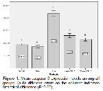
    Apoptosis and Autophagy Expressions: Caspase-3 and BCL-3 expression levels were presented in Figure 1 and 2, respectively. As seen on Figure 1, the lowest caspase-3 levels were in control and NRG groups and the highest caspase-3 levels were in MTX group. However these levels were significantly (P<0.05) lower in NRG50+MTX and NRG100+MTX treatment groups in dose dependent manner.


    Büyütmek İçin Tıklayın
    Figure 1: Mean caspase 3 expression levels among all groups. (a-d): different letters on the columns indicates statistical difference (P<0.05)


    Büyütmek İçin Tıklayın
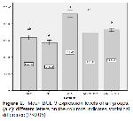
    Figure 2: Mean BCL 3 expression levels of all groups. (a-c): different letters on the columns indicates statistical difference (P<0.05)

    As presented in Figure 2, the most intense BCL-3 expression level was in MTX group and in NRG50+MTX and NRG100+MTX groups BCL-3 levels were significantly lower when compared to MTX alone group. The lowest BCL-3 expression levels were in control, NRG and NRG50+MTX groups. Also, there was no difference among NRG100+MTX, MRG50+MTX, and control groups when consider BCL-3 expression levels.

  • Başa Dön
  • Özet
  • Giriş
  • Materyal ve Metot
  • Bulgular
  • Tartışma
  • Kaynaklar
  • Tartışma
    MTX is one of the drugs that commonly used against some types of malignant cancers, rheumatoid arthritis 1, inflammatory bowel disease 2, and psoriasis 3. However, it is reported that the usage of MTX has increased ROS levels and thus decreased sperm quality and increased damage in testes 2,5. Under the normal condition there is a balance between oxidant and antioxidant compounds in the body. When ROS levels increased the more than the antioxidant defense capacity the balance between oxidant and antioxidant status is broken. In this situation the cellular defense system of the tissues cannot eliminate the increased ROS levels adequately. The target of ROS is macromolecules present in the cell membrane such as proteins and lipids. ROS cause disruption of these structures of molecules and thus cellular membrane integrity and finally damage of the cells. It is well known that the presence of polyunsaturated fatty acids in the membrane of testicular tissue cells makes it more sensitive against to oxidative stress 19-22. As proven previously, MTX increased the lipid peroxidation and thus oxidative stress. The usage of MTX can cause damage in reproductive system, cause apoptosis and degeneration in testes, decrease sperm quality. The side effects are generally attributed to increased lipid peroxidation and thus oxidative stress caused by MTX.

    The results of this study showed that MTX treatment increased to oxidative stress and decreased enzymatic and non-enzymatic antioxidant levels in male rats testes tissue. An elevation in lipid peroxidation is related to increase of free radicals. Increased ROS levels cause the damage in body cells by impairing the oxidant/antioxidant balance. As well known, increased oxidative stress causes the damage in body cells by destructing macromolecules such as proteins, lipoproteins in cell membrane and thus membrane integrity. Our results are supporting the results reported by other researchers that MTX increases the ROS level 4,5.

    However, in our study biochemical evaluations of testes showed that NRG treatment decreased MDA levels caused by MTX application and increased the SOD, CAT and GSH-Px activity and GSH levels when compared to MTX alone group in dose dependent manner. So, NRG treatment reduced the oxidative stress in testicular tissue by helping to increase enzymatic and non-enzymatic intracellular antioxidant levels for scavenging of free radicals. This protective effect can be attributed to antioxidant property of NRG.

    Gach et al. 23 suggested that increased ROS level stimulates the apoptosis process in somatic cells. Our findings are supporting this theory. According to our results MTX treatment increased ROS levels and decreased non-enzymatic antioxidant level and enzymatic antioxidant activities and also increased caspase-3 expression in testes tissue in MTX alone group when compared to control group. However, caspase-3 expression level was significantly lower in both NRG50+MTX and NRG100+MTX treatment groups when compared to MTX alone group. Also there is a statistical difference (P<0.05) between NRG50+MTX and NRG100+MTX groups for caspase-3 expression levels that means the higher dose of NRG protects more efficiently against apoptosis induced by MTX.

    Zhang et al. 24 reported that Bisphenol A-induced ROS levels stimulated the apoptosis and autophagy in goat testis Sertoli cells. They are reported that there was a relationship between increased ROS levels and stimulation of apoptosis and autophagy. Our data improves the relation between increased ROS caused by MTX and stimulation of apoptosis and autophagy. On the other hand, NRG treatment decreased these levels by helping the intracellular defense system via its antioxidant property. In our study the highest BCL-3 expression level was in MTX group when compared to control group. There was no difference in BCL-3 expression levels among control, NRG and NRG50+MTX groups. On the other hand, BCL-3 expression levels both in NRG50+MTX and NRG100+MTX groups were statistically lower than MTX alone group. These results indicate that MTX treatment stimulated autophagy in testes and NRG treatments decreased the expression levels of BCL-3. As mentioned above the possible reason for this situation can be the increased ROS levels induced by MTX treatment. However treatment with NRG can reduce the side effects of MTX induced reproductive damage by helping antioxidant defence system of cellular compounds.

    In conclusion NRG treatment could be beneficial for lightening or preventing of MTX-induced reproductive damage in dose dependent manner.

  • Başa Dön
  • Özet
  • Giriş
  • Materyal ve Metot
  • Bulgular
  • Tartışma
  • Kaynaklar
  • Kaynaklar

    1) O'Dell JR. Methotrexate use in rheumatoid arthritis Rheum. Dis Clin N Am 1997; 23: 779-796.

    2) Ley D, Jones J, Parrish J, Salih S, et al. Methotrexate reduces DNA integrity in sperm from men with inflammtory bowel disease. Gastroenterology 2018; 154: 2064-2067.

    3) Rademaker M, Gupta M, Andrews M, et al. The Australasian Psoriasis Collabaration view on methotrexate for psoriasis in the Australasian setting. Australas J Dermato 2017; 58: 166-170.

    4) Şener G, Ekşioğlu-Demiralp E, Çetiner M, Ercan F, Yeğen BÇ. β-glucan ameliorates methotrexate-induced oxidative organ injury via its antioxidant and immunomodulatory effects. Eur J Pharmacol 2006; 542: 170-178.

    5) Belhan S, Özkaraca M, Kandemir FM, et al. Effectiveness of Hesperidin on methotrexate-induced testicular toxicity. Kafkas Uni Vet Fak Derg 2017; 23: 789-796.

    6) Chen F, Zhang N, Ma X, et al. Naringin alleviates diabetic kidney disease through inhibiting oxidative stress and inflammatory reaction. PLoS One 2015; 10: e0143868.

    7) Ng TB, Liu F, Wang ZT. Antioxidative activity of natural products from plants. Life Sci 2000; 66: 709-723.

    8) Ozcelik B, Kartal M, Orhan I. Cytotoxicity, antiviral and antimicrobial activities of alkaloids, flavonoids, and phenolic acids. Pharm Biol 2011; 49: 396-402.

    9) Liu Y, Su WW, Wang S, Li PB. Naringin inhibits chemokine production in an LPS-induced RAW 264.7 macrophage cell line. Mol Med Rep 2012; 6: 1343-1350.

    10) Lee EJ, Kim DI, Kim WJ, Moon SK. Naringin inhibits matrix metalloproteinase-0 expression and AKT phosphorylation in tumor necrosis factor-alpha-induced vascular smooth muscle cells. Mol Nutr Food Res 2009; 53: 1582-1591.

    11) Malakul W, Pengnet S, Kumchoom C, Tunsophon S. Naringin ameliorates endothelial dysfunction in fructose-fed rats. Exp Ther Med 2018; 15: 3140-3146.

    12) Ikemura M, Sasaki Y, Giddings JC, Yamamoto J. Preventive effects of hesperidin, glucosyl hesperidin and naringin on hypertension and cerebral thrombosis in stroke-prone spontaneously hypertensive rats. Phytother Res 2012; 26: 1272-1277.

    13) Placer ZA, Cushmanni LL, Johnson BC. Estimation of products of lipid peroxidation (as malondialdehyde) in biochemical systems. Anal Biochem 1966; 16: 359-364.

    14) Sun Y, Larry WO, Ying L. A simple method for clinical assay of superoxide dismutase. Clin Chem 1988; 34: 497-500.

    15) Aebi H. Catalase. Methods in Enzymatic Analysis. New York: Academic Press, 1983.

    16) Sedlak J, Lindsay RHC. Estimation of total protein bound and nonprotein sulfhydryl groups in tissue with Ellmann’s reagent. Anal Biochem 1968; 25: 192-205.

    17) Lawrence RA, Burk RF. Glutathione peroxidase activity in selenium-deficient rat liver. Biochem Biophys Res Commun 1976; 71: 952-958.

    18) Lowry OH, Rosebrough NJ, Farr AL, Randall RJ. Protein measurement with folin reagent. J Biol Chem 1951; 193: 265.

    19) Aksu EH, Akman O, Özkaraca M, Ömür AD, Uçar Ö. Effect of Maclura pomifera extract on cisplatin-ınduced damages in reproductive system of male rats. Kafkas Univ Vet Fak Derg 2015; 21: 397-403.

    20) Aksu EH, Özkaraca M, Kandemir FM, et al. Mitigation of paracetamol-induced reproductive damage by chrysin in male rats via reducing oxidative stress. Andrologia 2016; 48: 1145-1154.

    21) Aksu EH, Kandemir FM, Altun S, et al. Ameliorative effect of carvacrol on cisplatin‐induced reproductive damage in male rats. J Biochem Mol Toxicol 2016; 30: 513-520.

    22) Aksu EH, Kandemir FM, Özkaraca M, et al. Rutin ameliorates cisplatin-induced reproductive damage via suppression of oxidative stress and apoptosis in adult male rats. Andrologia 2017; 49: e12593.

    23) Gach K, Dlugosz A, Janecka A. The role of oxidative stress in anticancer activity of sesquiterpene lactones. Naunyn Schmiedeberg’s Arch Pharmacol 2015; 388: 477-486.

    24) Zhang Y, Han L, Yang H, et al. Bisphenol A affects cell viability involved in autophagy and apoptosis in goat testis sertoli cell. Environ Toxicol Phar 2017; 55: 137-147.

  • Başa Dön
  • Özet
  • Giriş
  • Materyal ve Metot
  • Bulgular
  • Tartışma
  • Kaynaklar
  • [ Başa Dön ] [ Özet ] [ PDF ] [ Benzer Makaleler ] [ Yazara E-Posta ] [ Editöre E-Posta ]
    [ Ana Sayfa | Editörler | Danışma Kurulu | Dergi Hakkında | İçindekiler | Arşiv | Yayın Arama | Yazarlara Bilgi | E-Posta ]