Research and Publication Ethics: All experiments were approved by Adnan Menderes University Local Board of Ethics Committe for Animal Experiments (Number-64583101/2021/019).
Clinic records of 45 calves admitted to Adnan Menderes University Surgery Clinic for CCFT were received and analyzed. The calves with unilateral or bilateral CCFT underwent a conservative surgery via a unilateral or bilateral transection of the ulnaris lateralis tendon and flexor carpi ulnaris tendon was also studied in this research.
The affected limbs of the calves were divided into three consecutive classes in increasing severity depending on their initial classification of the flexural defect (Table 1).
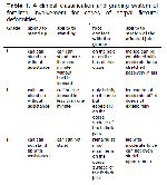
Büyütmek İçin Tıklayın |
Table 1: A clinical classification and grading system of forelimb involvement for cases of carpal flexural deformities |
The signalement, synchronized disease development and musculoskeletal conditions were obtained from medical records. The supplementary postoperative medical management and supportive treatment were documented.
Conventional Treatment: In grade I and II calves were treated with PVC bandage and oxytetracycline administration. Oxytetracycline was administered at a dose of 1mL/10 kg intramusculary (IM) once a day for 5 days. PVC bandage was applied to calves for 14 days.
Surgical Procedure: In grade III calves were treated with tenotomie. Procaine penicillin was given pre-operatively and continued for 3 days (22 mg/kg IM twice daily). Flunixine meglumine was given to healthy, well-hydrated calves at the time of pre-medication for the anesthesia (1.1 mg/kg IV once).
Surgery was performed under general anesthesia that was initiated and maintained with an intravenous detomidine-ketamine-midazolam combination and the calves were placed in lateral lying position with the affected limb at the upmost level. The tendons of insertion of the ulnaris lateralis and the flexor carpi ulnaris were cut through a lateral skin incision 5 cm long, pinpointed at the surface of the distal radial physis.
The two tendons of insertion of the ulnaris lateralis were marked, isolated by a blunt incision, and transected about 2 cm above the auxiliary carpal bone. A particular attention was given to avoid the the cut of lateral palmar vein and nerve. In the presence of a main part of muscular tissue at the performed tenotomy site, only the peripheral tendinous tissue was transected.
The underlying tissues were completely closed in a simple continuous design using 2-0 polydioxanone (PDS, Ethicon). Then the skin was stitched with horizontal sutures using 2-0 polydioxanone (PDS, Ethicon).
Postoperative Care: If the degree of extension was more than 50% postoperatively, the affected limb was covered by a fully padded limb bandage, and the calf was placed in a stall for 2 weeks.
In the case of less than 50% improvement of that recorded initially, a plastic-cast was administered into the limb (Figure 1C). The limb was then re-evaluated after 24 hours for the improvement of the CCFT and the strap was put on daily if necessary for up to 5 days.
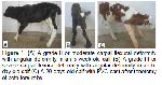
Büyütmek İçin Tıklayın |
Figure 1: (A) A grade II or moderate carpal flexural deformity with angular deformity in an 5-week-old calf (B) A grade III or severe carpal flexural deformity with angular deformity in an 6-day-old calf (C) A 20-days-old calf with PVC-cast after tenotomy of both forelimbs |
Follow-up: Change in the degree of carpal extension in the immediate post-surgical period was evaluated. If the flexural deformity had progressed less than 50% of that recorded initially, further strapping or casting was performed as explained in postoperative care above.
Further information was received at least 8 months after the operation by the phone with patients owners and/or breeders.


)
)
)
