 |
 |
| [ Ana Sayfa | Editörler | Danışma Kurulu | Dergi Hakkında | İçindekiler | Arşiv | Yayın Arama | Yazarlara Bilgi | E-Posta ] | |
| Fırat Üniversitesi Sağlık Bilimleri Veteriner Dergisi | |||||
| 2021, Cilt 35, Sayı 3, Sayfa(lar) 187-189 | |||||
| [ Özet ] [ PDF ] [ Benzer Makaleler ] [ Yazara E-Posta ] [ Editöre E-Posta ] | |||||
| İki Koyunda Bakır Zehirlenmesi Kaynaklı Patolojik Değişiklikler | |||||
| Yesari EROKSUZ1, Canan AKDENIZ INCILI1, Burak KARABULUT1, Ismet YILMAZ21, Zeynep YERLIKAYA3, Hatice EROKSUZ1 | |||||
| 1University of Fırat, Faculty of Veterinary Medicine, Department of Pathology, Elazığ, TURKEY 2University of İnönü, Faculty of Pharmacy, Department of Pharmacology, Malatya, TURKEY 3University of Fırat, Faculty of Veterinary Medicine, Department of Microbiology, Elazığ, TURKEY |
|||||
| Anahtar Kelimeler: Bakır toksikasyonu, patolojik bulgular, koyun | |||||
| Özet | |||||
Yüzaltmış Akkaraman koyunu bulunan sürüden iki hayvanda bakır intoksikasyonu bildirilmiştir. Makroskobik bulgular arasında belirgin sarılık, hemoglobinürik nefroz ve belirginliği artmış hepatik lobüler yapı vardı. Histopatolojik incelemede karaciğer ve böbrekte multifokal dejeneratif ve nekrotik değişiklikler görüldü. İmmunohistokimyasal olarak; rodanin pozitif sitoplazmik boyama, hepatositlerde ve tübüler epitel hücrelerinde multifokal olarak tespit edildi. Hepatik ve renal bakır konsantrasyonu ıslak dokularda 244.24 mg/1000 g ve 70.35 mg/1000 g idi. Genel olarak, veriler, böbrek hasarının hemoglobinüri ile ilişkili olduğu, hastalığın hemolitik sonrası bir fazda olduğunu göstermektedir. |
|||||
| Giriş | |||||
Copper is a fundamental mineral existing in all mammalian cells requiring it as a cofactor for specific enzymes and electron transport proteins. Its homeostasis is regulated through a complex system of copper transporters and chaperone proteins. When its concentration exceeds the body tolerance, copper might exert cytotoxic effects 1. Copper is absorbed from the small intestine by divalent metal transporter and then bound to transporters including ceruloplasmin, transcuprein, and albumin and carried to liver where it is sequestered in metallothionein or glutathione; and then excreted via bile or blood 2. Most of the incoming copper are rapidly transported into the hepatocytes while lesser amounts to the kidney. As the main reserve of copper is the liver, hepatocytes are the main target cells for copper toxicity 3. Unlike other minerals, copper absorption does not depend on animals daily requirements but in proportion to the concentration in their diet. Sheep are the most susceptible species to copper toxicity due to reduced biliary excretion, the avidity of liver for copper and their low dietary requirements. Sporadic outbreaks of primary copper toxicity may occur in sheep and farm animals 1,2. Chronic copper toxicity is characterized by an acute intravascular hemolysis due to the release of excess copper from the liver into the bloodstream 4. Stressful events, such as lambing, weather changes, poor nutrition, transportation or shearing, may exacerbate the copper intoxication. Primary copper poisoning occurs due to high intake from contamination of pasture or feed, whereas secondary copper poisoning arises from phytogenic and hepatogenic reasons 1,4. The aim of the present report is to describe the pathological and immunohistochemical findings in 2 sheep from an Akkaraman flock in Eastern Turkey, Elazığ. |
|||||
| Olgu Sunusu | |||||
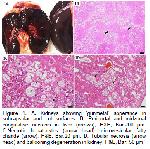
Firstly, certificate of approval was taken from the animal owner. Amongst 160 grazing Akkaraman sheep flock, 5 animal died and 20 animal exhibited the clinical signs in the last 7 days. In history, the animals grazed in apricot tree orchard and vineyard previously sprayed with fungicides known as Bordeaux mixture (CuSO4 and Ca(OH)2). The owner also stated that sheep consumed the chicken litter which was distributed to around the trees as organic fertilizer. Two sheep aged approximately 4-year-old were referred to the Veterinary Pathology Department with a history of icterus, abdominal pain, and hematuria. Animals were humanely euthanized and necropsied Gross and histological changes in both sheep were mostly identical. Tissue samples from the major organs and tissues were fixed in 10% formalin, processed routinely, embedded in paraffin and sectioned in 4-5 μm thickness and stained with hematoxylin-eosine (H&E). Selected tissue sections were stained by rhodamin, Ki-67 and Terminal deoxynucleotidyl transferase (TdT) dUTP Nick-End Labeling (TUNEL) methods. Ki-67 positive hepatocytes in the liver and tubulus epithelial cells in the kidney were counted in 10 different microscopic fields in a 40-magnification objective. TUNEL assays were performed using the In-Situ Cell Death Detection Kit (Roche Diagnostics, Indianapolis, IN, USA) according to manufacturers instructions. In brief, deparaffinized sections were washed 3 times for 5 min in PBS and permeabilized with 10 μg/mL proteinase K in PBS at room temperature for 30min. After rewashing 3 times in PBS, these sections were incubated in high pressure for 2 minutes in 10 mM citrate acid (pH 6.0). Subsequently, after thorough rinsing, the samples were incubated with a TUNEL reaction mixture for 60 min at 37°C in a humidifed atmosphere in the dark. Tissue samples from liver, kidneys, lungs were mechanically sectioned with a Polytron PT 1200 E (Kinematica, Switzerland) tissue mill. Total DNA isolation was performed using the BioRobot® EZ1 (Qiagen, Hilden, Germany) automated extraction device using the DNA Tissue Kit (Qiagen, Hilden, Germany) as recommended by the manufacturer. After the isolation, approximately 800 base pairs of the 16S rDNA region was amplified using GeneAmp PCR System 9700 (Applied Biosystems / USA) using primers p8FPL 5 'AGT TTG ATC ATG GCT CAG-3' and p806R 5'- GGA CTA CCA GGG TAT CTA AT- with a thermal cycler device. Amplification conditions were used for this purpose; followed by 94 °C/30 sec denaturation, 60 °C/30 sec binding and 72 °C/1 min elongation as 35 cycles, followed by a 3 min initial denaturation at 94 °C. Flame atomic absorption spectroscopy (Perkin Elmer, AAnalyst 800) was used to determine copper levels. Two-gram portions from the liver and kidney were wet-digested with 36 mL HNO3 and 4 mL HClO4 for 24 h in the tube. These tubes were then placed on a hot plate until the liquid was evaporated and the residue was suspended with 10 mL 0.1 M/L HNO3. Samples were then analyzed for copper with a flame atomic absorption. The carcasses were mild to moderately icteric with reference to yellow discolaration of conjunctiva, oral mucosa, adipose tissues, lungs and the great vessels. The livers and kidneys were swollen with round edges and bulging in cut surfaces. The livers had enhanced lobular (reticular) pattern with yellow-brown in color. The kidneys were soft and dark-black (gun metal) in color (Figure 1A). Abomasal mucosae were multifocally ulcerated measured by 3-4 mm in diameter. Urine was red in color containing blood. There were moderate echymotic hemorrhages in papillar muscle in the myocardium of one sheep.
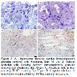
The hepatocytes showed necrosis, cloudy swelling, fatty change and apoptosis. Hepatocytes were expanded by numerous clear lipid vacuoles in zone-II, whereas the necrotic changes were multifocally distributed in zone II or rarely zone III. These hepatocytes had more eosinophilic cytoplasm and hypochromatic, karyorhectic or lytic nuclei. No inflammatory infiltrate was present related to necrosis, however periportal lymphohistiocytic infiltrations were detected. Also, dispersed hepatocytes showed apoptosis characterized by individualised and hypereosinophilic apoptotic bodies. The hepatocytic nuclei were vesicular with stripled chromatin. Additional findings were capsular fibrosis and multifocal bile pigment deposits in cytoplasm of hepatocytes. Renal tubular epithelial cells were multifocally necrotic or degenerative with pale vacuolated cytoplasm and swollen nuclei. Renal tubules in cortex and medulla contained eosinophilic casts, often with granules possessing a more pronounced bright red color (hemoglobinuric nephrosis), while others contain degenerating cells. The epithelium was attenuated in tubules containing the hemoglobunuric casts. Some of the tubules showed cellular swelling. Multifocally distributes tubular cells and hepatocytes have brown-gray cytoplasmic granules that were positive by rhodanine staining (Figure 2A and B).
The average number of Ki-67 positive cells was 22±10.84 in the liver and 41±7.95 in the kidneys in a microscopic area with high dry objective. The copper levels were 244.24 mg/1000 g and 70.35 mg/1000 g in wet tissues, in liver and kidney respectively. TUNEL analysis showed positive reaction in hepatocytes, and renal tubular epithelium approximately 20% and 15%. Hepatocytes showed more diffuse TUNEL positivity, however tubular epithelial cells showed multifocal immunopositivity. As a result of he obtained amplification products to 1.0% agarose gel electrophoresis, no band formation was present. |
|||||
| Tartışma | |||||
Copper sulphate spray and/or chicken litter referred in the history were hypothetically the source of copper sulfate as the cause of intoxication in the present report. Chicken litter and copper spraying were also blamed previously for copper poisoning in sheep 5,6. When the concentration of copper in the liver reaches 300 ppm in dry weight, lysosomal membrane integrity is disrupted and lysosomal hydrolases cause cellular destruction. Sheep with copper concentrations above 1000 ppm in the liver may be clinically and hematologically normal, and this continues as long as the copper is released from the dead cells. However, copper concentrations should not be above 230 mg/kg in liver, 12 mg/kg in kidney 3. In the present report, relatively low level of copper in the liver and the high level in the kidney indicated that the hemolysis has been completed. Similarly, TUNEL analysis showed that hepatocytes were more affected by apoptosis than renal tubular epithelium and the higher apoptotic index was compatible with copper analysis. Pyrrolizidine alkaloidosis might cause or exacerbate the copper poisoning by increasing copper uptake or decreasing its excretion in the body even in normal diet. However, the specific lesions of pyrrolizidine alkaloidosis 1,3 were not present. (karyomegaly, cytomegaly, intranuclear inclusions and megalocytosis) in this report. Nor high mitotic index in livers and kidneys is relevant to pyrrolizidine alkaloidosis. Hepatic damage might be explained by toxin and anoxia secondary to anemia, whereas renal changes could arise from cytotoxicity of myoglobin and reduction in renal blood flow. In conclusion, the data showed the presence ofa post-hemolytic phase of the disease, in which renal damage associated with hemoglobinuria was evident in sheep.
Conflict of Interest |
|||||
| Kaynaklar | |||||
1) Brown DL, Van Wettere AJ, Cullen JM. Hepatobiliary system and exocrine pancreas. In: Zachary JF. (Editor). Pathologic Basis of Veterinary Disease. 6th Edition, St. Louis, MO: Elsevier: 2017: 440-457.
2) Cullen JM, Stalker MJ. Liver and biliary system. In: Maxie MG. (Editor). Jubb, Kennedy, and Palmers Pathology of Domestic Animals. Vol 2, 6th Edition, St. Louis, MO: Elsevier, 2004: 342-343.
3) Simpson DM, Beynon RJ, Robertson DHL, Loughran MJ, Haywood S. Copper-associated liver disease: A proteomics study of copper challenge in a sheep model. Proteomics 2004; 4: 524-536.
4) Hoffman G. Copper-associated liver diseases. Vet Clin North Am Small Anim Pract 2009; 39: 489-511.
|
|||||
| [ Başa Dön ] [ Özet ] [ PDF ] [ Benzer Makaleler ] [ Yazara E-Posta ] [ Editöre E-Posta ] | |||||
 |
| [ Ana Sayfa | Editörler | Danışma Kurulu | Dergi Hakkında | İçindekiler | Arşiv | Yayın Arama | Yazarlara Bilgi | E-Posta ] |

