[ Ana Sayfa | Editörler | Danışma Kurulu | Dergi Hakkında | İçindekiler | Arşiv | Yayın Arama | Yazarlara Bilgi | E-Posta ]
Fırat Üniversitesi Sağlık Bilimleri Veteriner Dergisi
2023, Cilt 37, Sayı 1, Sayfa(lar) 001-007
[ Özet ] [ PDF ] [ Benzer Makaleler ] [ Yazara E-Posta ] [ Editöre E-Posta ]
İlçe çöp Atım Alanına Yakın ve Uzak Otlaklarda Otlatılan İki Farklı Sürüdeki Honamlı Irkı Keçilerin Süt Aflatoksin M1 Seviyeleri ile Serum Oksidatif Stres Düzeyleri Arasındaki İlişkinin Araştırılması
Kemal VAROL
Burdur Mehmet Akif Ersoy University, Food, Agriculture and Livestock Vocational College of Burdur, Department of Veterinary, Burdur, TÜRKİYE
Anahtar Kelimeler: AFM1, haptoglobin, keçi, OSİ, TAS, TOS, toksikasyon
Özet
Bu çalışmada, ilçe çöp atım merkezine yakın (Grup 1; n=20) ve ilçe çöp atım merkezine uzak (Grup 2; n=20) otlaklarda otlatılan iki farklı sürüde bulunan ve yaşları 2-4 yaş arasında değişen toplam 40 adet laktasyondaki Honamlı ırkı keçi değerlendirildi. Keçilerin süt serumlarından Aflatoksin M1 (AFM1) seviyeleri belirlendi. Kan serum örneklerinden ise, total oksidan seviyeleri (TOS), total antioksidan seviyeleri (TAS), oksidatif stres indeksleri (OSI), ve serum haptoglobin (Hp) seviyeleri belirlendi. Bulgularda; AFM1, TAS, TOS, OSI ve Hp değerlerinin gruplar arası karşılaştırılmasında istatistiksel olarak anlamlı farklılık (P<0.001) belirlendi. Koreleasyon bulgularında ise; AFM1, TAS ve Hp değerleri arasında pozitif yönde orta düzeyde anlamlı bir ilişki bulundu. AFM1, TOS ve OSI değerleri arasında ise negatif yönde orta düzeyde anlamlı bir ilişki bulundu. Sonuç olarak otlakların çöp atım merkezlerine olan yakınlığının AFM1 düzeyi üzerinde etkili olduğu, AFM1 seviyelerinin de serum TAS, TOS, OSI ve Hp seviyelerini etkilediği görülmüştür. Bu nedenle toksik ve küflenmiş ürünler bulunan ve AFM1 kaynağı olan çöp atım merkezlerinin kenarlarının tel örgü ile kapatılarak hayvanların bu vb alanlarda otlatılması engellenmelidir.
  • Başa Dön
  • Özet
  • Giriş
  • Materyal ve Metot
  • Bulgular
  • Tartışma
  • Kaynaklar
  • Giriş
    Aflatoxins are toxic, mutagenic, teratogenic and carcinogenic fungal metabolites synthesized by Aspergillus (A.) flavus, A. parasiticus, A. nomius and some other A. penicillium and Rhizopus species under suitable temperature and humidity conditions 1-3.

    Especially the toxins produced by A. flavus and A. parasiticus play an extremely important role in the formation of human and animal diseases. A. flavus produces only aflatoxin B1 (AFB1) and aflatoxin B2 (AFB2) A. parasiticus is responsible for the production of, AFB1, AFB2, aflatoxin G1 (AFG1) and aflatoxin G2 (AFG2) 4.

    Apart from AFB1, AFB2, AFG1 and AFG2, there are two more important aflatoxin derivatives called aflatoxin M1 (AFM1) and aflatoxin M2 (AFM2). When lactating animals consume feed contaminated with AFB1, it AFB1 is converted into a hydroxylated form called AFM1, a cytotoxic and genotoxic substance. AFB1 is biotransformed into AFM1 by hepatic microsomal cytochrome P450. AFM1 was isolated in milk and urine as a result of AFB1 contamination of feed used in feeding mammals in lactation period. AFM1 was isolated in milk and urine as a result of AFB1 contamination of feed used in feeding mammals in lactation period 5.

    Aflatoxins absorbed from the gastrointestinal tract are transported by serum albumin. They are mainly localized in the liver and soft tissues. After they settled, they cause serious toxicity by inhibiting DNA, RNA and protein synthesis in that region, reducing the efficiency of various enzymes in the region, and completely crashing glucose metabolism. In addition, they act by inhibiting the lipid synthesis formed, including triglycerides, cholesterol esters, phospholipids and free fatty acids. They also affect coagulation factor inhibition 6.

    Oxidants and antioxidants are active oxygen derivatives of free radicals in the organism. There is a balance between oxidants and antioxidants 7. Infections, inflammation, malabsorption, stress, heavy exercises, metabolic problems and environmental factors prevent the intake of antioxidants. As a result, this balance is disrupted in favor of oxidants and cellular damage occurs in living organisms. This situation is expressed as oxidative stres 8,9. Free radicals, a reactive molecule, are formed during the conversion of nutrients into energy using oxygen. Reactive oxygen and nitrogen species are among these reactive molecules. Reactive molecules damage the cell components such as nucleic acid, protein, carbohydrate and lipid 10,11. Various defense mechanisms develope in the organism to prevent the damage of free radicals to the cellular structures by reactive molecules. These mechanisms are called antioxidant defense systems. Oxidative stress is responsible for the pathogenesis of various diseases. It is known as a part of cellular and molecular tissue damage of living organism 12. Total antioxidant capacity (TAS) and total oxidant status (TOS) are frequently used to determine oxidative stress in ruminants 9,10,13,14.

    Acute phase response (APR) is a non-specific reaction that occurs shortly after tissue damage due to infective, immunological, neoplastic, traumatic, parasitic or other causes 15. APR is stimulated by proinflammatory cytokines such as interleukin 1 (IL-1), interleukin 6 (IL-6) and tumor necrosis factor - (TNF) released from activated leukocytes in the area of tissue damage 16,17. These cytokines are known to be responsible for the primary response management in infectious diseases and inflammation 18,19.

    Haptoglobin (Hp) is an important acute phase protein in ruminants 18,20,21. Hp is used to evaluate the inflammatory response in many inflammatory diseases (Mastitis, pneumonia, enteritis, peritonitis, endocarditis, abscess and endometritis, etc.) and other natural or experimental infections in cattle 20,22. Hp are also elevated in some conditions that are not associated with inflammation or tissue damage. For example, Hp concentration may increase in cattle with fatty liver syndrome, fasting and dexamethasone treatment, and stress during transportation 20,23.

    Aflatoxins have a wide range of toxic effects on the health of humans and animals 24. One of these effects is hepatotoxicity on the liver. It is thought that hepotoxicity caused by aflatoxins in liver cells is due to peroxidative damage by producing reactive oxygen 25. Waste disposal centers are generally established in mountainous and forested areas outside the city centers, which are far from people. However, these areas are very close to the wild nature and to the pastures of goats that graze freely in nature. It is inevitable that aflatoxins, the main source of which are moldy and spoiled foods, are high in waste disposal areas. These foods found in landfills are consumed by goats, who are curious for interesting objects. For this reason, in this study, it was determined whether feeding animals in nearby waste disposal centers increases aflatoxin exposure, as well as the extent to which aflatoxins cause oxidative stress in animals and how they affect the acute phase response.

  • Başa Dön
  • Özet
  • Giriş
  • Materyal ve Metot
  • Bulgular
  • Tartışma
  • Kaynaklar
  • Materyal ve Metot
    This study is subject to the permission of Burdur Mehmet Akif Ersoy University Animal Experiments Local Ethics Committee dated 14.04.2021 and numbered 88/749.

    In this study, a total of 40 lactating Honamlı goats at 2-4 years old from two different herds grazing on grasslands close to the district waste disposal area (Group 1; n=20) and grazing far from the district waste disposal area (Group 2; n=20) were evaluated. AFM1 levels were determined from milk serum of goats. TOS, TAS, oxidative stress indices (OSI) and Hp levels were determined from blood serum samples. Fifteen milliliter (mL) milk samples were taken from each goat into falcon tubes. 8.5 mL blood samples were also taken from the jugular vein of the animals into vacuuumed tubes with gel. In order to obtain the serum from the clotted blood, it was centrifujed at 3000 rpm for 10 minutes and the serum was extracted. The milk serum was extracted by applying centrifugation (3500 rpm/10 min) to the milk samples. Both serum samples were stored at -20°C until the ELISA test was performed.

    AFM1 levels were determined with a commercial ELISA kit (ELABSCIENCE KA36762ETS E0099Go) according to the procedure of this kit. TAS and TOS were determined using Erel's 26,27 methods. The formula [TOS (μmol H2O2 equivalent/L)/10 x TAS (mmol Trolox equivalent/L)] was used to determine OSI (28). Hp levels were determined using the ELISA kit (BT E0099Go) .

    Statistical Analysis:
    IBM SPSS 22.0 for Windows package program was used for statistical analysis of study data. The normal distribution of the groups in the analyzes was evaluated by using the Shapiro-Wilk test. Since the data were not normally distributed, Mann Whitney-U Test, one of the Non-Parametric tests, was used in paired group comparisons. Spearman correlation analysis was used to determine the relationship between the parameters because the data were not normally distributed.

  • Başa Dön
  • Özet
  • Giriş
  • Materyal ve Metot
  • Bulgular
  • Tartışma
  • Kaynaklar
  • Bulgular
    There was a statistically significant difference (P<0.05) in the mean AFM1 values between Groups 1 and 2, which were found to be 31.21±5.00 ng/L and 24.66±4.76 ng/L, respectively.

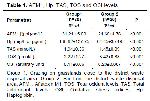
    There was a statistically significant difference (P<0.05) in the mean Hp values between Groups 1 and 2, which were found to be 188.62±137.75 μg/mL and 64.45±35.49 μg/mL, respectively. There was a statistically significant difference (P<0.05) in the mean TAS values between Groups 1 and 2, which were found to be 1.94±0.36 mmol/L and 1.45±0.09 mmol/L, respectively. There was a statistically significant difference (P<0.05) in the mean TOS values between Groups 1 and 2, which were found to be 2.22±1.27 μmol/L and 3.42±0.88 μmol/L, respectively. There was a statistically significant difference (P<0.05) in the mean OSI values between Groups 1 and 2, which were found to be 0.11±0.06 arbitrary units and 0.23±0.067 arbitrary units, respectively. (Table 1). In correlation findings; a moderately significant positive correlation was found between AFM1 and Hp values (r= 0.529; P<0.001). A moderately significant positive correlation was found between AFM1 and TAS values (r= 0.425; P= 0.006). In addition, a moderately significant negative correlation was found between AFM1 and TOS values (r= -0.517; P<0.001). A moderately significant negative correlation was found between AFM1 and OSI values (r= -0.613; P<0.001). A moderately significant positive correlation was found between TAS and Hp values (r= 0.461; P=0.003). In addition, a moderately significant negative correlation was found between TAS and TOS values (r= -0.452; P= 0.003). In addition, a highly significant negative correlation was found between TAS and OSI values (r= -0.711; P<0.001). A moderately significant negative correlation was found between Hp and TOS values (r= -0.497; P<0.001). A moderately significant negative correlation was found between Hp and OSI values (r= -0.631; P<0.001). A highly significant positive correlation was found between TOS and OSI values (r=0.915; P<0.001) (Table 1).


    Büyütmek İçin Tıklayın
    Table 1: AFM1, Hp, TAS, TOS and OSI levels


    Büyütmek İçin Tıklayın
    Table 2: Correlation findings between AFM1, Hp, TAS, TOS and OSI levels

  • Başa Dön
  • Özet
  • Giriş
  • Materyal ve Metot
  • Bulgular
  • Tartışma
  • Kaynaklar
  • Tartışma
    Mycotoxins are secondary metabolites of various fungi that have toxic effects on humans and animals and its effects are called "mycotoxicosis". The severity of mycotoxicosis also varies depending on factors such as age, gender, nutritional status in organisms exposed to this toxin 29. Among the studies on mycotoxins, aflatoxins take the first place which are known to have very strong hepatotoxic effects. They are mostly synthesized by A. flavus, A. nomius, A. parasiticus, A. bombycis and A. pseudotamarii 2-4. Mean and standard deviation (mean±standard deviation) AFM1 levels in goat milk according to country is 14.5±8.4 ng/L in Italy 30, 19±13.8 ng/L in Syria 31, in Pakistan 2.0±5.0 ng/L 32, 31.8±13.7 ng/L in Iran 33, 7.6±8.94 ng/L in Croatia 34. Ozdemir et al. 35 reported that the mean and minimum-maximum (mean (min-max)) AFM1 concentration in goat milk was 19.23 (5.16-116.78) ng/L in March and April in goat breeding enterprises in Kilis province. Karadal et al. 36 determined that mean (min-max) AFM1 concentration in goat milk was 3.07 (0.33-11.79) ng/L in goat breeding enterprises in Niğde province.

    In our study, the AFM1 concentration was found to 31.21±5.00 ng/L in the Group 1, and 24.66±4.76 ng/L in the Group 2. In also in our study, samples were taken in (April). Age-related changes were taken into account, therefore, healthy goats in lactation were selected in the close age range. Therefore, the changes that would occur due to these factors have been eliminated. Considering the nutritional status, the both groups of animals are given ground barley and then grazed in the bush. While the animals in Group 1 are grazed in the bush area close to the district waste disposal area, the animals in Group 2 are grazed in the normal bush area. Considering this information; it was observed that the AFM1 concentration in the milk of the goats in Group 1 was significantly higher (P<0.001) compared to the goats in Group 2. This situation believed to be associated with the consumption of mycotoxins produced by molds growing on the discarded food wastes. There are six types of aflatoxins: B1, B2, G1, G2, M1 and M2 37. and they are listed as AFB1 > AFG1 > AFB2 > AFG2 in terms of toxic effect. AFM1 and AFM2 are toxic metabolites of AFB1 and AFB2, respectively. Excretion occurs during lactation in animals fed with aflatoxins contaminated feed. This excretion occurs in the form of AFM1 and AFM2. In the studies, a linear relationship was found between the rate of AFB1 consumed by the animal and the rate of AFM1 excreted from its milk 38,39. It varies according to the breed of animal, milking period, milking interval and time. It has been reported that approximately 0.3-6.2% of AFB1 in milk is metabolized to AFM1 40. Feeding the offspring with milk and dairy products containing AFM1 can cause serious problems 14,41,42. In the present study, AFM1 levels were determined especially in milk due to the above concerns. Aflatoxin has been found in the ruminants milk and poultry eggs which were fed with aflatoxin-contaminated feeds 43. When feeds containing aflatoxin is taken into the gastrointestinal tract, it is converted to partially water-soluble conjugation products by the rumen microflora in the digestive tract of ruminants. They are easily absorbed and transported with a significant amount of serum albumin. It is distributed to soft tissues, mainly to liver. Aflatoxins in the circulating blood are highly retained in the liver. While some of the aflatoxins in the liver combine to large molecules such as hepatocytes, DNA, the binding surfaces of endoplasmic steroids and enzymes, another part is converted into oil- and water-soluble aflatoxins P1, Q1, B2a, aflatoxicol M1 and M2 in forms and rates that differ according to species. The shaped metabolism products are mostly excreted with bile 44. Aflatoxins are also metabolized by the urinary system. In most of the animal species, 50% of the amount excreted is with biliary secretion, while the amount removed by urinary elimination is about 15-25% 45. In the present study, low level of AFM1 was also observed in the animals grazed on the normal pasture and given the barley crumb, compared to the animals grazing on the edge of the waste, which indicates that aflatoxin can be seen in normal pasture as well. In addition, it was thought that the source of this low aflatoxin might be caused by cracking barley. Mortality rates from aflatoxicosis are reported to vary between 39% and 50%. Aflatoxicosis can be defined as acute or chronic due to aflatoxin’s dose and exposure time 46-48. The main toxic effects that occur in acute overdose exposure are hepatotoxicity, nephrotoxicity and sometimes death. In chronic exposure, genotoxicity, carcinogenicity and reproductive disorders are encountered 49. Animals such as ducks, sheep, turkeys, dogs, pigs and rats are most susceptible to AFB1. Monkeys, chickens, mice and ruminants are more resistant to AFB1 50.

    Regarding the immunostimulatory effects, there is an increasing evidence that aflatoxins elicit a biphasic immune response with a stimulating effect in the first phase and a suppressive effect in the second phase 51. According to Valtchev et al. 52, short-term exposure to low-dose aflatoxins stimulates the immune system, while prolonged exposure to high doses shows immunosuppressive effects. For example, upregulation of the expression of toll-like receptors has been observed in different immune cells in different organs when exposed to very low levels of aflatoxin. Cytokines are stimulated in response to aflatoxin exposure 50. With the stimulation of cytokinin, an acute phase response develops and leads to the formation of acute phase proteins. Hp is one of these acute phase proteins. In inflammatory conditions, stress, tissue damage, and some non-tissue damage-related conditions are also increased 23,53,54.

    In studies on Hp in goats, Hp values vary according to the measurements made with different ELISA kits. 19,55-58 It is also noteworthy that the heights in the standard deviation values of Hp values 55,56. Gonzalez et al. 55 reported that the mean and standard deviation values in healthy goats were 89±105 μg/mL. As it can be seen, the standard deviation value of the Hp value is higher than the mean and shows serious variability. In addition, in the same study, it was noted that the Hp value increased up to 4 times due to the disease 55. For this reason, comparison between groups would be more appropriate instead of comparison with reference values in goats. In the light of this information in our study, the Hp value was found to be 188.62±137.75 μg/mL in the group with high AFM1 level, and 24.66±4.76 μg/mL in the group with low AFM1 level. In addition, a moderately significant positive correlation was found between AFM1 and Hp values (r=0.374; P=0.017). It can be seen from the results that, as AFM1 increases, acute phase response develops, and Hp value increases.

    Free Radicals are reactive molecules containing one or more unpaired electrons in their outermost orbitals. Radicals containing oxygen in their main skeleton are called free oxygen radicals 59. Free radicals are produced in physiological amounts in all living cells. When they are overproduced, they cause cell and tissue damage. These effects of free radicals are eliminated by some enzymes and molecules called antioxidants. Oxidative stress is occur due to an imbalance between the production of free oxygen radicals and their elimination by antioxidants. Free radical reactions cause oxidation of lipids, proteins and polysaccharides and DNA damage, which has significant toxic biological effects 7,60,61.

    TOS can be used as an indicator for oxidants produced by the organism and taken up by environmental factors 62. It is reported that TOS levels will increase in inflammatory diseases 10. OSI is a key factor in determining oxidative stress 41. Various studies have been conducted to evaluate OSI in the determination of oxidative stress in farm animals. As a result of these studies, it has been reported that OSI values increase significantly in diseases where oxidative stress occurs 63.

    Antioxidants serve to protect cells from the destructive effects of oxidative stress. Considering the abundance of antioxidant substances and pathways; in the prevention of oxidative stress and in determining the overall antioxidant capacity, determining the quantitative antioxidant power or TAS level in biological samples is a serious point. Antioxidative status evaluation can also be done by measuring TAS levels 7. Antioxidants neutralize free radicals and protect the body against oxidative stress. The level of antioxidants decreases during oxidative stress 7,10,63.

    TAS value in healthy goats ranges from 1.18 to 1.79 mmol/L 64. In our study, the TAS value was found to be 1.94±0.36 mmol/L in the group with high AFM1 level, and as 1.45±0.09 mmol/L in the group with low AFM1 level. In addition, a moderately significant positive correlation was found between AFM1 and TAS values (r= 0.320; P= 0.044).

    The TOS value in healthy goats ranges from 1.05 to 1.36 mmol/L 64. In our study, the TOS value was found to be 2.02±1.31 mmol/L in the group with high AFM1 level, and 3.42±1.11 mmol/L in the group with low AFM1 level. In addition, a moderately significant negative correlation was found between AFM1 and TOS values (r= -0.388; P= 0.013).

    In our study, the OSI value was found to be 0.10±0.07 in the group with high AFM1 level, and 0.23±0.080 in the group with low AFM1 level. In addition, a moderately significant negative correlation was found between AFM1 and OSI values (r= - 0.419; P= 0.07).

    Radical damage that frequently occurs in the organism is lipid peroxidation. Fat radicals are formed as a result of the separation of a hydrogen from fatty acids in the cell membrane. As a result, aldehydes, which are cytotoxic products, form hydrocarbon gases such as pentane. Of these toxic products, malonaldehyde is the last step of aldehydes. MDA is used to determine lipid peroxidation. MDA is an indirect indicator of injury caused by reactive oxygen species 3. It is known that there is a positive relationship between milk AFM1 levels and blood malondialdehyde (MDA) levels 3,65-67. Therefore, the positive relationship between MDA and AFM1 is an indication that AFM1 leads to a significant increase in lipid peroxidation.

    However, its effect on other free radicals is not clear. Because AFM1, which is known to increase MDA, caused not an increase but even a decrease in TOS, which is an indicator of oxidative stress. It caused an increase in TAS. This has been attributed to the absence of an imbalance between the production of free oxygen radicals and their elimination by antioxidants.

    Another possible reason is although AFM1 increases MDA by increasing lipid peroxidation; in our study, it was observed that the oxidative stress was lower in the group with high AFM1 and the oxidative stress was higher in the group with low AFM1. However, in the findings of antioxidant capacity; it was observed that the antioxidant system was higher in the group with high AFM1 and the antioxidant system was lower in the group with low AFM1. This situation has been associated with the moderately high AFM1 level stimulating the immune system by stimulating antioxidant systems and suppressing oxidative stress.

    In conclusion, this study showed that AFM1 levels were found to be high in milk of goats grazing on the edge of the dump. It was observed that AFM1 caused an increase in TAS and Hp values, and a decrease in TOS and OSI values. It was observed that while AFM1 increased TAS and Hp values increased, it caused a decrease in TOS and OSI values It is thought that the moderately high AFM1 level can stimulate the immune system by suppressing oxidative stress by stimulating the antioxidant systems. In future studies, AFM1 level, oxidant and antioxidant capacity and acute phase response should be examined in more detail together with the immune system. In addition, the edges of waste disposal centers should be surrounded with wire to prevent animals from reaching toxic and moldy products.

  • Başa Dön
  • Özet
  • Giriş
  • Materyal ve Metot
  • Bulgular
  • Tartışma
  • Kaynaklar
  • Kaynaklar

    1) D’Mello JPF, Macdonald AMC. Mycotoxins. Anim Feed Sci 1997; 69: 155-166. 2. İşleyici Ö, Sancak YC, Morul F. Divle tulum peynirinde aflatoksin M1 düzeyi üzerine bir araştırma. Van Veterinary Journal 2011; 22: 105-110. 3. Güven A, Öztürk B, Deveci HA, Kaya İ. Investigation of the relationship between blood lipid peroxidation and the prevalence of aflatoxin M1 in milk samples from mothers and cows living in kars and surrounding villages. Kafkas Üniversitesi Fen Bilimleri Enstitüsü Dergisi 2020; 13: 67-75.

    4) Akdemir Ç, Altıntaş A. Ankara’da işlenen sütlerde aflatoksin M1 varlığının ve düzeylerinin HPLC ile araştırılması. Ankara Üniv Vet Fak Derg 2004; 51: 175-179.

    5) Özkaya Ş, Temiz A. Aflatoksinler: Kimyasal yapıları, toksisiteleri ve detoksifikasyonları. Orlab On-Line Mikrobiyoloji Dergisi 2003;1: 1-21.

    6) Hendrickse RG. Of sick turkeys, kwashiorkor, malaria, perinatal mortality, heroin addicts and food poisoning: Research on the influence of aflatoxins on child health in the tropics. Ann Trop Med Parasitol 1997; 91: 787-793.

    7) Akşit H, Kıral F, Yılmaz M, Ural M. Saanen keçilerinde erken ve geç laktasyon döneminde oksidatif durum. Balıkesir Sağlık Bilimleri Dergisi, 2014; 3: 74-78.

    8) Marreiro DN, Cruz KJC, Morais JBS, et al. Zinc and oxidative stress: Current mechanisms. Antioxidants 2017; 6: 24. 9. Kirmizigul AH, Ozcelik M, Öğün M, et al. Serum Cu, Mn and Zn levels and oxidative stress in cattle performing tongue-playing. Kafkas Univ Vet Fak Derg 2019; 25: 787-791.

    10) Bozukluhan K, Merhan O, Öğün M, Cihan M, Gökçe G. Determination of the levels of some oxidative stress parameters in calves with omphalitis. Fırat Üniversitesi Sağlık Bilimleri Veteriner Dergisi 2016; 30: 79-81.

    11) Sezer K, Keskin M. Role of the free oxygen radicals on the pathogenesis of the diseases. Fırat Üniversitesi Sağlık Bilimleri Veteriner Dergisi 2014; 28: 49-56.

    12) Özcan O, Erdal H, Çakırca G, Yönden Z. Oxidative stress and its impacts on intracellular lipids, proteins and DNA. J Clin Exp Invest 2015; 6: 331-336. 13. Varol K, Eser M. The Effects of toltrazuril administration on serum oxidative stress levels, serum haptoglobin levels and hematological parameters in the treatment of acute natural coccidiosis in honamli kids. Kocatepe Veterinary Journal 2022; 15: 355-362.

    14) Merhan O, Bozukluhan K, Kuru M, et al. Investigation of oxidative stress index and lipid profile in cattle with Brucellosis. Kafkas Univ Vet Fak Derg 2017; 23: 933-937.

    15) Eckersall PD, Young FJ, Nolan AM, et al. Acute phase proteins in bovine milk in an experimental model of Staphylococcus aureus subclinical mastitis. J Dairy Sci 2006; 89: 1488-1501. 16. Gruys E, Toussaint MJM, Niewold TA, Koopmans SJ. Acute phase reaction and acute phase proteins. J Zhejiang Univ SCI 2005; 11: 1045-1056. 17. Moshage HJ. Cytokines and the hepatic acute phase response. J Pathol 1997; 181: 257-266. 18. Eckersall PD. Recent advances and future prospects for the use of acute phase proteins as markers of disease in animals. Rev Med Vet 2000; 151: 577-584. 19. Ulutaş PA, Voyvoda H, Ulutaş B, Aypak S. Miks helmint enfeksiyonlu keçilerde haptoglobin, serum amiloid-A ve seruloplazmin konsantrasyonları. Türkiye Parazitololoji Dergisi 2008; 32: 229-233.

    20) Murata H, Shimada N, Yoshioka M. Current research on acute phase proteins in veterinary diagnosis. Vet J 2004; 168: 28-40.

    21) Chan JPW, Chu CC, Fung HP, et al. Serum haptoglobin concentration in cattle. J Vet Med Sci 2004; 66: 43-46.

    22) Çitil M. Puerperal infeksiyonlu ve abomasum deplasmanlı ineklerde Serum Amiloid A ve haptoglobin düzeyleri. Kafkas Üniv Vet Fak Derg 2003; 9: 147-51. 23. Coşkun A, Şen İ. Sığırlarda akut faz proteinleri ve klinik kullanım alanları. Sağlık Bilimleri Dergisi 2011; 20: 240-246.

    24) Alshannaq A, Yu JH. Occurence, toxicity and analysis of major mycotoxins in food. Int J Environ Res Public Health 2017; 14: 632.

    25) Yunus AW, Razzazi-Fazeli E, Bohm, J. Aflatoxin B1 in affecting broiler’s performance, immunity, and gastrointestinal tract: A review of history and contemporary ıssues. Toxins 2011; 3: 566-590.

    26) Erel O. A novel automated direct measurement method for total antioxidant capacity using a new generation, more stable ABTS radical cation. Clin Biochem 2004; 37(4): 277-285.

    27) Erel O. A new automated colorimetric method for measuring total oxidant status. Clin Biochem 2005; 38: 1103-1111.

    28) Karababa F, Yesilova Y, Turan E, et al. Impact of depressive symptoms on oxidative stress in patients with psoriasis. Redox Rep 2013; 18: 51-55.

    29) Peraica M, Radic B, Lucic A, Pavlovic M. Toxic effects of mycotoxins in humans. WHO 1999; 77: 754-766.

    30) Virdis S, Scarano C, Spanu V, et al. A survey on aflatoxin M1 content in sheep and goat milk produced in Sardinia Region, Italy (2005-2013). Ital J Food Saf 2014; 3: 4517.

    31) Ghanem I, Orfi M. Aflatoxin M1 in raw, pasteurized and powdered milk available in the Syrian market. Food Control 2009; 20: 603-605. 32. Hussain I, Anwar J, Asi MR, Munawar MA, Kashif M. Aflatoxin M1 contamination in milk from five dairy species in Pakistan. Food control, 2010; 21: 122-124.

    33) Rahimi E, Ameri M. A survey of aflatoxin M1 contamination in bulk milk samples from dairy bovine, ovine, and caprine herds in Iran. Bulletin of Environmental Contamination and Toxicology 2012; 89: 158-160. 34. Bilandžić N, Božić Đ, Đokić M, et al. Assessment of aflatoxin M1 contamination in the milk of four dairy species in Croatia. Food control 2014; 43: 18-21.

    35) Özdemir M. Determination of aflatoxin M1 levels in goat milk consumed in Kilis province. Ankara Univ Vet Fak Derg 2007: 54: 99-103. 36. Karadal F, Onmaz NE, Hızlısoy H, et al. Niğde ilindeki çiğ koyun, keçi ve inek sütlerinde aflatoksin M1 düzeyleri. Kocatepe Vet J 2018; 11: 119-125.

    37) Girgin B, Başaran N, Şahin G. Dünya’da ve Türkiye’de insan sağlığını tehdit eden mikotoksinler. Türk Hijyeni Deneysel Biyoloji Dergisi 2001; 58(3): 97-118.

    38) Kamkar A, Khaniki GRJ, Alavi S. Occurrence of aflatoxin M1 in raw milk produced in Ardebil of Iran. Iran J Environ Health Sci Eng 2011; 8: 133-140.

    39) Tavakoli HR, Kamkar A, Riazipour M, Mozaffari Nejad A, Rafati H. Assessment of aflatoxin M1 levels by enzyme-linked immunosorbent assay in yoghurt consumed in Tehran, Iran Asian J Chem 2013; 25: 2836-2838.

    40) Madali B, Ayaz A. Aflatoxin M1 in dairy products aflatoxin M1 in dairy products: Exposure and health risks. HÜ Sağlık Bilimleri Fakültesi Dergisi 2017; 4: 1-14.

    41) Celi P, Gabai G. Oxidant/antioxidant balance in animal nutrition and health: The role of protein oxidation. Front Vet Sci 2015; 2: 1-13.

    42) Aydoğdu U, Coşkun A, Başbuğ O, Ağaoğlu ZT. Parvoviral enteritisli köpeklerde total oksidan-antioksidan durum ile oksidatif stres indeksinin değerlendirilmesi. Fırat Üniversitesi Sağlık Bilimleri Veteriner Dergisi 2018; 32: 161-164.

    43) Arda M, Minbay A, Aydın N, Akay Ö, İzgür M. Özel Mikrobiyoloji; Epidemiyoloji, Bakteriyel ve Mikotik Enfeksiyonlar. 4th Edition, Erzurum: Atatürk Üniversitesi Yayınları, 1997.

    44) Şanlı Y. Mikotoksinler. In: Kaya S. (Editors). Veteriner Klinik Toksikoloji, Ankara: Medisan Yayınevi 1995: 283-306.

    45) Şener S, Yıldırım M. Veteriner Toksikoloji. İstanbul: Teknik Yayıncılık Akademik Eserler Serisi, 2000.

    46) Giovati L, Magliani W, Ciociola T, et al. AFM1 in milk: Physical, biological, and prophylactic methods to mitigate contamination. Toxins 2015; 7: 4330-4349.

    47) Wangia RN, Tang L, Wang JS. Occupational exposure to aflatoxins and health outcomes: A review. J Environ Sci Health C Environ Carcinog Ecotoxicol Rev 2019; 37: 215-234.

    48) Kebede H, Liu X, Jin J, Xing F. Current status of major mycotoxins contamination in food and feed in Africa. Food Control 2020; 110: 106975.

    49) Luo Y, Liu X, Li J. Updating techniques on controlling mycotoxins - A review. Food Control 2018; 89: 123-132. 50. Benkerroum N. Aflatoxins: Producing-molds, structure, health issues and incidence in Southeast Asian and Sub-Saharan African Countries. Int J Environ Res Public Health 2020; 17: 1215.

    51) Yunus AW, Razzazi-Fazeli E, Bohm J. Aflatoxin B1 in affecting broiler’s performance, immunity, and gastrointestinal tract: A review of history and contemporary issues. Toxins 2011; 3(6): 566-590.

    52) Valtchev I, Koynarski T, Sotirov L, Nikolov Y, Petkov P. Effect of aflatoxin B1 on moulard duck’s natural immunity. Pak Vet J 2015; 35: 67-70.

    53) Murata H, Shimada N, Yoshioka M. Current research on acute phase proteins in veterinary diagnosis. Vet J 2004; 168: 28-40.

    54) Issi M, Gul Y, Yilmaz S. Clinical, haematological and antioxidant status in naturally poxvirus infected sheep. Rev Med Vet 2008; 159: 54-58.

    55) Gonzalez FHD, Ruipérez FH, Sánchez JM, et al. Haptoglobin and serum amyloid A in subacute ruminal acidosis in goats. Rev Med Vet Zoot 2010; 57: 159-167.

    56) Denizhan V, Kozat S, Yilmaz A. Investigation of serum Amyloid A, haptoglobin and fibrinogen levels in sheep infected with Fasciola hepatica. J Livestock Sci 2019; 10: 53-58.

    57) Czopowicz M, Szaluś-Jordanow O, Mickiewicz M, et al. Agreement between commercial assays for haptoglobin and serum amyloid A in goats. Acta Vet Scand 2017; 59: 1-5.

    58) Odhah MN, Jesse FFA, Lawan A, et al. Responses of haptoglobin and serum amyloid A in goats inoculated intradermally with C. pseudotuberculosis and mycolic acid extract immunogen. Microb Pathog 2018; 117: 243-246.

    59) Cheeseman KH, Slater TF. An introduction to free radical biochemistry. Br Med Bull 1993; 49: 481-493

    60) Issi M, Gül Y, Başbuğ O, Ulutaş P. Haptoglobin, serum amyloid A and ceruloplasmin concentrations in cattle with suspicion of coryza gangrenosa bovum. Vet Arh 2017; 87: 703-712.

    61) Saugstad OD. Mechanisms of tissue injury by oxygen radicals: Implications for neonatal disease. Acta Paediatr 1996; 85: 1-4.

    62) Ertaş F, Kırmızıgül AH. Fasiyolozisli koyunlarda oksidatif stres ve metabolik profilin araştırılması. Atatürk Üniversitesi Veteriner Bilimleri Dergisi 2021; 16: 204-210.

    63) Hanedan B, Kirbas A, Kandemir FM, et al. Arginase activity and total oxidant/antioxidant capacity in cows with lung cystic Echinococcosis. Med Weter 2015; 71: 167-170.

    64) Yatoo M, Parray OR, Mir M, et al. Comparative evaluation of different therapeutic protocols for contagious caprine pleuropneumonia in Himalayan Pashmina goats. Trop Anim Health Prod 2019; 51: 2127-2137.

    65) Grintzalis K, Vernardis SI, Klapa M, Georgiou CD. Role of oxidative stress in sclerotial differentiation and aflatoxin B1 biosynthesis in Aspergillus flavus. Appl Environ Microbiol 2014; 80: 5561-5571.

    66) Shen HM, Shi CY, Lee HP, Ong CN. Aflatoxin B1-induced lipid peroxidation in rat liver. Toxicol Appl Pharmacol 1994; 127: 145-150.

    67) Souza MF, Tomé AR, Rao VS. Effects of aluminosilicates on lipid peroxidation and antioxidants in aflatoxin B1-induced tissue injury in chickens. J Pharm Pharmacol 1999; 51: 125-129.

  • Başa Dön
  • Özet
  • Giriş
  • Materyal ve Metot
  • Bulgular
  • Tartışma
  • Kaynaklar
  • [ Başa Dön ] [ Özet ] [ PDF ] [ Benzer Makaleler ] [ Yazara E-Posta ] [ Editöre E-Posta ]
    [ Ana Sayfa | Editörler | Danışma Kurulu | Dergi Hakkında | İçindekiler | Arşiv | Yayın Arama | Yazarlara Bilgi | E-Posta ]