[ Ana Sayfa | Editörler | Danışma Kurulu | Dergi Hakkında | İçindekiler | Arşiv | Yayın Arama | Yazarlara Bilgi | E-Posta ]
Fırat Üniversitesi Sağlık Bilimleri Veteriner Dergisi
2023, Cilt 37, Sayı 3, Sayfa(lar) 168-180
[ Özet ] [ PDF ] [ Benzer Makaleler ] [ Yazara E-Posta ] [ Editöre E-Posta ]
Rat İskelet Kası İskemi/Reperfüzyon Hasarında Morin Hidratın Koruyucu Etkisi
Halil Sezgin SEMİS1, Mustafa İLERİTÜRK2, Cihan GÜR3, Fatih Mehmet KANDEMİR4, Özgür KAYNAR 5
1Private Buhara Hospital, Department of Orthopedics and Traumatology, Erzurum, TÜRKİYE
2Atatürk University, Horasan Vocational College, Department of Animal Science, Erzurum, TÜRKİYE
3Atatürk University, Faculty of Veterinary Medicine, Department of Biochemistry, Erzurum, TÜRKİYE
4Aksaray University, Faculty of Medicine, Department of Medical Biochemistry, Aksaray, TÜRKİYE
5Kastamonu University, Faculty of Veterinary Medicine, Department of Biochemistry, Kastamonu, TÜRKİYE
Anahtar Kelimeler: İnflamasyon, iskelet kası, iskemi, morin, oksidatif stres, reperfüzyon
Özet
Reaktif oksijen türlerinin neden olduğu hücre hasarı iskemi/reperfüzyon (I/R) hasarında temel etiyolojik faktör olarak kabul edilmektedir. Bu nedenle sunulan çalışmada morin’in iskelet kası I/R hasarındaki etkilerinin incelenmesi amaçlandı. I/R modeli oluşturmak için Srague-Dawley ratların arka bacaklarına turnike aplikasyonu yapıldı ve hayvanlar rastgele dört gruba ayrıldı. Sham grubu ile karşılaştırıldığında malondialdehit (MDA) düzeylerinin I/R hasarında arttığı, morin uygulaması ile doza bağlı olarak azaldığı tespit edildi. Süperoksit dismutaz (SOD), glutatyon peroksidaz (GPx) ve katalaz (KAT) aktivitelerinin ve glutatyon (GSH) düzeylerinin I/R hasarında artmasına karşın morin uygulaması sonucu bu parametrelerin değerleri kontrol grubuna yaklaştı. Nrf-2 ve HO-1 gen ekspersyonları I/R hasarı durumunda baskılanmasına karşın morin uygulaması bu genlerin upregüle edilmesini sağladı. MPO ekspresyon düzeyleri I/R grubunda artarken morin uygulanan gruplarda azaldı. NF-κB, TNF-α, IL-1β, iNOS, COX-2, STAT3 ve MAPK 14 gen ekspresyonları I/R hasarı durumunda artarken özellikle I/R+Morin 100 grubunda anlamlı şekilde azaldığı tespit edildi (P<0.001). Morin’in caspase-3 ve Bax proteinlerinin ekspresyonlarını azaltırken Bcl-2 proteinin ekspresyonunu upregüle ettiği bulundu. I/R grubunda artan ICAM1 düzeyi ve beclin-1 aktivitesinin özellikle I/R+Morin 100 grubunda azaldığı tespit edildi. Sonuçlarımız, morin’in iskelet kası I/R hasarına karşı antioksidan savunma sistemini güçlendirerek ve apoptozu, otofajiyi ve inlamasyonu baskılayarak koruyabileceğini göstermektedir.
  • Başa Dön
  • Özet
  • Giriş
  • Materyal ve Metot
  • Bulgular
  • Tartışma
  • Kaynaklar
  • Giriş
    Ischemia-reperfusion (I/R) injury is a phenomenon of exacerbation of damage after restoring blood flow to tissues and organs whose blood flow has been interrupted for various reasons. Skeletal muscle is more susceptible to reperfusion injury after ischemia compared to other tissues, since it has a higher metabolic activity. Numerous clinical conditions such as trauma, limb or flap replantation, prolonged tourniquet application, primary thrombosis arterial grafting, and compartment syndrome can lead to severe ischemia, and subsequently, to reperfusion damage as blood flows back to the ischemic muscle1. I/R injury can cause necrosis, skeletal muscle fibrosis, and permanent damage that can affect limb function. In severe cases, patients may need an amputation. Furthermore, life-threatening multi-organ dysfunction syndromes may develop in patients with severe I/R injury2.

    The mechanism of skeletal muscle I/R injury is considerably complex and still not fully elucidated. Increasing the energy and oxygen supply to tissues and organs as well as lowering the energy consumption of harmed tissues or organs are two strategies for preventing I/R injury to skeletal muscle3,4. Besides, there is some evidence that oxidative stress and inflammation responses play a crucial role in protecting skeletal muscle from I/R injury5. Tissues and organs that are subjected to I/R damage have a significant elevation in the generation of reactive oxygen species (ROS). This increase in ROS amount results in lipid peroxidation and can inactivate proteins due to oxidative stress. As a consequence of this, the severity of I/R injury reaches significantly high levels6. Hence, the damage may lead to peroxidation of cell membranes7. Moreover, ROS-induced tissue damage can also cause the formation and release of many pro-inflammatory cytokines in skeletal muscle8.

    In previous studies, various protective agents were administered to minimize I/R injury in skeletal muscle9,10. Nonetheless, the results obtained in these studies are inadequate in terms of reducing the damage. Furthermore, this life-threatening situation still has unknown effect mechanisms. Recent studies have focused on natural chemicals produced from plants because of their potential antioxidant and anti-inflammatory characteristics in relation to skeletal muscle I/R damage11. Antioxidant compounds such as flavonoids lower the impact of I/R injury in tissues5. A natural polyphenolic antioxidant called morin is present in the majority of fruits, vegetables, nuts, seeds, other nutritional plants12. Morin, in addition to its antioxidant qualities, has been shown to be a non-toxic molecule with a variety of pharmacological activity such as neuroprotection, anti-carcinogenesis, anti-inflammatory, and anti-diabetic13.

    Although several medicines have been investigated for their potential in mitigating I/R damage, the precise underlying mechanism remains incompletely elucidated. Furthermore, the specific impact of morin on skeletal muscle in the setting of I/R damage has yet to be established. The primary objective of this investigation was to examine the possible impacts of morin on I/R-induced damage in the skeletal muscle of male Sprague-Dawley rats. The study primarily focused on evaluating the antiapoptotic, antioxidant, anti-inflammatory, and anti-autophagic capabilities of morin in this context.

  • Başa Dön
  • Özet
  • Giriş
  • Materyal ve Metot
  • Bulgular
  • Tartışma
  • Kaynaklar
  • Materyal ve Metot
    Research and Publication Ethics: The research received ethical clearance from the Animal Experiments Ethics Committee at Ataturk University, bearing the designated approval identifier 2020-12/193.

    Chemicals: (St. Louis, Missouri, United States). The researchers reached out to Santa Cruz Biotechnology, located in Dallas, United States, in order to obtain primary antibodies for nuclear factor kappa-B (NF-κB), intracellular adhesion molecule-1 (ICAM-1), B-cell lymphoma 2 (Bcl-2), Bcl-2-associated X protein (Bax), caspase-3, Beclin-1, and myeloperoxidase (MPO), as well as a secondary antibody of goat anti-mouse, for the purpose of conducting western blot analysis. Furthermore, the western blot analysis was conducted using an anti-Nrf2 primary antibody and an anti-rabbit secondary antibody, both of which were procured from Cell Signaling Technology, located in Massachusetts, United States.

    Animals: A set of 28 Sprague-Dawley rats, with an age range of 9 to 10 weeks and an average weight of 220-250 grams, was obtained from the Ataturk University Medical Experimental Application and Research Center in Ataturk, Türkiye. The experimental procedures were carried out only inside the premises of this facility. The rats were kept in a controlled environment that followed a 12-hour light and 12-hour dark cycle. The temperature was maintained at 24±1°C, and the humidity level was kept at 45±5%. The subjects were supplied with a typical diet for rodents and were given full access to water. Prior to the start of the experiment, in order to allow the animals to adoptto their environment, a period of one week was allotted for adaption, during which no experimental manipulations were implemented.

    I/R Injury Model Development in Rat Skeletal Muscle: Skeletal muscle I/R injury was performed following the previously established protocol14. Rats were anesthetized by intramuscular injection administering xylazine plus ketamine (0.1 mg/kg and 0.2 mg/kg) respectively, in accordance with the protocol. Ischemia was achieved for 2 hours by applying a rubber tourniquet as tightly as possible to the left hind limb of each rat. Following the ischemia, the tourniquet was removed, and blood flow was restored, and it was waited for 2 hours for the occurrence of reperfusion. Ischemia and reperfusion in the limb were followed by observing the color change in the paws.

    Experiment Design: Four groups of seven rats each were created at random from the rats;

    Group 1 (Sham Group): The rats were injected intraperitoneally with 1 mL of physiological saline after 4 hours of anesthesia without ischemia/reperfusion.

    Group 2 (I/R Group): In order to induce ischemia, a tourniquet was applied around the pubic region of the left hind limb with a rubber band as tightly as possible. 30 minutes before the tourniquet was removed, 1 mL of saline was administered intraperitoneally. It was left for 2 hours for reperfusion after the tourniquet was taken off.

    Group 3 (I/R + Morin 50 Group): Ischemia was created in the same process as Group 2. 30 minutes before the tourniquet was removed, an intraperitoneal injection of 50 mg/kg/bw morin hydrate was administered15. 2 hours were allowed for reperfusion after the tourniquet was removed.

    Group 4 (I/R + Morin 100 Group): Ischemia was created in the same process as Group 2. 30 minutes before the tourniquet was removed, an intraperitoneal injection of 100 mg/kg/bw morin hydrate was administered15. 2 hours were allowed for reperfusion after the tourniquet was removed.

    Collection of The Tissues: Following 2 hours of reperfusion, animals were anesthetized with mild isoflurane (IsoFlo; Abbott, Queenborough, UK) and decapitated, and their gastrocnemius muscles were taken. The muscles that were acquired underwent a rinsing process using an isotonic solution. Subsequently, they were homogenized with the use of liquid nitrogen and preserved at a temperature of -80 °C until they were ready for analysis.

    Preparation of The Homogenates: The skeletal muscle tissues were diluted in a 1:10 (w/v) ratio using 1.15% KCl and homogenized using the TissueLyser. The homogenates were subjected to centrifugation at a speed of 1000xg at a temperature of +4°C for a duration of 15 minutes. This centrifugation process was performed in order to assess the levels of malondialdehyde (MDA), as well as the activities of superoxide dismutase (SOD) and catalase (CAT). The homogenates used for the examination of glutathione (GSH) levels and glutathione peroxidase (GPx) activity were subjected to centrifugation at a speed of 9000xg and a temperature of +4°C for a duration of 20 minutes. Analyses were conducted promptly after to the preparation of homogenates.

    Determination of the Oxidative Stress State: Lipid peroxidation assessment was performed by quantifying MDA levels following the procedure detailed in the work of Placer et al.16. The results were expressed in nanomoles per gram of tissue. SOD activity was determined in units per gram of protein using the methodology developed by Sun et al.17. CAT activity was assessed in accordance with the technique established by Aebi18 and reported as catal/g of protein. GPx activity was measured using the method developed by Lawrence and Burk19 and quantified in units per gram of protein. GSH levels were assessed following the protocol detailed by Sedlak and Lindsay20 and expressed in units of nmol/g of tissue. Total protein content was comprehensively studied using the method described by Lowry et al.21, with BSA serving as the reference standard for result calculations.

    Western Blot Analysis: We began the enelysis by extracting the muscles from each left hind limb and subsequently homogenizing them in a lysis buffer known as radioimmunoprecipitation, maintained at 4°C. Following homogenization, the samples underwent centrifugation at 12,000 x g for a duration of 20 minutes, leading to the collection of the supernatant that was later utilized for the western blot analysis. Following that, we solubilized a quantity of 30 μg of protein in Laemmli sample buffer and then conducted their separation on a 10% SDS-PAGE. After the separation process, the proteins were then deposited onto PVDF membranes.

    After the transfer, the membranes were blocked. Once the blocking step was complete, we performed five consecutive 5-minute washes of the membranes in PBST. Following this, the membranes were subjected to overnight incubation at a temperature of 4°C with specified primary antibodies, namely β-actin, NF-κB, ICAM-1, caspase-3, Bax, Bcl-2, Beclin-1, Nrf2, and MPO, which were of the mouse monoclonal type.

    After the primary antibody application, we performed five additional 5-minute washes of the membranes in PBST. Following this, the membranes were exposed to a secondary antibody for a duration of 1.5 hours.

    Real Time-PCR Analysis: The extraction of total RNA from the tissues followed the manufacturer's protocol, employing QIAzol Lysis Reagent (Qiagen, Cat: 79306, Germany). Subsequently, total RNA quantification was conducted using a NanoDrop spectrophotometer. For cDNA synthesis from the total RNA, Qiagen's ROTOR-GENE Q equipment (Germany) was utilized. The iScript ™ cDNA Synthesis Kit from BIO-RAD (United States) was employed for this purpose, adhering to the manufacturer's provided instructions.

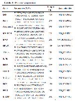
    The mRNA transcript levels of several genes, namely Caspase-3, Bax, Bcl-2, MAPK 14, STAT3, NF-κB, IL-1β, TNF-α, iNOS, COX-2, Nrf2, HO-1, and MPO, were quantified using the same instrument. β-actin was employed as an internal control for normalization purposes. The primer sequences corresponding to these genes are shown in Table 1. The determination of relative fold expressions was conducted using the 2-ΔΔCT technique, as described in reference22.


    Büyütmek İçin Tıklayın
    Table 1: Primer sequences

    Statistical Analysis: Shapiro-Wilk test was done to evaluate the samples as the sample number was under 50. The analytical findings were represented as the mean along with the standard deviation. The data analysis utilized SPSS software (version 20.0; SPSS, Chicago, IL). Multiple comparisons were executed through Tukey's post hoc test and one-way analysis of variance (ANOVA). Statistical significance was determined at a P-value below 0.05.

  • Başa Dön
  • Özet
  • Giriş
  • Materyal ve Metot
  • Bulgular
  • Tartışma
  • Kaynaklar
  • Bulgular
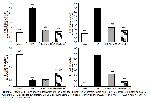
    Assessing Oxidative and Antioxidant Markers: Figure 1 and Figure 2 depict the impact of I/R damage and the administration of morin on oxidant and antioxidant markers in skeletal muscle.


    Büyütmek İçin Tıklayın
    Figure 1: Effects of I/R and Morin administrations on MDA levels, GPx, SOD, and CAT activities in skeletal muscle of rats. MDA:Malodialdhyde, GPx: Glutathione peroxidase, SOD: Superoxide dismutase, CAT: Catalase. Values are given as mean ± SD. ***P<0.001, **P<0.01, *P<0.05 Sham vs Others; ###P<0.001, ##P<0.01 I/R vs Others; ΔΔp˂0.01 I/R+Morin 50 vs I/R+Morin 100, ΔΔΔp˂0.001 I/R+Morin 50 vs I/R+Morin 100.


    Büyütmek İçin Tıklayın
    Figure 2: GSH levels in rat skeletal muscle after receiving I/R and morin treatments. GSH: Glutathione. Values are given as mean ± SD. ***P<0.001 Sham vs Others; ###P<0.001, ##P<0.01 I/R vs Others; ΔΔp˂0.01 I/R+Morin 50 vs I/R+Morin 100.

    The levels of MDA were shown to have increased in the I/R group as compared to the Sham group, with a statistically significant difference (P<0.01). Additionally, it has been demonstrated that the activities of SOD, GPx, and CAT, along with the levels of reduced GSH, experienced a decrease following the imposition of I/R. However, the administration of morin resulted in an augmentation of the antioxidant defense mechanism. A notable disparity was seen across the morin dosages in many parameters, with the exception of CAT. The group treated with IR+Morin 100 exhibited greater efficacy (P<0.01).

    Analysis of mRNA transcripts using RT-PCR has shown that Nrf2 and HO-1 expressions are downregulated during I/R injury. The administration of morin was observed to stimulate these genes and provide significant protection against oxidative stress. Moreover, it was clear that the high dose of morin demonstrated markedly superior effectiveness compared to the low dose of morin (P<0.05). The results for Nrf2 and HO-1 mRNA expression can be found in Figure 3. Nrf2 protein levels were quantified through western blot analysis, and the corresponding outcomes are presented in Figure 4. This data suggests that administering morin at a dosage of 100 mg/kg was notably more effective than the I/R injury group (P<0.001). Furthermore, when analyzing the disparity between the dosages, it was ascertained that the administration of high dose morin exhibited statistically superior efficacy compared to the administration of low dose (P<0.05).


    Büyütmek İçin Tıklayın
    Figure 3: Rat skeletal muscle Nrf2, HO-1, and MPO mRNA transcript levels in response to I/R and morin treatments. Nrf2: Nuclear factor erythroid 2-related factor 2, HO-1: Heme oxygenase-1, MPO: Myeloperoxidase. Values are given as mean ± SD. ***P<0.001 Sham vs Others; ###P<0.001 I/R vs Others; Δp˂0.05 I/R+Morin 50 vs I/R+Morin 100, ΔΔp˂0.01 I/R+Morin 50 vs I/R+Morin 100.


    Büyütmek İçin Tıklayın
    Figure 4: Protein levels of NF-κB, ICAM1, caspase-3, Bax, Bcl-2, Beclin-1, Nrf2, and MPO were evaluated through western blot analysis. NF-κB: Nuclear factor kappa-B, ICAM1: Intracellular adhesion molecule-1, Bax: Bcl-2-associated X protein, Bcl-2: B-cell lymphoma 2, Nrf2: Nuclear factor erythroid 2-related factor 2, MPO: Myeloperoxidase.

    Evaluation of MPO Expression Results: The study revealed that the levels of MPO expression, a heme protein known for its pivotal involvement in inflammation and oxidative stress and released by leukocytes, exhibited a statistically significant increase in the I/R group as compared to the Sham group. In contrast, it was shown that the injection of morin resulted in a considerable reduction in MPO expression as compared to the Sham group (P<0.001). Moreover, it was shown that the administration of a dosage of 100 mg/kg yielded greater efficacy among the morin groups, exhibiting a statistically significant disparity (P<0.01). All results are presented in Figure 3. In addition, blots of MPO are presented in Figure 4 and a bar graph of these blots in Figure 5. Based on the provided data, it can be shown that the group subjected to I/R exhibited elevated protein levels in comparison to the control group. However, administration of morin was associated with a reduction in MPO protein levels. A noteworthy distinction in outcomes was observed (P<0.05) between the I/R+Morin 50 and 100 groups following the administration of morin.


    Büyütmek İçin Tıklayın
    Figure 5: Effects of I/R and morin administrations on Nrf2 and MPO protein levels in skeletal muscle of rats. Nrf2: Nuclear factor erythroid 2-related factor 2, MPO: Myeloperoxidase. Values are given as mean ± SD. ***P<0.001, **P<0.01 Sham vs Others; ###P<0.001, ##P<0.01, #P<0.05 I/R vs Others; Δp˂0.05 I/R+Morin 50 vs I/R+Morin 100.

    The Assessment of Inflammatory Markers: The study assessed the expression levels of key inflammation markers, including iNOS, NF-κB, IL-1β, TNF-α, COX-2, STAT-3, and MAPK 14, in rat skeletal muscle using RT-PCR. The results from the research indicate that I/R-induced damage led to an upregulation of these inflammation markers compared to the sham group. Nevertheless, the treatment of morin exhibited strong anti-inflammatory properties, leading to a considerable reduction in the elevation of inflammation to levels comparable to those reported in the sham group (P<0.001).

    Furthermore, when examining the expression of IL-1β, COX-2, TNF-α, STAT-3, and MAPK 14 genes, it was evident that the I/R + Morin 100 group exhibited a more pronounced reduction in I/R injury (P<0.001). The mRNA transcript levels are depicted in Figure 6 and Figure 7 for reference.


    Büyütmek İçin Tıklayın
    Figure 6: Influence of I/R and morin treatments on the mRNA transcript levels of TNF-α, NF-κB, IL-1β, and iNOS in rat skeletal muscle. NF-κB: Nuclear factor kappa-B, TNF-α: Tumor necrosis factor alpha, IL-1β: Interleukin-1 beta, iNOS: Inducible nitric oxide synthase. Values are given as mean ± SD. ***P<0.001, **P<0.01, *P<0.05 Sham vs Others; ###P<0.001, ##P<0.01, #P<0.05 I/R vs Others; Δp˂0.05 I/R+Morin 50 vs I/R+Morin 100, ΔΔp˂0.01 I/R+Morin 50 vs I/R+Morin 100, ΔΔΔp˂0.001 I/R+Morin 50 vs I/R+Morin 100.


    Büyütmek İçin Tıklayın
    Figure 7: Effects of I/R and morin administrations on COX-2, STAT3, and MAPK 14 mRNA transcript levels in skeletal muscle of rats. COX-2: Cyclooxygenase-2, STAT3: Signal transducer and activator of transcription 3, MAPK14: Mitogen-activated protein kinase 14. Values are given as mean±SD. ***P<0.001 Sham vs Others; ###P<0.001, ##P<0.01 I/R vs Others; ΔΔΔp˂0.001 I/R+Morin 50 vs I/R+Morin 100.

    The Assessment of Apoptotic Markers: Bax, Apoptosis may be detected by caspase-3 and Bcl-2. Thus, the aforementioned parameters were assessed using RT-PCR and western blot analysis, and the results are shown in Figures 4, 8, and 9. Upon evaluation of the findings, it was ascertained that the process of I/R led to apoptosis by the upregulation of Bax and caspase-3 expression, while concurrently downregulating Bcl-2 expression. It was determined that this situation caused the I/R injury to aggravation. On the other hand, as compared with the Sham group, it was determined that morin significantly reduced apoptosis in the groups administered and contributed significantly to the protection of cells from I/R injury (P<0.001). Moreover, as the Bax/Bcl-2 ratio was examined, it was detected that apoptosis decreased significantly with morin administration.


    Büyütmek İçin Tıklayın
    Figure 8: Effects of I/R and morin administrations on Caspase-3, Bax, and Bcl-2 mRNA transcript levels in skeletal muscle of rats. Bax: Bcl-2-associated X protein, Bcl-2: B-cell lymphoma 2. Values are given as mean±SD. ***P<0.001 Sham vs Others; ###P<0.001 I/R vs Others; ΔΔΔp˂0.001 I/R+Morin 50 vs I/R+Morin 100.


    Büyütmek İçin Tıklayın
    Figure 9: Effects of I/R and morin administrations on Caspase-3, Bax, Bcl-2 protein levels and Bax/Bcl-2 ratio in skeletal muscle of rats. Bax: Bcl-2-associated X protein, Bcl-2: B-cell lymphoma 2. Values are given as mean±SD. ***P<0.001, *P<0.05 Sham vs Others; ###P<0.001 I/R vs Others.

    Evaluation of Autophagic Marker: In the study, Beclin-1 levels, one of the important indicators of autophagy, were analyzed and the results are presented in Figure 4 and Figure 10. The evidence presented in this study indicates that the application of I/R resulted in the stimulation of autophagy. Furthermore, a notable rise in Beclin-1 levels was seen in comparison to the Sham group. In contrast, it was seen that the administration of morin resulted in significant protection against autophagy as compared to the I/R group. This protection was evident by a reduction in Beclin-1 levels, with a decrease of around 12% in the I/R+Morin 50 group and 20% in the I/R+Morin 100 group. Furthermore, it was discovered via the analysis that the administration of a high dosage of morin exhibited a higher level of effectiveness, with statistical significance at a significance level of P<0.05.


    Büyütmek İçin Tıklayın
    Figure 10: Influence of I/R and morin interventions on NF-κB, ICAM1, and Beclin-1 protein concentrations in rat skeletal muscle. NF-κB: Nuclear factor kappa-B, ICAM1: Intracellular adhesion molecule-1. Values are given as mean±SD. ***P<0.001, **P<0.01 Sham vs Others; ###P<0.001, ##P<0.01 I/R vs Others.

    Evaluation of ICAM1 Protein Levels: As ICAM1 levels, which have crucial functions such as cell signaling, leukocyte-endothelial transmigration, tissue stability, and cell-cell interaction, were examined, it was determined that the levels of ICAM1 increased significantly (P<0.001) in the presence of I/R injury in skeletal muscle, and this situation was reversed with antioxidant administration and approached the control group levels. Furthermore, a notable difference was seen between the groups treated with I/R + Morin 50 and I/R + Morin 100, with statistical significance (P<0.05). The results are presented in Figure 4 and Figure 10.

  • Başa Dön
  • Özet
  • Giriş
  • Materyal ve Metot
  • Bulgular
  • Tartışma
  • Kaynaklar
  • Tartışma
    I/R, which is defined as the reduction of oxygen to tissues and subsequent restoration of blood flow, can lead to irreversible damage. Since skeletal muscle is the predominant tissue in the limb, it is more vulnerable to I/R injury. Although I/R generally occurs in acute situations and treatment with chemical agents is less preferred, in recent years, researchers have carried out numerous studies aimed at reducing I/R injury in skeletal muscle with various compounds23. The current investigation aimed to assess the efficacy of morin, a compound with established antioxidant, anti-inflammatory, and anti-apoptotic properties, in mitigating skeletal muscle I/R damage. The obtained results demonstrate that morin significantly reduces I/R injury.

    The balance of the oxidant state in the body is greatly influenced by antioxidant enzymes and substances24. Cells experience oxidative stress when there is a reduction in antioxidant activity and a depletion of antioxidant molecules, resulting in an elevation of oxidants25. I/R leads to a decrease in tissue antioxidant levels by causing excessive ROS formation. Ultimately, this decrease causes the antioxidant defense mechanism to be ineffective against oxidative tissue damage8. The susceptibility of membrane lipids in skeletal muscle to oxidation is significant. Ischemia and the accompanying reperfusion process give rise to the generation of reactive oxygen species, resulting in oxidative damage by upsetting the equilibrium between oxidants and antioxidants. The current study established that the concentration of MDA, a crucial marker of lipid peroxidation, increased following the initiation of I/R injury in skeletal muscle. Conversely, the administration of morin resulted in a reduction of MDA levels, with the extent of drop being contingent upon the dosage administered. Enzymatic antioxidants, including as SOD, CAT, and GPx, as well as the non-enzymatic antioxidant GSH, are integral components of the defense system against oxidative stress26. In a prior investigation, scholars unveiled that I/R injury instigated oxidative stress by means of free radical assault, resulting in substantial damage to skeletal muscle tissue. This detrimental effect was attributed to the diminished enzymatic activities of SOD, CAT, and GPx, as well as reduced amounts of reduced GSH27. The current investigation revealed that the administration of morin in rats with I/R damage resulted in a rise in GSH levels, with the extent of increase being dependent on the dosage supplied. The study revealed a reduction in the activities of SOD, CAT, and GPx enzymes in the group experiencing ischemia/reperfusion (I/R) damage. Conversely, the groups treated with morin exhibited an increase in the activities of these enzymes. The results of this research indicate that morin possesses the capacity to alleviate oxidative stress triggered by I/R injury in skeletal muscle. Furthermore, it highlights morin's potential to shield cells from harm by boosting the activities of antioxidant enzymes and increasing the levels of GSH.

    A basic leucine zipper protein called Nrf2 may control the production of antioxidant proteins which protect against oxidative damage brought on by injury and inflammation. The substance is mostly distributed throughout the cytoplasm and forms a combination with Keap1. Keap1 serves as the inhibitory regulatory factor for Nrf2 and facilitates the degradation of Nrf2 via the process of ubiquitination. This mechanism enables the maintenance of Nrf2's physiological state at a low level of activity28. Concerning oxidative stress, Nrf2 migrates to the nucleus after undergoing phosphorylation and dissociation from Keap1. This translocation leads to the activation of downstream mechanisms that regulate the expression of genes such as HO-1, GPx, and NQO1. These genes, along with the associated antioxidant response element (ARE) sequence in their promoters, collectively contribute to the essential role of Nrf2 in safeguarding cells against inflammatory processes. HO-1 serves as the principal enzyme responsible for antioxidative and anti-inflammatory processes, which are initiated by the activation of Nrf229. In research conducted by Gendy et al.30, it was demonstrated that there was a decrease in Nrf2 and HO-1 mRNA expression levels in the context of I/R injury. Another research has shown that the injection of Fisetin, a kind of flavonoid, during I/R injury leads to an elevation in Nrf2 levels and an enhancement of HO-1 activity, thereby providing protection against oxidative damage. Hence, this research aimed to examine the potential antioxidant properties of morin in mitigating skeletal muscle I/R injury, focusing on its modulation of the Nrf2/HO-1 signaling pathway. The findings of the study indicate that the expression of Nrf2 and HO-1 was downregulated in skeletal muscle subjected to ischemia and subsequent reperfusion. However, the administration of morin resulted in an increase in Nrf2 expression, particularly in the I/R + Morin 100 group (P<0.001). This upregulation of Nrf2 led to an elevation in HO-1 levels, hence suggesting a reduction in oxidative stress.

    MPO belongs to the peroxidase superfamily and is mostly expressed in monocytes and neutrophils. Reactive species formed by MPO are principally involved in the process of phagocytosis, hence playing a crucial role in the antimicrobial action directed against a wide range of pathogens. Therefore, elevated MPO activity serves as a reliable marker for the presence of neutrophil infiltration in I/R damage31. Elevated levels of circulating MPO are associated with inflammation and oxidative stress32. The results of our study show that I/R injury upregulates MPO gene expression, but morin administration reduces MPO expressions by regulating neutrophil infiltration in skeletal muscle.

    Besides oxidative stress, there is an increasing body of data suggesting that inflammation and cytokine activity play a crucial role in the development of skeletal muscle I/R damage33. NF-κB, a transcription factor of considerable importance, governs a multitude of biological processes such as cell cycle regulation, cellular migration, programmed cell death, and inflammatory responses. The investigation of the NF-κB pathway has significant therapeutic importance due to its ability to induce transcription of proinflammatory mediators, including COX-2, IL-1β, and TNF-α34. This phenomenon is one of the compelling evidence, which supports the link between oxidative stress and inflammation in the progression of ischemia/reperfusion.

    In conjunction with proinflammatory cytokines, nitric oxide (NO) serves as a significant molecule involved in the mediation of tissue damage in the context of I/R injury. Inflammatory triggers, such as endotoxins, cytokines, and lipid mediators, result in an increase in iNOS expression35. Ince et al.36 have reported that skeletal muscle is impacted by NO produced by iNOS. Flavonoids play a crucial role in regulating modulation of inflammation pathways and cellular functions such as cell cycle signals. Similar to the studies in the literature, our results show that inflammatory markers are increased in I/R injury, and morin administration protects skeletal muscle by inhibiting cytokine activities. Based on these results, it can be said that the inflammation process in reperfused skeletal muscle after ischemia was suppressed by morin administration and the damage caused by inflammation was reduced.

    Mitogen-activated protein kinases (MAPKs) have important activities as cellular signaling mediators37. The activation of MAPKs leads to an upregulation of gene expression, therefore inducing a diverse range of biological reactions, including the production of pro-inflammatory substances. MAPK 14, sometimes referred to as P38α, is well recognized as a pivotal modulator of the inflammatory response across several cellular lineages38. In their study, Qin et al.39 observed an elevation in the level of MAPK 14 after I/R damage. In a similar vein, our findings demonstrate that the expression of MAPK 14 is upregulated as a consequence of the heightened activation of NF-κB in the context of skeletal muscle ischemia-reperfusion damage. Conversely, the injection of morin has a suppressive effect on such activity while concurrently providing cellular protection against potential injury.

    An integral part in several pathophysiological diseases, including inflammation, is played by STAT3, a transcription factor and member of the STAT family40. STAT3 translocation in the nucleus as a result of activation leads to increased NF-κB transcriptional activity, which contributes to the increased expression of cytokine genes41. Information on STAT3 gene expression in skeletal muscle I/R injury is limited in the literature. Pottecher et al.42 found out an increase in STAT3 activation in the case of skeletal muscle exposure to I/R. Similarly, the results obtained in the present study also indicate that in I/R injury, STAT3 gene expression increases depending on the transcriptional activity of NF-κB but decreases with the administration of morin depending on the dose.

    Apoptosis, commonly referred to as programmed cell death, is a natural process activated by various physiological and pathological factors. Bax and Bcl-2 are recognized as pivotal markers of apoptosis. Disturbance in the equilibrium between Bax and Bcl-2 results in an upsurge of cytochrome C levels within the cytoplasm, thereby initiating the activation of caspase-3 43. Caspase-3 is known as a good marker of apoptosis and initiates the apoptotic cascade by activating other caspase enzymes25. In a research conducted by Cheng et al.44, it was revealed that caspase-3 levels increased in skeletal muscle subjected to I/R injury. Additionally, a separate investigation reported that the limb I/R model resulted in elevated Bax levels and reduced Bcl-2 levels45. Our study's findings demonstrate that during I/R conditions, there is an upregulation in Bax and caspase-3 expressions, accompanied by a downregulation of Bcl-2 expression, ultimately leading to cellular apoptosis. Administration of morin at doses of 50 mg/kg b.w. and 100 mg/kg b.w. has shown significant efficacy in inhibiting cell death, preserving mitochondrial homeostasis, and maintaining membrane potential.

    Excessive production of ROS leads to the irreversible oxidation of cellular macromolecules, including DNA, resulting in cellular damage. Autophagy is a crucial cellular degradation mechanism that facilitates the lysosomal breakdown of misfolded proteins and damaged or superfluous organelles 13. Beclin-1 is a skeletal protein of the PI3K complex and a good marker of autophagy. Based on the analyzes by Western blotting, I/R injury increases autophagy by damaging cells, as seen in Fig. 5. However, morin administration prevents this impact by reducing autophagy. Consistent with our results, researchers have found in the previous studie on the skeletal muscle that I/R injury increases autophagy46.

    The pathophysiological process of ischemia/reperfusion injury is complex and significantly regulated by the oxidative stress response and the acute inflammatory response5. Inflammatory mediators such as ICAM-1 can aggravate and block micro vessels. Moreover, this phenomenon might result in the production of inflammatory mediators and cytokines via the activation of leukocytes. Additionally, the release of free radicals can lead to neuronal injury47. NF-κB can modulate gene expression levels of chemotactic factors, including ICAM, thus playing a critical role in cell inflammation and apoptosis. Calcium overload and oxidative stress can be induced by reperfusion following ischemia for further NF-κB activation. ICAM-1 plays a role in the inflammatory pathological process, as the expression level of ICAM-1 can be increased by various inflammatory factors48. Xiang et al.49 have shown an elevation in ICAM-1 levels in the context of skeletal muscle I/R damage. The findings obtained in our investigation also shown an elevation in ICAM-1 levels in the context of I/R damage, consistent with the existing literature. It was determined that the damage was reduced through the administration of morin, and ICAM-1 levels approached the control group levels.

    In conclusion, the findings of the current research provide evidence that morin has protective properties against I/R damage in rat skeletal muscle. The protective effect of morin against I/R damage may be attributed to its anti-apoptotic, antioxidant, anti-autophagic, and anti-inflammatory qualities. However, before a therapeutic application is advised, further research must be carried out to validate these results.

  • Başa Dön
  • Özet
  • Giriş
  • Materyal ve Metot
  • Bulgular
  • Tartışma
  • Kaynaklar
  • Kaynaklar

    1) Li Y, Jiang J, Tong L, et al. Bilobalide protects against ischemia/reperfusion-induced oxidative stress and inflammatory responses via the MAPK/NF-κB pathways in rats. BMC Musculoskelet Disord 2020; 21: 449.

    2) Kobayashi J, Murata I. Nitrite as a pharmacological intervention for the successful treatment of crush syndrome. Physiol Rep 2018; 6: e13633.

    3) Paradis S, Charles A-L, Meyer A, et al. Chronology of mitochondrial and cellular events during skeletal muscle ischemia-reperfusion. Am J Physiol Cell Physiol 2016; 310: C968-C982.

    4) Zhang Y, Li H, Wang M, et al. Vagus nerve stimulation attenuates acute skeletal muscle injury ınduced by ischemia-reperfusion in rats. Oxid Med Cell Longev 2019; 2019: 9208949.

    5) Ergün Y, Kılınç M, Aral M, et al. Protective effect of epigallocatechin gallate in ischemia-reperfusion injury of rat skeletal muscle. J Surg Res 2020; 247: 1-7.

    6) Sheikholeslami MA, Ghafghazi S, Pouriran R, et al. Attenuating effect of paroxetine on memory impairment following cerebral ischemia-reperfusion injury in rat: The involvement of BDNF and antioxidant capacity. Eur J Pharmacol 2021; 893: 173821.

    7) Sangodele JO, Inuwa Z, Lawal B, et al. Proxeed plus salvage rat testis from ischemia- reperfused injury by enhancing antioxidant’s activities and inhibition of iNOS expression. Biomed Pharmacother 2021; 133: 111086.

    8) Cheng F, Zhang Q, Yan F-F, et al. Lutein protects against ischemia/reperfusion injury in rat skeletal muscle by modulating oxidative stress and inflammation. Immunopharmacol and Immunotoxicol 2015; 37: 329-334.

    9) Tetsi L, Charles A-L, Georg I, et al. Effect of the phosphodiesterase 5 inhibitor sildenafil on ischemia-reperfusion-induced muscle mitochondrial dysfunction and oxidative stress. Antioxidants 2019; 8: 93.

    10) Boroujeni MB, Khayat ZK, Anbari K, et al. Coenzyme Q10 protects skeletal muscle from ischemia-reperfusion through the NF-kappa B pathway. Perfusion 2017; 32: 372-377.

    11) Ekinci Akdemir FN, Gülçin İ, Karagöz B, Soslu R. Quercetin protects rat skeletal muscle from ischemia reperfusion injury. J Enzyme Inhib Med Chem 2016; 31: 162-166.

    12) Gur C, Kandemir FM, Darendelioglu E, et al. Morin protects against acrylamide-induced neurotoxicity in rats: An investigation into different signal pathways. Environ Sci Pollut Res 2021; 28: 49808-49819.

    13) Kandemir FM, Yıldırım S, Kucukler S, et al. Protective effects of morin against acrylamide-induced hepatotoxicity and nephrotoxicity: A multi-biomarker approach. Food Chem Toxicol 2020; 138: 111190.

    14) Abd El-Mottaleb NA, Mahmoud GS, Negm EA, Abdel Maksoud FM. Garlic antagonizes skeletal muscle ischemia reperfusion injury through regulating inflammation, apoptosis and desmin expression in adult male rats. Int J Physiol Pathophysiol Pharmacol 2019; 11: 126-137.

    15) Tian Y, Li Z, Shen B, et al. Protective effects of morin on lipopolysaccharide/ d-galactosamine-induced acute liver injury by inhibiting TLR4/NF-κB and activating Nrf2/HO-1 signaling pathways. Int Immunopharmacol 2017; 45: 148-155.

    16) Placer ZA, Cushman LL, Johnson BC. Estimation of product of lipid peroxidation (malonyl dialdehyde) in biochemical systems. Anal Biochem 1966; 16: 359-364.

    17) Sun Y, Oberley LW, Li Y. A simple method for clinical assay of superoxide dismutase. Clin Chem 1988; 34: 497-500.

    18) Aebi H. Catalase in vitro. Methods Enzymol 1984; 105: 121-126

    19) Lawrence RA, Burk RF. Glutathione peroxidase activity in selenium-deficient rat liver. Biochem Biophys Res Commun 1976; 71: 952-958.

    20) Sedlak J, Lindsay RH. Estimation of total, protein-bound, and nonprotein sulfhydryl groups in tissue with Ellman’s reagent. Anal Biochem 1968; 25: 192-205.

    21) Lowry OH, Rosebrough NJ, Farr AL, et al. Protein measurement with the Folin phenol reagent. J Biol Chem 1951; 193: 265-275.

    22) Livak KJ, Schmittgen TD. Analysis of relative gene expression data using real-time quantitative PCR and the 2(−ΔΔC(T)) method. Methods 2001; 25: 402-408.

    23) Tiryakioglu O, Erkoc K, Tunerir B, et al. The effect of ıloprost and n-acetylcysteine on skeletal muscle ınjury in an acute aortic ıschemia-reperfusion model: An experimental study. BioMed Res Int 2015; 2015: 1-8.

    24) Ileriturk M, Dogan T, Kandemir Ö. Investigation of the effect of berberine with arginase activity and oxidant-antioxidant parameters on bortezomib-ınduced spleen ınjury in rats. Kocatepe Veterinary Journal 2020; 14: 6-15.

    25) Kandemir FM, Ileriturk M, Gur C. Rutin protects rat liver and kidney from sodium valproate-induce damage by attenuating oxidative stress, ER stress, inflammation, apoptosis and autophagy. Mol Biol Rep 2022; 49: 6063-6074.

    26) Semis HS, Gur C, Ileriturk M, et al. Investigation of the anti-inflammatory effects of caffeic acid phenethyl ester in a model of λ-Carrageenan–induced paw edema in rats. Hum Exp Toxicol 2021; 40: S721-S738.

    27) Akbari G. Role of Zinc Supplementation on Ischemia/Reperfusion Injury in Various Organs. Biol Trace Elem Res 2020; 196: 1-9.

    28) Yardım A, Kucukler S, Özdemir S, et al. Silymarin alleviates docetaxel-induced central and peripheral neurotoxicity by reducing oxidative stress, inflammation and apoptosis in rats. Gene 2021; 769: 145239.

    29) Yardim A, Kandemir FM, Ozdemir S, et al. Quercetin provides protection against the peripheral nerve damage caused by vincristine in rats by suppressing caspase 3, NF-κB, ATF-6 pathways and activating Nrf2, Akt pathways. Neurotoxicology 2020; 81: 137-146.

    30) Gendy A, Soubh A, Al-Mokaddem A, et al. Dimethyl fumarate protects against intestinal ischemia/reperfusion lesion: Participation of Nrf2/HO-1, GSK-3β and Wnt/β-catenin pathway. Biomed Pharmacother 2021; 134: 111130.

    31) Ye Y, Shan Y, Bao C, et al. Ginsenoside Rg1 protects against hind-limb ischemia reperfusion induced lung injury via NF-κB/COX-2 signaling pathway. Int Immunopharmacol 2018; 60: 96-103.

    32) Kandemir FM, Yildirim S, Kucukler S, et al. Therapeutic efficacy of zingerone against vancomycin-induced oxidative stress, inflammation, apoptosis and aquaporin 1 permeability in rat kidney. Biomed Pharmacother 2018; 105: 981-991.

    33) Guo YX, Wang GY, Cheng W, et al. Activation of Opioid Receptors Attenuates Ischemia/Reperfusion Injury in Skeletal Muscle Induced by Tourniquet Placement. Mediators Inflamm 2021; 2021: 1-10.

    34) Caglayan C, Temel Y, Kandemir FM, et al. Naringin protects against cyclophosphamide-induced hepatotoxicity and nephrotoxicity through modulation of oxidative stress, inflammation, apoptosis, autophagy, and DNA damage. Environ Sci Pollut Res 2018; 25: 20968-20984.

    35) Yesildag K, Gur C, Ileriturk M, Kandemir FM. Evaluation of oxidative stress, inflammation, apoptosis, oxidative DNA damage and metalloproteinases in the lungs of rats treated with cadmium and carvacrol. Mol Biol Rep 2022; 49: 1201-1211.

    36) İnce I. Renoprotect ve effect of edaravone n acute l mb ischemia/reperfusion injury. Turk J Thorac and Cardiovasc Surg 2020; 28: 274-281.

    37) Caglayan C, Kandemir FM, Yildirim S, et al. Rutin protects mercuric chloride‐induced nephrotoxicity via targeting of aquaporin 1 level, oxidative stress, apoptosis and inflammation in rats. Journal of Trace Elements in Medicine and Biology 2019; 54: 69-78.

    38) Caglayan C, Kandemir FM, Darendelioğlu E, et al. Rutin ameliorates mercuric chloride-induced hepatotoxicity in rats via interfering with oxidative stress, inflammation and apoptosis. Journal of Trace Elements in Medicine and Biology 2019; 56: 60-68.

    39) Qin SQ, Zhang ZS, Wang XY, et al. MiR-24 protects cardiomyocytes against hypoxia/reoxygenation-induced injury through regulating mitogen-activated protein kinase 14. International Heart Journal 2020; 61: 806-814.

    40) Fouad AA, Abdel-Aziz AM, Hamouda AAH. Diacerein downregulates NLRP3/Caspase-1/IL-1β and IL-6/STAT3 pathways of inflammation and apoptosis in a rat model of cadmium testicular toxicity. Biological Trace Element Research 2020; 195: 499-505.

    41) Fan Y, Mao R, Yang J. NF-κB and STAT3 signaling pathways collaboratively link inflammation to cancer. Protein & Cell 2013; 4: 176-185.

    42) Pottecher J, Adamopoulos C, Lejay A, et al. Diabetes worsens skeletal muscle mitochondrial function, oxidative stress, and apoptosis after lower-limb ischemia-reperfusion: Implication of the RISK and SAFE pathways? Front Physiol 2018; 9: 579.

    43) Ilerituk M, Kandemir O, Akaras N, et al. Hesperidin has a protective effect on paclitaxel-induced testicular toxicity through regulating oxidative stress, apoptosis, inflammation and endoplasmic reticulum stress. Reprod Toxicol 2023; 118: 108369.

    44) Cheng Y, Di S, Fan C, et al. SIRT1 activation by pterostilbene attenuates the skeletal muscle oxidative stress injury and mitochondrial dysfunction induced by ischemia reperfusion injury. Apoptosis 2016; 21: 905-916.

    45) Huang T, Wang W, Tu C, et al. Hydrogen-rich saline attenuates ischemia–reperfusion injury in skeletal muscle. Journal of Surgical Research 2015; 194: 471-480.

    46) Zhang S, Jiang S, Wang H, et al. SIRT6 protects against hepatic ischemia/reperfusion injury by inhibiting apoptosis and autophagy related cell death. Free Radical Biology and Medicine 2018; 115: 18-30.

    47) Ji K, Xue L, Cheng J, Bai Y. Preconditioning of H 2 S inhalation protects against cerebral ischemia/reperfusion injury by induction of HSP70 through PI3K/Akt/Nrf2 pathway. Brain Research Bulletin 2016; 121: 68-74.

    48) Chien CT, Jou MJ, Cheng TY, et al. Exendin-4-loaded PLGA Microspheres relieve cerebral ischemia/reperfusion ınjury and neurologic deficits through long-lasting bioactivity-mediated phosphorylated Akt/eNOS signaling in rats. Journal of Cerebral Blood Flow & Metabolism 2015; 35: 1790-1803.

    49) Xiang Y, Ye S, Cai C, et al. Salvianolic acid a attenuates limb ischemia/reperfusion injury in skeletal muscle of rats. Biomedicine & Pharmacotherapy 2018; 97: 551-556.

  • Başa Dön
  • Özet
  • Giriş
  • Materyal ve Metot
  • Bulgular
  • Tartışma
  • Kaynaklar
  • [ Başa Dön ] [ Özet ] [ PDF ] [ Benzer Makaleler ] [ Yazara E-Posta ] [ Editöre E-Posta ]
    [ Ana Sayfa | Editörler | Danışma Kurulu | Dergi Hakkında | İçindekiler | Arşiv | Yayın Arama | Yazarlara Bilgi | E-Posta ]