[ Ana Sayfa | Editörler | Danışma Kurulu | Dergi Hakkında | İçindekiler | Arşiv | Yayın Arama | Yazarlara Bilgi | E-Posta ]
Fırat Üniversitesi Sağlık Bilimleri Veteriner Dergisi
2024, Cilt 38, Sayı 1, Sayfa(lar) 070-077
[ Özet ] [ PDF ] [ Benzer Makaleler ] [ Yazara E-Posta ] [ Editöre E-Posta ]
Bisfenol A’ya Maruz Kalan Sıçanların Pankreas Dokusunda Koenzim Q10, Fulleren C60 ve Alfa-Lipoik Asit’in Histomorfometrik, Antiapoptotik ve İnsülin Ekspresyonu Üzerine Etkileri
Songül ÇERİBAŞI1, Abdurrauf YÜCE2, Özgür BULMUŞ3
1Firat University, Faculty of Veterinary Medicine, Department of Pathology, Elazığ, TÜRKİYE
2Firat University, Faculty of Veterinary Medicine, Department of Physiology, Elazığ, TÜRKİYE
3Balıkesir University, Faculty of Medicine, Department of Physiology, Balıkesir, TÜRKİYE
Anahtar Kelimeler: Alpha-lipoic acid, bisphenol A, coenzyme Q10, fulleren C60, pancreas
Özet
Bu çalışmada, erken pubertal gelişim döneminden itibaren düşük dozda BPA’ya maruz kalan erkek sıçanlarda pankreasta meydana gelen morfometrik değişimler, apoptozis temelli pankreatik doku hasarı ile insülin ekspresyonundaki farklılıkların ortaya konulması ve BPA’nın olumsuz etkilerine karşı KoQ10, FUL, ALA’in muhtemel koruyucu özelliklerinin belirlenmesi amaçlandı. Çalışmada; 1 aylık, 60 adet Sprague-Dawley ırkı erkek sıçan kullanıldı. Hayvanlar, canlı ağırlıkları dikkate alınarak rastgele toplam 8 gruba ayrılarak özel kafeslere yerleştirildi. Gruplara, 7 hafta süreyle uygulamalar oral gavaj yöntemiyle BPA, KoQ10, FUL, ALA 0.2 mL zeytinyağına katılarak gerçekleştirildi. Tüm gruplarda Langerhans adacıklarında yapılan morfometrik ölçümler sonucu ortalama adacık sayı, adacık çap ve alanlarının BPA grubunda azaldığı dikkati çekti. BPA ile birlikte KoQ10 ve ALA uygulamasının adacık sayılarında artışa neden olduğu, FUL’un ise etkili olmadığı gözlendi. KoQ10 ve FUL’un adacık çapı ve alanlarında BPA’ya karşı etkisiz olduğu ancak ALA’nın olumlu etkisinin olduğu belirlendi. İnsülin pozitifliğinin BPA uygulanan tüm gruplarda kontrol grubuna göre azaldığı, adacıklarda insülin ekspresyonu KoQ10, FUL ve ALA’nın, BPA’ya karşı anlamlı bir etkisinin olmadığı saptandı. BPA’nın öncülük ettiği apoptotik değişimlere karşı KoQ10 ve ALA’nın etkili olduğu ancak FUL’in olumlu etkisinin olmadığı izlendi. Sonuç olarak sunulan çalışmada sıçanlarda uzun süreli BPA uygulaması ile endokrin pankreasta histopatolojik değişimler gözlenmemekle birlikte morfometrik açıdan önemli değişimler ile adacık hücrelerinde insülin salınımının azaldığı, apoptozisin ise tetiklendiği belirlenmiş; BPA’nın endokrin pankreastaki etkisine karşı ise KoQ10 ve ALA’in kısmen koruyucu etkilerinin olduğu ortaya konmuştur.
  • Başa Dön
  • Özet
  • Giriş
  • Materyal ve Metot
  • Bulgular
  • Tartışma
  • Kaynaklar
  • Giriş
    Endocrine-disrupting chemicals (EDCs) are natural or synthetic compounds that bind to specific receptors and mimicking biological activities1,2. Bisphenol A (BPA) is classified into the xenoestrogen group due to its action on the estrogen receptor (ER) and is classified into the endocrine-disrupting chemical category because of its effects on hormones and receptors3. Humans are primarily exposed to BPA through food and food-contact materials, and to a lesser extent through epoxy coatings of metal cans, kitchen utensils, medical devices, and dental composites and adhesives4. BPA has been found to play a role in the development of obesity, diabetes, endocrine and immune system disorders, polycystic ovary syndrome, hypertension, atherosclerosis, liver damage, irregular thyroid hormone secretion, and breast and prostate cancers in both humans and experimental animals5-7. Furthermore, BPA has been reported to increase insulin secretion from the pancreas 8, disrupt blood glucose homeostasis9, and induce insulin resistance10.

    While it has long been recognized that BPA exhibits estrogen-like effects, it has been stated that this effect occurs not only through classical ERs and mechanisms but also through different pathways 11. In experimental studies, BPA has been shown to mimic the effects of 17β-estradiol, leading to excessive insulin stimulation and insulin resistance12. In mice, both BPA and estrogen have been found to increase insulin secretion at low doses and decrease plasma glucose concentration. Prolonged exposure to BPA has been observed to cause chronic hyperinsulinemia and subsequent insulin resistance10. Furthermore, BPA has been found to reduce the number of macrophages in the pancreas of mice and increase suppressive T lymphocytes and apoptotic cell counts13,14. Additionally, BPA has been shown to decrease the number of β cells and their proliferative capacity in the pancreas15, as well as reduce the viability of insulin-secreting cells16. Moreover, BPA has been reported to slowdown the body's energy metabolism and disrupt insulin signaling in skeletal muscle. Therefore, BPA is considered a risk factor for the development of diabetes17. On the other hand, it has been determined that BPA weakens Ca2+ signals in α cells of the pancreas10 and increases glucagon expression18.

    Coenzyme Q10 (CoQ10) is an important coenzyme with strong antioxidant effects. It is required for the activities of enzyme systems involved in redox reactions in the electron transfer chain and is located in the hydrophobic parts of all membranes and the inner mitochondrial membrane19-21. It is found at the highest concentrations in the heart, liver, kidney, and pancreas. CoQ10 is a potent antioxidant in the mitochondrial respiratory chain, serving as a membrane stabilizer and an essential co-factor, facilitating the production of adenosine triphosphate (ATP), the main cellular energy source. CoQ10 acts directly by protecting cellular components from free radicals and indirectly by regenerating other antioxidants such as α-tocopherol and ascorbate22. Besides maintaining membrane stability, CoQ10 is also involved in cellular signaling, gene expression, cell growth, and apoptosis control23. Fullerene C60 (FUL) has been recognized as a biocompatible agent that does not exhibit toxic effects on normal tissues at low concentrations and possesses strong free radical scavenging and antioxidant properties24,25. Studies have revealed its anticancer, neuroprotective, anti-inflammatory, antiatherogenic, and radioprotective properties26,27. Alpha-lipoic acid (ALA) is an antioxidant molecule with both hydrophilic and lipophilic properties, synthesized in various tissues of living organisms and found in certain vegetables, mainly broccoli and spinach28,29. Due to its antioxidant properties, ALA has been reported to be used as a supportive therapy in conditions such as diabetes, ischemia, heavy metal poisoning, radiation exposure, neurodegeneration, and HIV infection30.

    In this study, we aimed to identify the morphometric changes in the pancreas, apoptosis-based pancreatic tissue damage, and differences in insulin expression in male rats exposed to low doses of BPA during the early pubertal period. Additionally, the potential protective properties of CoQ10, FUL, and ALA against the adverse effects of BPA were investigated.

  • Başa Dön
  • Özet
  • Giriş
  • Materyal ve Metot
  • Bulgular
  • Tartışma
  • Kaynaklar
  • Materyal ve Metot
    Research and Publication Ethics: The study was conducted with the approval of the Fırat University Local Ethics Committee for Animal Experiments (dated 12.10.2022, approval number 2022/17-10).

    Laboratory Animals: Sixty male Sprague-Dawley rats with an average weight of approximately 100 grams and aged 1 month were used. The animals were placed in special cages and the light schedule was adjusted to 12 hours of light and 12 hours of darkness. The rats were also provided with ad libitum access to food and water.

    Experimental Procedure: The animals were randomly divided into eight groups based on their live weights and placed in cages. The administrations to the groups were carried out for seven weeks, every day, using the oral gavage method. BPA, CoQ10, FUL, and ALA were administered by mixing them with 0.2 ml of olive oil. Accordingly, the groups received the following treatments: Control group: 0.2 mL of olive oil; BPA group: 25 mg/kg of BPA; CoQ10 group: 40 mg/kg; FUL group: 8 μg/kg of FUL in aqueous form; ALA group: 70 mg/kg of ALA; BPA + CoQ10 group: 25 mg/kg of BPA with 40 mg/kg of CoQ10; BPA + FUL group: 25 mg/kg of BPA with 8 μg/kg of FUL in aqueous form; BPA + ALA group: 25 mg/kg of BPA with 70 mg/kg of ALA.

    Collection of Tissue Samples: Pancreas samples were taken from the decapitated rats and fixed in a formaldehyde solution for 48 hours. Following fixation, the samples underwent routine processing to prepare paraffin blocks. Sections with a thickness of 5 microns (μm) were obtained from the paraffin blocks and stained using the Hematoxylin and Eosin (H&E) method. Subsequently, the stained sections were examined under a light microscope31.

    Histomorphometric Analysis: For histological examinations, Langerhans islets, which are the endocrine components of the pancreas, were examined in H&E-stained pancreas sections. The number of islets was determined by counting them in ten different randomly selected microscopic fields at 10x magnification. Additionally, in H&E-stained sections, the number of islets in the pancreas and the diameter and area of 20 different randomly selected islets at 40x magnification were calculated using the Image J (Wayne Rasband and contributors, National Institutes of Health, USA, Version 1.53t) analysis program.

    Immunohistochemical Analysis: The Avidin-Biotin-Peroxidase method was used for immunohistochemical analysis 32. For this purpose, the universal immunoperoxidase kit (Ultravision Detection System, Antipolyvalent, HRP/DAB, Thermo Scientific, Cat No: TP-015-HD) was employed. Sections with a thickness of 4 microns obtained from paraffin blocks were deparaffinized with xylene and dehydrated through a graded series of alcohols to remove water. To expose the antigenic receptors, the sections were incubated in 0.01 M sodium citrate for 20 minutes, then washed with PBS, and incubated in 3% H2O2 prepared with methanol for 10 minutes to block endogenous peroxidase activity. To prevent nonspecific binding, the sections were incubated in 1% normal goat serum for 1 hour. Subsequently, the sections were incubated overnight at 4°C with primary antibodies for insulin (diluted at 1/100, 1/200; Abcam, Kimera Medical Laboratory Supplies, Cat No: ab-63820) and caspase 3 (Bioss Inc Cat No: bs-0081R). After washing with PBS, the sections were incubated with biotinylated secondary antibodies (Thermo Scientific, Cat No: TP-015-BNS) for 10 minutes. Then, the sections were incubated with streptavidin peroxidase (Thermo Scientific, Cat No: TS-015-HRS) for 10 minutes. The color-developing substrate used was 3-amino-9-ethylcarbazole (AEC) (Thermo Scientific, Cat No:001122). After applying AEC to the sections, the reaction was stopped as soon as the color change began. In the final step, the sections were counterstained with Mayer's Hematoxylin (MH) for 2 minutes, washed in tap water, covered with immu-mount, and examined under a light microscope at appropriate magnifications.

    Immunohistochemical Evaluation: The images of 20 randomly selected islets from the pancreatic sections of each animal in the microscope field at 40x magnification were captured with a camera. Subsequently, using the Image J program, the positive cells for insulin and caspase 3 were counted, and the obtained group mean values were used as the β-cell index and apoptotic cell index.

    Statistical Analyses: The data were statistically evaluated using IBM SPSS software (IBM Corp. Released 2012. IBM SPSS Statistics for Windows, Version 22.0. Armonk, NY, USA). Shapiro–Wilk and Levene's tests were performed to analyze the normality of the data and the homogeneity of the variances, respectively. The data were tested using one-way analysis of variance (ANOVA). Post hoc comparisons between the groups were conducted using Tukey's test. The data were presented as mean ± standard error, and a P-value of less than 0.05 was considered significant.

  • Başa Dön
  • Özet
  • Giriş
  • Materyal ve Metot
  • Bulgular
  • Tartışma
  • Kaynaklar
  • Bulgular
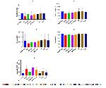
    Histomorphometric Analysis: The endocrine pancreas regions in all groups maintained normal histological appearance. The average values of morphometric measurements (count, diameter, and area of islets) in Langerhans islets are summarized in Table 1 for all groups. When comparing the groups based on the mean islet count, the BPA group had the lowest number of islets. There was a statistically significant difference in islet count between the control, CoQ10, FUL, and ALA groups compared to the BPA group (P≤0.001). A similar difference was also observed between the BPA group and the BPA+CoQ10 and BPA+ALA groups. Although there was no statistical difference between the BPA and BPA+FUL groups, there was a noticeable numerical difference (Figure 1a). Except for in the BPA and BPA+FUL groups, the mean islet counts in the other experimental groups were found to be similar to the control group (P≥0.05).


    Büyütmek İçin Tıklayın
    Table 1: Mean Langerhans islet number, diameter, area values, and β cell and apoptotic cell indexes in the control and experimental groups


    Büyütmek İçin Tıklayın
    Figure 1: a. Number of islets, b. Islet diameter, c. Islet area, d. β Cell Index, e. Apoptotic Cell Index

    The average islet diameters were found to be similar in the control, CoQ10, FUL, and ALA groups. Additionally, the diameter values in the BPA+ALA group were statistically similar to those in the control, CoQ10, FUL, and ALA groups. The smallest diameters were found in the BPA, BPA+CoQ10, and BPA+FUL groups, respectively. While there was no difference between the BPA and BPA+CoQ10 groups in terms of diameter (p>0.05), a significant difference was detected between the BPA and BPA+FUL groups (Figure 1b). A similar difference was also observed between the BPA+ALA and BPA+CoQ10 groups (P≤0.001).

    The islet areas were similar in the control, CoQ10, FUL, ALA, and BPA+ALA groups. However, the islet areas were significantly decreased in the BPA, BPA+CoQ10, and BPA+FUL groups compared to the aforementioned groups (P≤0.001). There was no statistical difference in islet areas between the BPA, BPA+CoQ10, and BPA+FUL groups (Figure 1c).

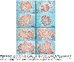
    β cell index and apoptotic index values in islets of Langerhans in control and experimental groups are presented in Table 1. It was observed in immunostaining that insulin positivity was homogeneously distributed in the islets except for the peripheral parts of the islets in the BPA group, and it was cytoplasmic and granular in the islet cells. In the BPA group, areas devoid of insulin expression were observed near the islet center (Figure 2). According to the groups, the β cell index was found to be lowest in BPA, BPA+FUL, BPA+ALA, BPA+CoQ10 groups and highest in control, FUL, ALA, CoQ10 groups. There was no statistical difference between BPA and BPA treatment groups. β cell index values were similar in control and CoQ10, FUL, and ALA groups (Figure 1d).


    Büyütmek İçin Tıklayın
    Figure 2: Insulin positivity in β cells of Langerhans islets A. Control group, B. The appearance of insulin positive cells (arrow) in islets in BPA group, C. BPA+CoQ10 group, D. BPA+FUL group, E.BPA+ALA group, F. CoQ10 group, G. FUL group, H. ALA group; MH × 100, Bar= 50 μ.

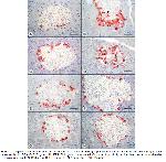
    Caspase 3 positivity was generally cytoplasmic and granular in the cells located at the periphery of the islets except for the BPA and BPA+FUL groups. In BPA and BPA+FUL groups, positivity was also observed towards the center of the islets (Figure 3). Apoptotic index was highest in BPA+FUL, BPA, BPA+ALA, and BPA+CoQ10 groups. It was noteworthy that there was a statistically significant difference between the aforementioned groups. The groups with the lowest apoptotic index were determined as CoQ10, control, ALA, and FUL. Apoptosis was significantly increased in the FUL group compared to the CoQ10 and control groups. A similar difference was observed between the CoQ10 and ALA groups. It was noteworthy that both BPA and FUL treatment alone increased apoptosis (Figure 1e).


    Büyütmek İçin Tıklayın
    Figure 3: Caspase 3 positivity in islets of Langerhans A. Control, B. BPA group, increase in Caspase 3 positive cell density in islets (arrowhead), C. BPA+CoQ10 group, D. BPA+FUL group pronounced Caspase 3 positivity in the islet center (arrowheads), E.BPA+ALA group, F. CoQ10 group, G. FUL group, H. ALA group; MH × 100, Bar= 50 μ.

  • Başa Dön
  • Özet
  • Giriş
  • Materyal ve Metot
  • Bulgular
  • Tartışma
  • Kaynaklar
  • Tartışma
    In recent years, it was shown that BPA can adversely affect the endocrine system, even at low doses11. Previous studies have shown that BPA can induce excessive insulin stimulation in the endocrine pancreas and contributing to insulin resistance12,33. It has been reported that BPA may increase the risk of developing type II diabetes by directly affecting pancreatic cells, impairing insulin and glucagon secretion, inhibiting cell growth, triggering cell apoptosis, affecting muscle, liver, and fat cell function, and activating insulin resistance14,33,34. Similarly, it was reported that 100 mg/kg intraperitoneal injection of BPA into female mice decreased the size and cell number of islets of Langerhans35. In the present study, statistical differences were determined between the groups in terms of islet number, diameter, and area. Accordingly, we determined that the number of islets decreased by 50.29%, 17.34%, 37.68% and 20.81% in BPA, BPA+CoQ10, BPA+FUL, and BPA+ALA groups, respectively, compared to the control group. In the BPA, BPA+CoQ10, BPA+FUL, and BPA+ALA groups, islet diameters decreased by 28.42%, 20.08%, 14.51%, and 6.09% and islet areas decreased by 40.79%, 28.61%, 25.41% and 5.27%, respectively. These observed changes in islets of Langerhans were evaluated as an indication that BPA can directly affect pancreatic cells and lead to the development of type I diabetes. It was reported that insulitis developed at the end of 11 weeks and type 1 diabetes developed at the end of 28 weeks in offspring born from pregnant mice administered BPA at a dose of 10 mg/l in daily drinking water14.

    A previous study determined that no histopathological changes occurred in the islets of Langerhans of male rats exposed to 5, 50, or 500 μg/kg/day oral doses of BPA for 8 weeks. However, in the same study, the percentage of insulin-positive β cells in the islets of Langerhans increased significantly in the group administered BPA at a dose of 500 μg/kg/day compared to the other groups2. Another study in which rats were administered Bisphenol F (BPF) at 20, 100, or 500 mg/kg/day for 28 days, insulin positive β cell index values decreased in BPF groups compared to the control group33. In the present study, no histologic changes were observed in the pancreas of male rats after oral administration of 25 mg/kg/day BPA for 7 weeks, and insulin positive β cell index was significantly decreased in BPA-treated groups compared to control and antioxidant-only groups. It was determined that β cell index decreased by 9.72%, 6.47%, 8.77%, and 6.52% in BPA, BPA+CoQ10, BPA+FUL, and BPA+ALA groups, respectively, compared to the control group. BPA dose, animal species, and exposure time seem to be the main factors determining whether histopathologic changes will occur in the pancreas.

    It has been reported that BPA causes oxidative stress and has detrimental effects on organs such as the pancreas, liver, brain, kidney, and testis35. In addition, it was reported that BPA injection at a dose of 2mg/kg/day in male rats caused a decrease in antioxidative effective enzymes in the pancreas at the end of four weeks and an increase malondialdehyde levels, a product indicative of oxidative stress36. In the present study, caspase 3 positivity increased by 265.37%, 141.30%, 321.89%, and 223.16% in the BPA, BPA+CoQ10, BPA+FUL, and BPA+ALA groups, respectively, compared to the control group. In the islets of Langerhans in rats, β-cells make up 60-80% of the total number of cells, α-cells 15-20%, δ-cells less than 10%, and PP-cells approximately 1% (37-39). It was reported that a significant increase in caspase 3 positivity was observed in the islets of Langerhans in 11-week-old offspring of female mice exposed to BPA (3000 μg/kg/day) during pregnancy14. In the present study, we interpreted that the decreases in the number, diameter, and area of islets detected in the BPA-exposed groups compared to the control group may be due to oxidative stress-based apoptotic cell deaths in islets, especially β cells.

    CoQ10 is a fat-soluble compound that acts as a coenzyme in important enzymatic reactions during energy production in cells and can be found in every cell40,41. Although CoQ10 is at a lower concentration than other antioxidants in plasma, it is the first antioxidant to react with plasma oxidants. It is also known to play a role in the regeneration of other antioxidants40,42. When CoQ10 was administered with metformin in diabetic rats, it was found to reduce the number of apoptotic cells in the pancreas and provided a significant improvement in insulin staining intensity in islets43. In addition, it was reported that CoQ10 reduced changes such as interstitial edema, inflammatory cell infiltration, and acinar necrosis in an experimental pancreatitis model in rats44. In rats with diabetes induced by sirolimus, CoQ10 administration at a dose of 20 mg/kg/day for two weeks caused a decrease in the number of TUNEL positive cells in islets of Langerhans43. In another study, there was no significant difference observed in terms of β cell area determined by histopathological and immunohistochemical insulin positivity between the group administered 100 mg/kg/day dose of BPA alone and the group administered 10 mg/kg/day dose of CoQ10 with the same dose of BPA for 14 days45. In the present study, CoQ10 administration at a dose of 40 mg/kg/day significantly ameliorated the apoptotic effects of BPA on the endocrine pancreas.

    FUL treatment was found to be effective for the prevention of oxidative stress against increased lipid peroxidation and changes in antioxidant enzyme activities in doxorubicin-treated male rats46. In male rats with acute cholangitis, development of acute pancreatitis characterized by necrosis, degeneration, edema, and thrombosis in the acinus was observed, and degenerative changes in the pancreas were suppressed by oral or intraperitoneal 0.5 mg/kg FUL administration for 3 days47. In the present study, it was surprisingly noted that FUL administration at a dose of 8 μg/kg/day together with 20 mg/kg/day BPA exacerbated the apoptotic effect of BPA in islets. In addition, it was found to have no effect on morphometric changes. Consistent with the present study, the cell number and volume in the islets decreased by approximately 50% at the end of 5 days in the endocrine pancreas of rats with streptozotocin (STZ)-induced diabetes; 25.00 mg/kg FUL treatment daily with STZ injection did not improve the condition in the pancreas and caused further deterioration of periductal inflammatory changes48.

    It has been emphasized that ALA has suitable properties to treat tissue damage caused by free oxygen radicals due to its strong antioxidative effects49. It was reported that administration of 100 mg/kg/day ALA for 8 weeks to STZ-induced diabetic rats prevented necrotic and degenerative destruction in endocrine pancreatic cells50. Consistent with previous studies, we found in the present study that administration of 70 mg/kg/day ALA together with 25 mg/kg/day BPA for 7 weeks significantly improved BPA-induced morphometric changes in the pancreas and decreased the number of apoptotic cells in islets. In previous studies, ALA was shown to prevent oxidative damage and cell death by scavenging oxygen radicals, metal ion chelates, and various mechanisms protective against lipid peroxidation50-52.

    In conclusion, while long-term BPA administration in rats did not result in observable histopathological changes in the endocrine pancreas, significant morphometric alterations, decreased insulin secretion, and apoptosis in islet cells were identified in the present study. Notably, CoQ10 and ALA exhibited partial protective effects against the impact of BPA on the endocrine pancreas. Given the absence of studies exploring the effects of ALA and FUL on BPA-induced pancreatic tissue damage, it is suggested that future research should focus on unraveling the detailed interaction of these antioxidant substances with BPA.

  • Başa Dön
  • Özet
  • Giriş
  • Materyal ve Metot
  • Bulgular
  • Tartışma
  • Kaynaklar
  • Kaynaklar

    1) Liu J, Yu P, Qian W, et al. Perinatal bisphenol A exposure and adult glucose homeostasis: Identifying critical windows of exposure. PLOS ONE 2013; 8: e64143.

    2) Ozaydın T, Oznurlu Y, Sur E, et al. Effects of bisphenol A on antioxidant system and lipid profile in rats. Biotech Histochem. 2018; 93: 231-238.

    3) Lang IA, Galloway TS, Scarlett A, et al. Association of urinary bisphenol A concentration with medical disorders and laboratory abnormalities in adults. JAMA 2008; 300: 1303-1310.

    4) Andujar N, Ontiveros YG, Gomez AZ, et al. Bisphenol A Analogues in Food and Their Hormonal and Obesogenic Effects: A Review. Nutrients 2019; 11: 2136.

    5) Deb P, Bhan A, Hussaın I, et al. Endocrine disrupting disrupting chemical, bisphenol-A, induces breast cancer associated gene HOXB9 expression in vitro and in vivo. Gene 2016; 590:234-243.

    6) Melzer D, Rice NE, Lewis C, et al. Association of urinary bisphenol a concentration with heart disease: Evidence from NHANES 2003/06. PLoS One 2010; 5: e8673.

    7) Kandaraki E, Chatzigeorgiou A, Livadas S, et al. Endocrine disruptors and polycystic ovary syndrome (PCOS): Elevated serum levels of bisphenol A in women with PCOS. The Journal of Clinical Endocrinology and Metabolism 2011; 96: E480-E484.

    8) Adachi T, Yasuda K, Mori C, et al. Promoting insulin secretion in pancreatic islets by means of bisphenol A and nonylphenol via intracellular estrogen receptors. Food Chem Toxicol 2005; 43: 713-719.

    9) Ding S, Fan Y, Zhao N, et al. High-fat diet aggravates glucose homeostasis disorder caused by chronic exposure to bisphenol A. J Endocrinol 2014; 221: 167-179.

    10) Alonso-Magdalena P, Morimoto S, Ripoll C, et al. The estrogenic effect of bisphenol A disrupts pancreatic beta-cell function in vivo and induces insulin resistance. Environ Health Perspect 2006; 114: 106-112.

    11) Vandenberg LN, Hauser R, Marcus M, et al. Human exposure to bisphenol A (BPA). Reproductive Toxicology 2007; 24: 139-177.

    12) Nadal A, Alonso-Magdalena P, Soriano S, et al. The pancreatic beta-cell as a target of estrogens and xenoestrogens: Implications for bloodglucose homeostasis and diabetes. Mol Cell Endocrinol 2009; 304(1-2): 63-68.

    13) Bodin J, Bølling AK, Samuelsen M, et al. Long-term bisphenol A exposure accelerates insulitis development in diabetes-prone NOD mice. Immunopharmacol Immunotoxicol 2013; 35: 349-58.

    14) Bodin J, Bølling AK, Becher R, et al. Transmaternal bisphenol A exposure accelerates diabetes type 1 development in NOD mice. Toxicol Sci 2014; 137: 311-323.

    15) Alonso-Magdalena P, García-Arévalo M, Quesada I, et al. Bisphenol-A treatment during pregnancy in mice: a new window of susceptibility for the development of diabetes in mothers later in life. Endocrinology 2015; 156: 1659-1670.

    16) Weldingh NM, Jørgensen-Kaur L, Becher R, et al. Bisphenol A is more potent than phthalate metabolites in reducing pancreatic β-cell function. Biomed Res Int 2017; 2017: 4614379.

    17) Batista TM, Alonso-Magdalena P, Vieira E, et al. Short-term treatment with bisphenol-A leads to metabolic abnormalities in adult male mice. PLOS ONE 2012; 7(3): e33814.

    18) Whitehead R, Guan H, Arany E, et al. Prenatal exposure to bisphenol A alters mouse fetal pancreatic morphology and islet composition. Horm Mol Biol Clin Investig 2016; 25: 171-179.

    19) Kowaltowski AJ, Anıbal EV. Mitochondrial damage induced by conditions of oxidative stres, Free Rad Biol Med 1999; 26: 463-471.

    20) Ishrat T, Khan MB, Hoda MN, et al. Coenzyme Q10 modulates cognitive impairment against intracerebroventricular injection of streptozotocin in rats. Behav Brain Res 2006; 171: 9-16.

    21) Yaşar S, Yur F, Ceylan E. Effects of CoQ10 and vitamin E on blood lipid peroxidation, some antioxidant and biochemical parameters in doxorubicin administered rats. FU Vet J Health Sci 2021; 35: 103-108.

    22) Sattarinezhad E, Shafaroodi H, Sheikhnouri K, et al. The effects of coenzyme Q10 on seizures in mice: The involvement of nitric oxide. Epilepsy Behav 2014; 37: 36-42.

    23) Crane FL. Biochemical functions of coenzyme Q10. Journal of the American College of Nutrition 2001; 20: 591-598.

    24) Harhaji L, Isakovic A, Zogovic NS, et al. Multiple mechanisms underlying the anticancer action of nanocrystalline fullerene. European Journal of Pharmacology 2007; 568: 89-98.

    25) Kolosnjaj TJ, Baati T, Szwarc H, et al. Toxicity studies of [60] fullerene and carbon nanotubes: State of the art. In: D’Souza F, Kadish KM. (Editors). Handbook of Carbon Nano Materials 1st Edition, Singapore: World Scientific 2012: 49-75.

    26) Andrievsky GV, Bruskov VI, Tykhomyrov AA, et al. Peculiarities of the antioxidant and radioprotective effects of hydrated C60 fullerene nanostuctures in vitro and in vivo. Free Radical Biology and Medicine 2009; 47: 786-793.

    27) Tykhomyrov AA, Nedzvetsky VS, Klochkov VK, et al. Nanostructures of hydrated C60 fullerene (C60HyFn) protect rat brain against alcohol impact and attenuate behavioral impairments of alcoholized animals. Toxicology 2008; 246: 158-165.

    28) Lodge JK, Youn H, Handelman G. Natural sources of lipoic acid: Determination of lipoyllysine released from protease-digested tissues by high performance liquid chromatography incorporating electrochemical detection. J Appl Nutr 1997; 49: 3-11.

    29) Rochette, L. Ghibu, S. Richard, C, et al. Direct and indirect antioxidant properties of α-lipoic acid and therapeutic potential. Mol Nutr Food Res 2013; 57: 114-125.

    30) Packer L, Witt EH, Tritschler HJ. Alpha-Lipoic acid as a biological antioxidant. Free Radical Biology and Medicine 1995; 19: 227-250.

    31) Luna L. Manual of Histologic Staining Methods of the Armed Forces Institute of Pathology. 3rd Edition, New York: McGraw-Hill, 1968.

    32) Bourne JA. Handbook of Immunoperoxidase Staining Methods. Immunochemistry Laboratory: Dako Corporation USA, 1983; 6-34.

    33) Doğan E, Kaptaner B, Doğan A. Bisfenol F’nin sıçan pankreası üzerindeki toksik etkileri. Yüzüncü Yıl Üniversitesi Fen Bilimleri Enstitüsü Dergisi 2022; 27: 436-451.

    34) Provvisiero DP, Pivonello C, Muscogiuri G, et al. Influence of bisphenol A on type 2 diabetes mellitus. International Journal of Environmental Research & Public Health 2016; 13: 989.

    35) Arroyo-Salgado B, Garcia-Espiñeira M, Olivero-Verbel J. Effects of bisphenol A on streptozotocin treated female mice. Indian Journal of Experimental Biology 2018; 56: 419-429.

    36) Moghaddam HS, Samarghandian S, Farkhondeh T. Effect of bisphenol A on blood glucose, lipid profile and oxidative stress indices in adult male mice. Toxicology Mechanisms and Methods 2015; 25: 507-513.

    37) Brissova M, Fowler MJ, Nicholson WE, et al. Assessment of human pancreatic islet architecture and composition by laser scanning confocal microscopy. Journal of Histochemistry & Cytochemistry 2005; 53: 1087-1097.

    38) Cabrera O, Berman DM, Kenyon NS, et al. The unique cytoarchitecture of human pancreatic islets has implications for islet cell function. Proc Natl Acad Sci USA. 2006; 14,103: 2334-2339.

    39) Karan M, Timurkaan S, Atalar Ö. The localization of insulin, glucagon, somatostatin immunoreactive cells in the pancreas of the martes foina. FÜ Sağ Bil Vet Derg 2008: 22: 13-16.

    40) Overvad K, Diamant B, Holm L, et al. Review Coenzyme Q10 in health and disease, European Journal of Clinical Nutrition 1999; 53: 764-770.

    41) Stocker R. “Coenzyme Q10.Reviewed, Linus Pauling Institute Micronutrient Research for Optimum Health”. http://lpi.oregonstate.edu/infocenter/othernuts/coq10/2007/ 25.09.2023.

    42) Bhagavan HN, Chopra RK, Craft NE. Chitchumroonchokchai C, Failla ML. Assesment of coenzyme Q10 absorbtion using in vitro digestion-Caco-2 cell model. Int Journ Pharmace 2017; 333: 112-117.

    43) In O Sun, Long Jin, Jian Jin, et al. The effects of addition of coenzyme Q10 to metformin on sirolimus-induced diabetes mellitus. Korean J Intern Med 2019; 34: 365-374.

    44) Mirmalek SA, Boushehrinejad AG, Yavari H, et al. Antioxidant and anti-inflammatory effects of coenzyme Q10 on L-arginine-induced acute pancreatitis in rat. Oxid Med Cell Longev 2016; 5818479.

    45) Yıldız M, Güleş O, Sandıkçı M, et al. The effects of coenzyme Q10 on bisphenol a-induced pancreatic changes in rats. Journal of Harran University Medical Faculty 2020; 17: 335-341.

    46) Srdjenovic B, Torres VM, Grujic N, et al. Antioxidant properties of fullerenol C60(OH)24 in rat kidneys, testes, and lungs treated with doxorubicin. Toxicology Mechanisms and Methods 2010; 20: 298-305.

    47) Kuznietsova H, Dziubenko N, Lynchak OV, et al. Efects of pristine C60 fullerenes on liver and pancreas in α-naphthylisothiocyanate-induced cholangitis. Digestive Diseases and Sciences 2020; 65: 215-224.

    48) Yue You, Jinxia Li, Mei Wang, et al. The effect of fullerenols on streptozotocin-induced diabetic rats, Fullerenes, Nanotubes and Carbon Nanostructures 2022; 30: 438-451.

    49) Van Dam PS, Van Asbeck BS, Van Oirschot JF et al. Glutathione and alpha-lipoate in diabetic rats: Nerve function, blood flow and oxidative state. European journal of clinical investigation 2001; 31: 417-424.

    50) Budin SB, Kee KP, Eng MYS, et al. Alpha Lipoic Acid prevents pancreatic islet cells damage and dyslipidemia in streptozotocin-induced diabetic rats. Malaysian Journal of Medical Sciences 2007; 14: 47-53.

    51) Ghoneim FM, Alrefai H, Elsamanoudy AZ, et al. The protective role of prenatal alpha lipoic acid supplementation against pancreatic oxidative damage in offspring of valproic acid-treated rats: Histological and Molecular Study. Biology 2020; 9: 239.

    52) Packer L, Kreamer K, Rimbach G. Molecular aspect of lipoic acid in the prevention of diabetes complications. Nutrition 2001; 17: 888-895.

  • Başa Dön
  • Özet
  • Giriş
  • Materyal ve Metot
  • Bulgular
  • Tartışma
  • Kaynaklar
  • [ Başa Dön ] [ Özet ] [ PDF ] [ Benzer Makaleler ] [ Yazara E-Posta ] [ Editöre E-Posta ]
    [ Ana Sayfa | Editörler | Danışma Kurulu | Dergi Hakkında | İçindekiler | Arşiv | Yayın Arama | Yazarlara Bilgi | E-Posta ]