[ Ana Sayfa | Editörler | Danışma Kurulu | Dergi Hakkında | İçindekiler | Arşiv | Yayın Arama | Yazarlara Bilgi | E-Posta ]
Fırat Üniversitesi Sağlık Bilimleri Veteriner Dergisi
2024, Cilt 38, Sayı 1, Sayfa(lar) 082-091
[ Özet ] [ PDF ] [ Benzer Makaleler ] [ Yazara E-Posta ] [ Editöre E-Posta ]
İshalli Buzağılarda Nötrofil/Lenfosit Oranı, Lenfosit/Monosit Oranı ve Yangısal Marker Olarak Serum Demir Düzeylerinin Değerlendirilmesi
Ömer AYDIN
Atatürk University, Faculty of Veterinary Medicine, Department of Internal Medicine, Erzurum, TÜRKİYE
Anahtar Kelimeler: Calves diarrhea, lymphocyte/monocyte ratio, neutrophil/lymphocyte ratio, serum iron level
Özet
Bu çalışmada ishalli buzağılarda hematolojik kriter olan nötrofil-lenfosit oranı (NLO), lenfosit-monosit oranı (LMO) ve demir (Fe) düzeylerinin araştırılması ve birbirleriyle ilişkilerinin belirlenmesi amaçlandı. Çalışma materyalini 0-10 gün yaş arasında olan toplam 171 adet Brown Swiss, Simmental ve bunların melez dişi ve erkek buzağıları oluşturmuştur. Çalışma kontrol grubu (Grup-1, n=15), E. coli grubu (Grup-2, n=32), Coronavirüs grubu (Grup-3, n=27), Rotavirüs grubu (Grup-4, n=32), Giardia spp. grubu (Grup-5, n=14), Cryptosporidium spp. grubu (Grup-6, n=15), Rota-Coronavirus grubu (Grup-7, n=21), E. coli- Rotavirus grubu (Grup-8, n=15) olmak üzere sekiz gruptan oluştu. Total lökosit sayısı, nötrofil sayısı, NLO ve Fe düzeyleri ishal gruplarında kontrol grubuna göre anlamlı olarak farklı bulundu. E. coli grubunun NLO değerleri diğer ishal gruplarına ve kontrol grubuna göre yüksek, lenfosit, monosit ve Fe değerleri ise daha düşüktü. E. coli kaynaklı ishal grubunda inflamatuar düzeyin diğer ishal gruplarına göre daha yüksek olduğu ve ishalli buzağılarda inflamatuar durumun belirlenmesinde NLO, LMO ve Fe düzeylerinin anlamlı değerler verdiği sonucuna varıldı.
  • Başa Dön
  • Özet
  • Giriş
  • Materyal ve Metot
  • Bulgular
  • Tartışma
  • Kaynaklar
  • Giriş
    Neonatal calf diarrhea is a disease that affects calves in the pre-weaning period. This disease can result in death in many cases due to hypovolemia and acidosis in young calves1. Neonatal calf diarrhea can be infectious and noninfectious. While non-infectious calf diarrhea occurs due to vitamin deficiencies and mistakes in milk-feeding, infectious diarrhea is caused by viral, bacterial, and protozoal pathogens2.

    Inflammation is a reaction to cellular or tissue damage. This reaction manifests itself by reducing or eliminating the effects of agents that damage the organism. Later, this inflammatory response contributes to the restructuring of damaged cells and tissues3. Acute inflammation is a condition that may develop within hours or even minutes, depending on the intensity and type of tissue damage4. Chronic inflammation is a type of response that occurs when the organism’s response to acute inflammatory responses or autoimmune reactions is insufficient and lasts longer (weeks or even months) than acute inflammatory conditions. This type of inflammation is characterized by the activation of lymphocytes, macrophages, and other inflammatory cells3.

    The evaluation of hematological data is of great importance for cattle health. With the correct interpretation of hematological data, important information can be obtained both in terms of the early diagnosis of diseases and the prognosis5. Neutrophil-lymphocyte ratio (NLR) and platelet-lymphocyte ratio (PLR), mean platelet volume to platelet ratio (MPV/PLT) are obtained from hemogram analysis. It has been shown as a prognostic marker in many diseases such as NLR, PLR, MPV/PLT thromboemboli diseases, sepsis in human medicine6-8. In veterinary medicine, NLR and PLR have mostly been evaluated in certain disease states. Lymphocyte-monocyte ratio (LMR) has been evaluated as a prognostic marker in cancer or lymphoproliferative diseases in human medicine9. NLR has been extensively investigated in oncology in oropharyngeal tumors10. In veterinary medicine, it has been stated that LMR shows a poor prognosis in high-grade lymphomas11.

    Iron (Fe) is an essential trace element for living organisms and bacterial pathogens, and also has important functions in many enzymatic reactions12. Serum Fe concentration decreases rapidly in response to inflammation. It has been reported that this situation is resulted in by the retention of Fe, which is necessary for bacterial virulence and replication, for host defense13. It has been reported that serum Fe concentration can be used as an acute inflammatory marker in different animal species13-15.

    In this study, it was aimed to determine the levels of NLR, LMR, and Fe in calves with diarrhea caused by different etiological pathogens and also to investigate the relationship between these hematological parameters and Fe.

  • Başa Dön
  • Özet
  • Giriş
  • Materyal ve Metot
  • Bulgular
  • Tartışma
  • Kaynaklar
  • Materyal ve Metot
    Research and Publication Ethics: The study was approved by the Local Ethics Committee of Atatürk University (Ethics Committee Decision No: 2023/05).

    Animals: The study material consisted of calves brought to Atatürk University Faculty of Veterinary Medicine, Animal Hospital with diarrhea and a total of 171 Brown Swiss, Simmental, and their crossbred female and male calves aged 0-10 days. The study consisted of eight groups: control group (Group-1, n=15) and E. coli group (Group-2, n=32), Coronavirus group (Group-3, n=27), Rotavirus group (Group-4, n=32), Giardiasis spp. group (Group-5, n=14), Cryptosporidium group (Group-6, n=15), Rota-Coronavirus group (Group-7, n=21), and E. coli- Rotavirus group (Group-8, n=15).

    Recording of Clinical Findings: In the inclusion of calves with diarrhea in the study, fecal scoring, and clinical examination findings (body temperature, degree of dehydration) were determined according to the criteria determined by Larson et al. 16. Calves with excessive watery diarrhea, a body temperature above 39.3°C, and a dehydration degree of 8-10% were included in the study due to these parameters. In addition, in determining the severity of advanced dehydration, criteria such as advanced enophthalmos (6 mm and over), decreased cervical skin elasticity (7 s and above), white mucous membranes, cooling in the extremities, remaining in a lying position and inability to stand up were taken into account17. Health status of the animals in the control group was confirmed by clinical examination, hematological finding based on hematological analysis (Abacus Junior Vet 5®, Hungary).

    Sampling: Hematological analyses were performed by taking blood samples into K2EDTA tubes (Vacutainer, K2E 3.6 mg, BD, UK) from the vena jugularis of the animals (Abacus Junior Vet 5®, Hungary), and the blood taken in serum tubes (Vacutainer, BD, UK) was kept at room temperature for 30 minutes. Afterward, serum tubes were +4 °C centrifuged at 3000 rpm for 10 minutes in a refrigerated centrifuge device (Bechman Coulter, USA) to obtain serum samples. The obtained serum samples were transferred to Eppendorf tubes and stored in a refrigerator at -80°C until further processing.

    The etiological agents of diarrhea were determined by using a immunochromatographic test (BoviD-5 Ag Test Kit, Korea). This test detects Rotavirus Ag, Coronavirus Ag, E. coli K99, Cryptosporidium Ag, and Giardia Ag from the stool samples. Blood and fecal samples were collected only once within 24 hours from the onset of diarrhea. In addition, animals in the diarrhea group were included in the study after being examined for elimination of diseases such as respiratory system diseases, aspiration pneumonia, omphalitis and arthritis.

    Hematological Analysis and Biochemical Analysis: Hematological analysis incuding total leukocytes (WBC), lymphocytes (LYM), monocytes (MON), LMR (LMR was calculated the absolute lymphocyte count divided by the absolute monocyte count), neutrophils (NEU), and NLR (NLR was obtained by dividing the absolut neutrophil count by the absolute lymphocyte count) were determined by using a desktop hematology analyzer (Abacus Junior Vet® brand hemogram instrument, Diatron MI Ltd., Hungary). Serum Fe levels were measured using an autoanalyzer device (Randox Monaco, UK) by the colorimetric method.

    Statistical Analysis: The analysis of the data was conducted using IBM SPSS Statistics software version 27.0.1. Normality measures of numerical values were obtained by determining the Shapiro-Wilk normality test. A homogeneity test was applied for normally distributed data (WBC, LYM, MON, NEU, NLR, Temperature, Pulsation ratio, Respiration ratio). For data showing normal distribution but not homogeneity (WBC, LYM, MON, NEU, NLR), the Brown-Forsythe test was used, and the Gamel-Howell test was used as the post hoc test. One-way analysis of variance was used for normally and homogeneously distributed data (Temperature, Respiration ratio and Pulsation ratio), and LSD (for temperature) and Tukey HSD (for respiration ratio and pulsation ratio) tests were applied as post hoc tests. Since LMR and Fe values did not show a normal distribution, the Kruskal-Wallis test was performed, and the Dunn-Bonferroni test was applied as a post hoc test. Spearman's Rank Correlation Coefficient was applied to the correlation between the data. As stated by Chan18, Spearman's correlation coefficients <0.3 were considered as a weak correlation, 0.3-0.5 as a moderately strong correlation, 0.6-0.8 as a strong correlation, and a value of at least 0.8 and above as a very strong correlation. Linear regression analysis was applied to normally distributed data. P<0.05 was considered to be as statistically significant.

  • Başa Dön
  • Özet
  • Giriş
  • Materyal ve Metot
  • Bulgular
  • Tartışma
  • Kaynaklar
  • Bulgular
    Clinical Findings: When the groups were examined clinically, it was determined that the body temperature and respiratory frequency were within the normal reference range, and there were no signs of dehydration in the calves in the control group. In calves with diarrhea, T>39.3 was determined, and the degree of dehydration was 8-10%16. Clinical findings between the groups are shown in Table 1. When evaluated in terms of clinical findings, it was determined that the highest value among the groups in terms of temperature was in group-2 (E. coli group) (P<0.001). It was observed that the other diarrheal group values were higher than the control group values (P<0.001). It was determined that the lowest value for pulsation rate was in the control group and was significantly higher in all other diarrheal groups than in the control group (P<0.001). It was determined that the lowest value for respiration rate was in the control group and that the values of all other diarrheal groups were higher than the control group (P<0.05). It was determined that the values of groups 3, 6 and 7 were significantly higher than the values of group 4 and group 8 (P<0.05).


    Büyütmek İçin Tıklayın
    Table 1: Clinical findings in diarrheal and control group calves

    Hematological Findings and Biochemical Findings: The mean data of the hematological and serum Fe values between the groups are shown in Table 2.


    Büyütmek İçin Tıklayın
    Table 2: Hematological index values among groups

    In the evaluation between groups in terms of WBC values, it was determined that all other diarrhea group data were significantly higher than the Group-1 value (P<0.001). In addition, Group-3 and Group-6 values were found to be significantly higher than Group-4 values (P<0.05). It was determined that the Group-5 value was higher than the Group-2 value, which has the lowest numerical value in terms of LYM values (P<0.05). In the comparison between groups in terms of MON values, Group-5 and Group-7 values were found to be statistically higher than Group-2 values (P<0.05). It was determined that the Group-5 value was higher than the Group-3, 6, and 8 values, and this increase was significant (P<0.05). In terms of NEU values, it was observed that all diarrhea group values had a significant difference compared to the control group (P<0.001). In terms of NLR value, it was determined that the Group-1 value was the lowest value and all diarrhea group data were significantly higher (P<0.05). Similarly, for the NLR value, Group-2 data was found to be significantly higher than all other diarrhea group data, except for Group-8 data (P<0.05). In terms of LMR values, it was noticed that the Group-5 value was significantly lower than all diarrheal group values except for the value of Group-7 (P<0.05). For Fe values, the Group-1 value was found to be significantly higher than all diarrheal group values except the Group-5 group (P<0.01).

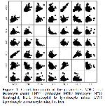
    Correlation Findings: Data obtained from the whole blood and serum of animals are shown in Table 3. There was a moderate positive correlation between WBC data and LYM values (rho= 0.533; P<0.001), and a weak positive correlation between WBC and MON values (rho=0.191; P<0.05). A very strong correlation was obtained between the WBC and NEU value (rho=0.815; P<0.001), while a weak correlation was observed between the WBC and NLR (rho=0.169; P<0.05) and LMR (rho=0.176; P<0.05) values. There was a moderate correlation between LYM and MON value (rho=0.393; P<0.001), while a weak correlation was obtained between LYM and LMR (rho=0.285; P<0.001) and Fe (rho=0.287; P<0.001). A strong correlation was observed between LYM and NLR (rho=-0.676; P<0.001). There was a weak correlation between MON and NLR values (rho=-0.289; P<0.001) and a strong correlation between MON and LMR values (rho=-0.723; P<0.001). A strong correlation was found between NEU and NLR (rho=0.626; P<0.001), while a weak correlation was obtained between NEU and Fe (rho=-0.165; P<0.05). A weak correlation was observed between NLR and LMR (rho=-0.194; P<0.05) and a moderately strong correlation was observed between NLR and Fe (rho=-0.390; P<0.001). The correlation between the values is shown in Figure 1.


    Büyütmek İçin Tıklayın
    Table 3: Results of the correlation analyses among hematological parameters


    Büyütmek İçin Tıklayın
    Figure 1: Correlation graph of the parameters. WBC:Total leukocyte count; LMY: Lymhocyte; MON: Monocyte; NEU: Neutrophil; NLR: Neutrophil to lymphocyte ratioe; LMR: Lymphocyte to monocyte ratio; Fe: Iron

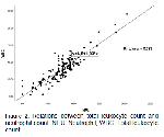
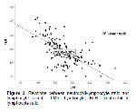
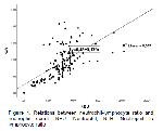
    Regression Analysis: Since P<0.05 was obtained in all numerical parameters investigated for linear regression, the established regression model was found to be significant. Data obtained from whole blood are shown in Table 4. According to the results of the regression analysis, it was found that 28% of the WBC value was shaped by LYM and the effect was positive (R=0.53; R2=0.280; P<0.001). It has been shown that 0.05% of WBC is formed by MON and this effect is positive (R=0.22; R2=0.05; P<0.01). It was found that 78% of WBC was caused by NEU and the effect between the two parameters was positive (R=0.53; R2=0.78; P<0.001) (Figure 2). A negative effect was observed between NLR and LYM (R=0.65; R2=0.43; P<0.001) (Figure 3). A positive effect was observed between NLR and NEU (R=0.59; R2=0.35; P<0.001) (Figure 4).


    Büyütmek İçin Tıklayın
    Table 4: Linear regression among hematological parameters


    Büyütmek İçin Tıklayın
    Figure 2: Relations between total leukocyte count and neutrophil count. NEU: Neutrophil; WBC: Total leukocyte count


    Büyütmek İçin Tıklayın
    Figure 3: Relations between neutrophil-lymphocyte ratio and lymphocyte count. LMY: Lymhocyte; NLR: Neutrophil to lymphocyte ratio


    Büyütmek İçin Tıklayın
    Figure 4: Relations between neutrophil-lymphocyte ratio and neutrophil count. NEU: Neutrophil; NLR: Neutrophil to lymphocyte ratio

  • Başa Dön
  • Özet
  • Giriş
  • Materyal ve Metot
  • Bulgular
  • Tartışma
  • Kaynaklar
  • Tartışma
    In this study, it was aimed to investigate NLR, LMR, and serum Fe levels and the relationship between these data in calves with different diarrhea etiologies. Many inflammatory biomarkers have been investigated in calf diarrhea, but there are barely any studies in which the levels of NLR and LMR were investigated in calves with diarrhea.

    It has been reported that depending on the severity of the infection, clinical symptoms such as reluctance to move, anorexia, increased body temperature, increased respiratory rate and skin fold test time, and decreased sucking reflex may develop in calves with diarrhea19. In this study, higher body temperature, respiration and pulsation rates were obtained in calves in the diarrheal groups than in the control group calves. It is thought that the reason for the increase in body temperature in calves with diarrhea may be due to the effects of infectious pathogens and the severity of dehydration20. It is thought that the reason for high respiration in calves with diarrhea may be due to a compensatory polypnea to return the pH to normal levels, and tachycardia may occur due to faster heart working to ensure more intense on pumping of blood to the periphery in case of hypovolemia caused by dehydration21.

    The main factors of infectious causes of calf diarrhea can be listed as viruses (rotavirus, bovine viral diarrhea virus, and coronavirus), bacteria (Escherichia coli and Salmonella spp.), and protozoa (like Eimeria zuernii), as well as non-infectious factors such as stress, sudden ration changes, and imbalances in ambient humidity22,23.

    Early confirmation of inflammatory conditions is of utmost importance for cattle herd health, welfare, and early diagnosis of diseases. Nowadays, WBC, differential WBC counts, and acute phase protein levels are mostly used for the detection of inflammatory conditions in cattle24. WBC counts are higher in calves less than 6 months old as compared to adult cattle. However, this level returns to normal levels after 3 years of age 25. Neutrophilia occurs primarily during the recovery period of intense inflammatory conditions or during mild to moderate inflammatory conditions. Neutrophilia has also been reported to occur in infectious diseases, neoplastic conditions, non-inflammatory conditions, or tissue damage. Neutropenia occurs in cattle during acute, intense inflammatory conditions such as sepsis, mastitis, metritis, salmonella infections, pneumonia, and peritonitis5. In a study in which Fe levels were investigated for the confirmation of inflammatory status in cattle, it was reported that band NEU levels increased significantly in mastitis and RPT disease compared to cattle in the control group14. In a study in which a bovine respiratory disease model was established for viral and bacterial infections, it was reported that WBC and NEU counts were significantly increased after experimental intratracheal exposure of Mannheimia haemolytica (M. haemolytica) agent compared to the group that did not receive the M. haemolytica exposure26. The reason for this situation was attributed to the fact that intratracheal M. haemolytica can rapidly initiate acute reactions within 24 hours after exposure, as stated by Corrigan et al.27. In study conducted on calf diarrhea, it was reported that higher WBC and NEU values were obtained in the diarrhea groups compared to the control group 28.

    In this study, it was observed that the WBC and NEU values in the diarrhea group were significantly higher than those in the control group (P<0.001). This is believed to be due to the rapid onset of acute inflammatory reactions, as stated by Corrigan et al.27. In addition, the strong correlation (rho=0.815; P<0.01) and regression findings (R=0.88; R2=0.78) between WBC and NEU support this finding.

    In human medicine, it is stated that NLR is a useful biomarker in determining sepsis29. In the field of veterinary medicine, studies on the NLR ratio are mostly investigated in tumoral diseases and different disease states in dogs10,30,31. It has been understood in the literature review that studies on NLR are less common in cattle. It has been reported that an increase in the level of WBC, NEU, LYM and NLR in calves infected with M. haemolytica infection is caused by an increase in the infection load that triggers an inflammatory response32. It has been reported that the higher NLR level in cattle with acute toxic mastitis compared to the control group may be due to the increased number of neutrophils with the effect of increased acute inflammatory response33.

    In this study, similar to the above studies, higher WBC, NEU, and NLR levels were found in the diarrhea groups compared to the control group. This may be due to the increase in the number of NEUs as a result of the increase in the acute inflammatory response caused by infectious agents as mentioned by Braun et al. 33. The highest NLR value was found in group-2 (E. coli group). In addition, the strong correlation between WBC and NEU (rho=0.815; P<0.01) and the strong degree regression data between WBC and NEU (R=0.88; R2=0.78) proved that acute inflammation was more severe in group-2 compared to the other groups.

    Lymphocytosis is not commonly seen in cattle, but it has been reported to occur under conditions such as chronic viral and pyogenic infections. It has been stated that lymphopenia occurs in corticosteroid administration and stress or diseases and pathological conditions such as acute viral and bacterial infections, and rarely immunodeficiency34. Circulating leukocytes show an increase in the number of neutrophils in the face of stressful situations, while the number of lymphocytes decreases35. LMR is an important marker of systemic inflammatory response 36. To the best of the author’s knowledge, there have been very few studies investigating LMR in cattle. It has been reported that NLR was decreased and LMR was increased with the administration of hydrolyzed tannin extract in a study on calf health and this may be attributed to the anti-inflammatory and immunomodulatory properties of tannin extract37. In inflammatory bowel disease, low level of LMR has been reported to be positively correlated with advanced disease38. Similarly, low level of LMR has been reported to be associated with poor prognosis in lymphomas in dogs and cats39,40. It has been reported that the presence of moderately pronounced lymphopenia haematologically is directly proportional to the severity of the disease and the damage caused by this severity 41. It has been reported that monocytes have an active role in the control of infection as well as in the pathogenesis of inflammatory diseases42. In a study on breast cancer in human medicine, it was reported that elevated Fe-Monocyte-Lymphocyte ratio and low LMR ratio associated with low Fe in individuals with Coronavirus-19 (COVID-19) disease indicate poor prognosis43,44. Different studies on giardiosis have reported that increased activation of macrophages plays an active role in the digestion of giardia trophozoites45,46.

    In this study, it was determined that group-5 (Giardia spp.) had the highest value in terms of MON numbers (P<0.05). It was observed that the group with the lowest LMR values was formed in group-5, and this value created a significant difference (P<0.05). This may be due to the highly active role of macrophages (hence monocytes) against Giardia spp. infections as stated by Hill and Pohl45. In addition, the statistically insignificant correlation between the level of LMR, which is an inflammatory marker, and Fe also supports this finding (rho=0.135; P>0.05).

    Since acute phase proteins such as haptoglobin or serum amyloid A require special equipment for use in clinical practice, it is extremely important to be able to determine the inflammation status more quickly in cattle health and to use alternative biomarkers with less cost, since it is time consuming and impractical. Fibrinogen is an acute phase protein that can be easily measured manually in blood14. The levels of Fe, which is one of the trace elements, decrease rapidly in inflammatory diseases in cattle. Moreover, Fe can be easily measured together with other parameters with standard biochemical devices47. Fe stimulates immune system activation by affecting the function of immune system cells. This effect is manifested by activation of NEU, LYM and macrophages in the presence of sufficient levels of Fe. In inflammatory conditions, the storage of Fe in reticuloendothelial cells increases with the effect of acute phase proteins such as hepcidin, alpha-1 antitrypsin and cytokines and blood Fe levels decrease accordingly. This is a protective mechanism that inhibits the proliferation of pathogenic microorganisms that utilize Fe48. Serum Fe concentration has been used to confirm inflammatory status in horses13. However, in terms of cattle health, it is seen that relatively limited data are obtained for the confirmation of inflammatory conditions of Fe. It has been reported that serum level of Fe is significantly decreased in various inflammatory diseases47, cattle with traumatic peritonitis and mastitis14,49, bovine respiratory disease (BRD) and Fe can be used as an inflammatory marker50. Tsukano et al.50 also reported that the number of experiments was not sufficient for Fe to be used as an inflammatory marker and the etiology and severity of BRD were not determined. In contrary to these studies, it was stated that there was no significant change in serum levels of Fe within 24 hours in cattle with endotoxemia model and its use as a prognostic marker was not significant. The reason for this situation has been shown as not knowing the beginning of the infection period in animals with endotoxemia and the small number of animals used in the research51.

    In this study, although similar results were obtained to the studies mentioned above, different results were also observed. First of all, the reason for this difference may be the differences in experimental design between the above-mentioned studies and this study, the inability to create a possible sample in terms of disease severity in the experimental group animals of the above-mentioned studies, and the differences in the immune status of the animals depending on their age. In addition, one of the most important difference is that the animals in the experimental group in this study were a suitable sample in accordance with the diarrhea protocol and the number of animals used in the study was sufficient. The formation of significant data between the diarrhea and control groups in terms of serum Fe levels in this study supports the importance of the number of subjects used in the study as stated by Tsukano et al.50.

    Boyuk et al.52 reported that the NLR rate was higher and Fe levels were lower in the patient groups in Helicobacter pylori infection and a negative correlation was observed between NLR and Fe. This negative correlation has been suggested to occur since Fe increases the proliferation of immune system cells53. In severe COVID-19 infection, low level of Fe, which negatively correlates with high NLR with excessive increase in inflammatory response, has been reported to be indicators of increased rate of mortality54. In the present study, the levels of Fe were found to be significantly lower in all diarrhea groups compared to the control group (P<0.01). The lowest Fe level was observed in group-2 (E. coli group). In addition, when the correlation findings were examined, a negative correlation (rho=-0.390; P<0.01) was obtained between the NLR and Fe levels. Therefore, it was concluded that the inflammatory response was significantly higher in group-2, which had the highest NLR level and the lowest Fe level, compared to the other diarrhea groups. This may be explained by the enhancing effect of Fe on the proliferation of immune system cells53 or by the severity of the inflammation (high NLR level) in the E. coli group54.

    The present study has several limitations. First, although the groups were selected according to broadly different aetiologies, it is evident that more sensitive analyses are needed to determine the etiology with certainty. However, Rotavirus, Coronavirus, E. coli (K99), and Cryptosporidium spp. are reported as the causative agents of calf diarrhea in the first 30 days. The prevalence of these agents was reported to be 6.69% for Cryptosporidium spp., 2.84% for bovine coronavirus, and 1.64% for Enterotoxigenic E. coli55. Therefore, in the current study, a rapid diagnosis kit was used to determine the most common factors in neonatal calf diarrhea. Secondly, blood and stool samples were taken from the calves only once. It is seen that the prognostic evaluation of the study with repeated measurements can yield meaningful results. Finally, it is predicted that valuable results can be obtained with studies investigating hematological inflammatory markers (NLR, LMR), Fe, inflammatory cytokines, and acute phase biomarkers together.

    In this study, it was observed that NLR, LMR, and Fe levels may be found at different levels according to the etiological agents in calves with diarrhea and may explain the inflammatory state. It was determined that NLR levels in calves with E. coli diarrhea were higher than in other calves with diarrhea, but LYM, MON, and Fe levels were lower. Therefore, it was concluded that the inflammatory response was more intense in calves with E. coli diarrhea according to NLR and Fe levels used as inflammatory markers. However, in order to verify these data, it is extremely important to investigate the reality of this situation with repeated measurements and experimental studies by forming patient groups on larger scales.

  • Başa Dön
  • Özet
  • Giriş
  • Materyal ve Metot
  • Bulgular
  • Tartışma
  • Kaynaklar
  • Kaynaklar

    1) Maier GU, Breitenbuecher J, Gomez JP, et al. Vaccination for the prevention of neonatal calf diarrhea in cow-calf operations: A scoping review. Vet Anim Sci 2022; 15: 100238.

    2) Cho YI, Han JI, Wang C, et al. Case-control study of microbiological etiology associated with calf diarrhea. Vet Microbiol 2013; 166: 375-385.

    3) Baron S, Lee C. Host response to injury, acute inflammatory response. In: Baron S, Lee C. (Editors). General Pathology, Lange Pathology. New York: McGraw-Hill 2006: 70-75.

    4) Kumar RK, Wakefield D. Inflammation: chronic. In: Lönn J, Nakka S, Olsson H, Bengtsson T, Almer S, Nayeri F. (Editors). Encyclopedia of Life Sciences. Chichester: John Wiley & Sons Ltd. 2010: 1-7.

    5) Jones ML, Allison RW. Evaluation of the ruminant complete blood cell count. Vet Clin North Am Food Anim Pract 2007; 23: 377-402.

    6) Qin B, Ma N, Tang Q, et al. Neutrophil to lymphocyte ratio (NLR) and platelet to lymphocyte ratio (PLR) were useful markers in assessment of inflammatory response and disease activity in SLE patients. Mod Rheumatol 2016; 26: 372-376.

    7) Shen Y, Huang X, Zhang W. Platelet-to-lymphocyte ratio as a prognostic predictor of mortality for sepsis: Interaction effect with disease severity-a retrospective study. BMJ Open 2019; 9: e022896.

    8) Wang S, Liu H, Wang Q, et al. Neutrophil-to-lymphocyte ratio and platelet-to-lymphocyte ratio are effective predictors of prognosis in patients with acute mesenteric arterial embolism and thrombosis. Ann Vasc Surg 2018; 49: 115-122.

    9) Yokus O, Saglam EN, Goze H, et al. Prognostic role of lymphocyte/monocyte ratio in chronic lymphocytic leukemia. J Hematol 2020; 9: 116-122.

    10) Rejec A, Butinar J, Gawor J, et al. Evaluation of complete blood count indices (NLR, PLR, MPV/PLT, and PLCRi) in healthy dogs, dogs with periodontitis, and dogs with oropharyngeal tumors as potential biomarkers of systemic inflammatory response. J Vet Dent 2017; 34: 231-240.

    11) Davies O, Szladovits B, Polton G, et al. Prognostic significance of clinical presentation, induction and rescue treatment in 42 cases of canine centroblastic diffuse large B-cell multicentric lymphoma in the United Kingdom. Vet Comp Oncol 2018; 16: 276-287.

    12) Ong ST, Ho JZ, Ho B, et al. Iron-withholding strategy in innate immunity. Immunobiology 2006; 211: 295-314.

    13) Borges AS, Divers TJ, Stokol T, et al. Serum iron and plasma fibrinogen concentrations as indicators of systemic inflammatory diseases in horses. J Vet Intern Med 2007; 21: 489-494. 14. Baydar E, Dabak M. Serum iron as an indicator of acute inflammation in cattle. J Dairy Sci 2014; 97: 222-228.

    15) Değirmençay Ş, Kirbaş A, Aydin H, et al. Evaluation of serum iron and ferritin levels as inflammatory markers in calves with bovine respiratory disease complex. Acta Veterinaria 2022; 72: 59-75.

    16) Larson LL, Owen FG, Albright JL, et al. Guidelines toward more uniformity in measuring and reporting calf experimental data. J Dairy Sci 1977; 60, 989-991.

    17) Constable PD, Walker PG, Morin DE. Clinical and laboratory assessment of hydration status of neonatal calves with diarrhoea. J Am Vet Med Assoc 1998; 212: 991-996.

    18) Chan YH. Biostatistics 104: Correlational analysis. Singapore Med J 2003; 44: 614-619.

    19) Constable PD, Hinchcliff KW, Done S, et al. Veterinary medicine book: A textbook of the diseases of cattle, horses, sheep, pigs and goats. 11th Edition, Elsevier Health Sciences, 2016.

    20) Torche S, Boussena S, Beroual K, et al. Physiopathology of diarrhea in young calves: Clinical signs and metabolic disturbances. J New Sci Agric Biotechnol 2020; 76: 4443-4451.

    21) Bleul U, Gotz E. The effect of lactic acidosis on the generation and compensation of mixed respiratory-metabolic acidosis in neonatal calves. Vet Rec 2013; 172: 528.

    22) Bartels CJ, Holzhauer M, Jorritsma R, et al. Prevalence, prediction and risk factors of enteropathogens in normal and non-normal faeces of young Dutch dairy calves. Prev Vet Med 2010; 93: 162-169.

    23) Tsuchiaka S, Masuda T, Sugimura S, et al. Development of a novel detection system for microbes from bovine diarrhea by real-time PCR. J Vet Med Sci 2016; 78: 383-389.

    24) Ceciliani F, Ceron JJ, Eckersall PD, et al. Acute phase proteins in ruminants. J Proteomics 2012; 75: 4207-4231.

    25) Kramer JW. Normal hematology of cattle, sheep, and goats. In: Feldman BF, Zinkl JG, Jain NC (Editors). Schalm’s Veterinary Hematology. 5th Edition, Philadelphia: Lippincott Williams and Wilkins 2000: 1075-1084.

    26) Burciaga-Robles LO, Step DL, Krehbiel CR, et al. Effects of exposure to calves persistently infected with bovine viral diarrhea virus type 1b and subsequent infection with Mannheima haemolytica on clinical signs and immune variables: model for bovine respiratory disease via viral and bacterial interaction. J Anim Sci 2010; 88: 2166-2178.

    27) Corrigan ME, Drouillard JS, Spire MF, et al. Effects of melengestrol acetate on the inflammatory response in heifers challenged with Mannheimia haemolytica. J Anim Sci 2007; 85: 1770-1779.

    28) Aydin O, Ulas N, Genc A, et al. Investigation of hemogram, oxidative stress, and some inflammatory marker levels in neonatal calves with Escherichia coli and coronavirus diarrhea. Microb Pathog 2022; 173: 105802.

    29) Martins EC, Silveira LDF, Viegas K, et al. Neutrophil-lymphocyte ratio in the early diagnosis of sepsis in an intensive care unit: A case-control study. Rev Bras Ter Intensiva 2019; 31: 64-70.

    30) Dinler Ay C. Neutrophil to lymphocyte ratio as a prognostic biomarker in puppies with acute diarrhea. J Vet Emerg Crit Care (San Antonio) 2022; 32: 83-89.

    31) Dourmashkin LH, Lyons B, Hess RS, et al. Evaluation of the neutrophil-to-lymphocyte and platelet-to-lymphocyte ratios in critically ill dogs. J Vet Emerg Crit Care (San Antonio) 2023; 33: 52-58.

    32) Sharon KP, Liang Y, Sanchez NCB, et al. Pre-weaning plane of nutrition and Mannheimia haemolytica dose influence inflammatory responses to a bovine herpesvirus-1 and Mannheimia haemolytica challenge in post-weaning Holstein calves. J Dairy Sci 2019; 102: 9082-9096.

    33) Braun U, Gerspach C, Riond B, et al. Haematological findings in 158 cows with acute toxic mastitis with a focus on the leukogram. Acta Vet Scand 2021; 63: 1-11.

    34) House JK, Smith BP, Maas J, et al. Hemochromatosis in Salers cattle. J Vet Intern Med 1994; 8: 105-111.

    35) Pierini A, Gori E, Lippi I, et al. Neutrophil-to-lymphocyte ratio nucleated red blood cells and erythrocyte abnormalities in canine systemic inflammatory response syndrome. Res Vet Sci 2019; 126: 150-154.

    36) Çekici Y, Yılmaz M, Seçen Ö. New inflammatory indicators: Association of high eosinophil-to-lymphocyte ratio and low lymphocyte-to-monocyte ratio with smoking. J Int Med Res 2019; 47: 4292-4303.

    37) Serri MA, Mahdavi AH, Riasi A, et al. The addition of hydrolyzable tannin extract to milk affects calves' performance, health, blood metabolites, and pathogen shedding. Anim Feed Sci Technol 2022; 292: 115451.

    38) Gao L, Zhan Y, Hu X, et al. Platelet-lymphocyte ratio and lymphocyte-monocyte ratio in inflammatory bowel disease and disease activity: A systematic review and meta-analysis. Scott Med J 2023; 68: 101-109.

    39) Tagawa M, Shimbo G, Matsumoto K, et al. Prognostic value of lymphocyte-to-monocyte ratio in canine high-grade lymphoma cases. World Vet J 2019; 9: 218-229.

    40) Tagawa M, Shimbo G, Miyahara K. Prognostic role of lymphocyte to monocyte ratio in feline high-grade lymphomas. Can Vet J 2021; 62: 1095-1103.

    41) Vilá LM, Alarcón GS, McGwin G, et al. Systemic lupus erythematosus in a multiethnic US cohort, XXXVII: Association of lymphopenia with clinical manifestations, serologic abnormalities, disease activity, and damage accrual. Arthritis Rheum 2006; 55: 799-806.

    42) Shi C, Pamer EG. Monocyte recruitment during infection and inflammation. Nat Rev Immunol 2011; 11: 762-774.

    43) Biamonte F, Botta C, Mazzitelli M, et al. Combined lymphocyte/monocyte count, D-dimer and iron status predict COVID-19 course and outcome in a long-term care facility. J Transl Med 2021; 19: 79.

    44) Duan F, Zhong M, Ye J, et al. The iron-inflammation axis in early-stage triple-negative breast cancer. Front Cell Dev Biol 2022; 10: 784179.

    45) Hill DR, Pohl R. Ingestion of Giardia lamblia trophozoites by murine Peyer's patch macrophages. Infect Immun 1990; 58: 3202-3207.

    46) Owen RL, Allen CL, Stevens DP. Phagocytosis of Giardia muris by macrophages in Peyer's patch epithelium in mice. Infect Immun 1981; 33, 591-601.

    47) Murakami Y, Tsukano K, Hirata H, et al. Evaluation of blood serum iron concentration as an alternative biomarker for inflammation in dairy cows. Biol Trace Elem Res 2023; 201: 4710-4717.

    48) Weiss G. Modification of iron regulation by the inflammatory response. Best Pract Res Clin Haematol 2005; 18: 183-201.

    49) Tsukano K, Suzuki K. Serum iron concentration is a useful biomarker for assessing the level of inflammation that causes systemic symptoms in bovine acute mastitis similar to plasma haptoglobin. J Vet Med Sci 2020; 82: 1440-1444.

    50) Tsukano K, Fukuda T, Ikeda K, et al. Serum iron concentration is candidate inflammatory marker for respiratory diseases in beef cows. J Vet Med Sci 2021; 83: 824-828.

    51) Tsukano K, Shimamori T, Suzuki K. Serum iron concentration in cattle with endotoxaemia. Acta Vet Hung 2020; 68: 53-58.

    52) Boyuk B, Saydan D, Mavis O, et al. Evaluation of Helicobacter pylori infection, neutrophil–lymphocyte ratio and platelet–lymphocyte ratio in dyspeptic patients. Gastroenterol Insights 2020; 11: 2-9.

    53) Beard JL. Iron biology in immune function, muscle metabolism and neuronal functioning. J Nutr 2001; 131: 568S-580S.

    54) Zhou S, Zhang F, Chen F, et al. Micronutrient level is negatively correlated with the neutrophil-lymphocyte ratio in patients with severe COVID-19. Int J Clin Pract 2022; 2022: 6498794.

    55) Brunauer M, Roch FF, Conrady B. Prevalence of worldwide neonatal calf diarrhoea caused by bovine rotavirus in combination with bovine coronavirus, Escherichia coli K99 and Cryptosporidium spp.: A meta-analysis. Animals 2021; 11: 1014.

  • Başa Dön
  • Özet
  • Giriş
  • Materyal ve Metot
  • Bulgular
  • Tartışma
  • Kaynaklar
  • [ Başa Dön ] [ Özet ] [ PDF ] [ Benzer Makaleler ] [ Yazara E-Posta ] [ Editöre E-Posta ]
    [ Ana Sayfa | Editörler | Danışma Kurulu | Dergi Hakkında | İçindekiler | Arşiv | Yayın Arama | Yazarlara Bilgi | E-Posta ]