[ Ana Sayfa | Editörler | Danışma Kurulu | Dergi Hakkında | İçindekiler | Arşiv | Yayın Arama | Yazarlara Bilgi | E-Posta ]
Fırat Üniversitesi Sağlık Bilimleri Veteriner Dergisi
2024, Cilt 38, Sayı 1, Sayfa(lar) 092-094
[ Özet ] [ PDF ] [ Benzer Makaleler ] [ Yazara E-Posta ] [ Editöre E-Posta ]
Bir Akkaraman Koyununda Timik T Hücreli Lenfosarkom
Canan AKDENİZ İNCİLİ1, Yesari ERÖKSÜZ1, Hasan ABAYLI2, Hatice ERÖKSÜZ1
1Fırat University, Faculty of Veterinary Medicine, Department of Patoloji, Elazığ, TÜRKİYE
2Fırat University, Faculty of Veterinary Medicine, Department of Viroloji, Elazığ, TÜRKİYE
Anahtar Kelimeler: Pathology, sheep, thymic lymphoma
Özet
Timik T hücreli lenfosarkom, hayvanlarda timustan köken alan ve diğer organlara yayılabilen nadir bir malignitedir. Bu olgu sunumunda 11 aylık Akkaraman koyunun da timik lenfosarkom olgusunu sunuyoruz. Sığır lösemi virüsü (BLV) enfeksiyonu olmayan koyunlarda ve diğer hayvanlarda timik lenfosarkom oluşumu hala tam olarak anlaşılamamıştır. PCR analizimiz, tümörün BLV ile ilişkili olmadığını gösterdi. Sonuç olarak, bulgularımız koyunlarda BLV enfeksiyonu olmayan timik T hücreli lenfosarkom olduğunu göstermektedir. Koyun ve diğer hayvanlarda timik lenfosarkomun etiyolojisini ve patogenezini anlamak için daha fazla araştırmaya ihtiyaç vardır.
  • Başa Dön
  • Özet
  • Giriş
  • Olgu Sunusu
  • Tartışma
  • Kaynaklar
  • Giriş
    Lymphosarcoma is a type of malignancy that arises from lymphocytic cells and can occur in any tissue. While lymphoid organs are the most commonly affected, including lymph nodes, spleen, and bone marrow, lymphosarcoma can also arise in other tissues1.

    Thymic lymphoma is a malignant neoplasm arising from thymus and may spread to mediastinum and other organs2. It is a rare form of malignancy that arises from the thymus and can spread to other organs, including the mediastinum. In cattle, thymic lymphosarcoma is commonly found in young feeder breeds. While there have been two reports of ovine thymic lymphoma in the literature, its occurrence is still relatively rare. Based on the literature review, 2 reports of ovine thymic lymphoma is available in the literature3,4. Five out of 4.3 million sheep had thymic lymphoma in an abattoir study3. In another study, amongst 38 ovine lymphomas, one case was diagnosed as thymic lymphoma4. The goat had lymphoma after injected of sheep lymphocytes containing bovine leukemia virus5.

    The objective of this report is to describe a case of ovine thymic lymphosarcoma spreading into submandibular and mediastinal lymph nodes.

  • Başa Dön
  • Özet
  • Giriş
  • Olgu Sunusu
  • Tartışma
  • Kaynaklar
  • Olgu Sunusu
    Case History: An 11-month-old Akkaraman sheep, one of 240 animal in a flock, underwent necropsy at the Department of Veterinary Pathology, Fırat University.The sheep were free-ranging and fed on pasture grass, barley, and hay. The animal had two weeks of history of weakness, loss of appetite, weight loss and submandibular swelling.

    Gross Findings: Gross examination showed the tumoral masses in the thymus extending anteriorly to the submandibular region and posteriorly to the mediastinal lymph nodes. The tumor was grayish in color with an irregular yellowish appearance, and it was circumscribed and non-encapsulated (Figure 1a). Typical swelling and congestion were also present in the submandibular region of the neck. The cross-sectional areas of the tumors were gray with irregular yellow color (Figure 1b). The tumoral mass extend into posterior mediastineum and spread into lymph nodes (Figure 1c).


    Büyütmek İçin Tıklayın
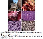
    Figure 1: Morphological features of thymic lymphoma.
    a. Enlargement due to tumor development in submandibular area, primary tumor (arrow) and submandibular lymph nodes(arrow heads).
    b. The cut surfaces of tumoral lymph nodes and primary tumor in cervical region.
    c. Thymic tumor and mediastinal lymph nodes.
    d. Dense lymphocytic proliferations divided by thin fibrous septa.
    e. The lymphocytes had intermediate nuclear size, dispersed chromatin, indistinct nucleoli, and minimal cytoplasm; T-cell lymphoblastic lymphoma.
    f. CD20 immunoreactivity in tumor cells.

    Histopathological and Immunohistochemical Studies: Tissue samples collected during necropsy were fixed in a solution of formalin, processed routinely, and stained with hematoxylin and eosin (H-E) for light microscopic examination. Immunohistochemistry (IHC) was also performed using an autostainer (Dako Autostainer Universal Staining System; Dako, Carpinteria, CA) following standard procedures. BDTM Retrieval A solution was used for high-temperature antigen retrieval, and methanol containing 3.0% H2O2 was used to block endogenous peroxidase activity. Mouse monoclonal against human CD3 and rabbit polyclonal against human CD20 (Dako M7051 and A0452) were used as primary antihuman antibodies and applied for 30 minutes at room temperature.

    Microscopic examination of the tissue samples revealed a diffuse population of neoplastic cells with a polymorphic morphology and of lymphoid origin. These cells formed sheets, and were mainly composed of lymphocytes. Thin, irregular collagenous septums were present within the tumoral mass (Figure 1d). The cells had an irregular nucleus, anisokaryosis, fine chromatin, a marked nucleolus, and a scant cytoplasm (Figure 1e). The cells had a high nucleus/cytoplasm ratio, with frequent mitotic cells and apoptotic bodies. Lymphoblastic cells were also observe.

    The immunohistochemical staining results indicated that the tumor cells were positive for CD20 (Figure 1f), and negative for CD3.

    Molecular Studies: Nested PCR procedure for detecting enzootic bovine leukosis virus (EBLV) was performed as previously described (6).

    DNA Extraction and PCR: The template DNA was extracted from mediastinal lymph nodes using the DNA/RNA purification kit (Zymo-research, CA, USA) according to the manufacturer’s instructions First and the second PCR steps were performed using 2X PCR master mix (Thermo Fisher, MA, USA) with env5032/5608r external primers and env5099/5521r internal primers, respectively. The amplification procedure for both PCR steps was performed as follows: 40 cycles, denaturation at 94 °C for 30 sec, annealing at 62 °C for 30 sec, and elongation at 72 °C for 1 min to amplify 598 and 444 bp products. Glyceraldehyde 3-phosphate dehydrogenase (GAPDH) housekeeping primers were used as an extraction control7. A blank reaction consisting of primers but no DNA template was included to serve as a reagent control.

    PCR results, with env5032/5608r and env5099/5521r, showed occurrence of no amplicons. Similarly, no amplicon was detected in the negative control. Approximately 700 bp product was obtained with the GAPDH primers. Therefore, the tissue samples were negative for EBLV.

  • Başa Dön
  • Özet
  • Giriş
  • Olgu Sunusu
  • Tartışma
  • Kaynaklar
  • Tartışma
    This case report describes a rare case of thymic T-cell lymphosarcoma in an Akkaraman sheep. The sheep was presented for necropsy due to a two-week history of weakness, loss of appetite, weight loss, and submandibular swelling. Gross examination showed tumoral masses in the thymus extending anteriorly to the submandibular region and posteriorly to the mediastinal lymph nodes. Histopathological and immunohistochemical analyses confirmed the presence of neoplastic T-cell lymphocytes in the thymus and lymph nodes. PCR analysis showed that the tumor was not associated with BLV.

    Amongst the ruminants, thymic lymphosarcoma is the most common in cattle8,9. Thymic tumors are sometimes associated with BLV infection in cattle and sheep9. In cats, 4 out of 8 thymic lymphosarcoma cases were also positive for feline leukaemia virus10. Although metastatic thymic lymphoma cases have been reported in cattle with the involvement of intestineum, brain9, liver, intestine, heart, and kidney10, the only involvement in the present report was submandibular and mediastinal lymph nodes. Some authors consider thymic lymphosarcoma as a form of bovine sporadic lymphosarcoma. The exact cause of thymic lymphosarcoma is unknown, however familial thymic lymphosarcoma cases have been documented in the Holstein calves11,12. Similar to this finding, thymic lymphoma due to C57BL/6J p53 gene deficient mice was well-established experimentally13. Thymic lymphoma is a rare form of malignancy that arises from the thymus and can spread to other organs, including the mediastinum.

    Differentiating thymic lymphoma from thymoma is of paramount importance. Despite both having their origins in the thymus, they represent distinct tumor types, necessitating varied treatment modalities. Thymoma predominantly emerges from the thymic epithelial cells, often accompanied by variable amounts of benign lymphoid tissue. Conversely, thymic lymphoma, categorized as a lymphatic system cancer, originates from thymic lymphocytes1. Distinctions in cellular morphology, growth patterns, and various histological features serve as criteria for distinguishing between these two conditions. Collectively; thymic T-cell lymphoblastic lymphoma has been reported in an Akkaraman sheep not due to bovine lymphoma virus.

  • Başa Dön
  • Özet
  • Giriş
  • Olgu Sunusu
  • Tartışma
  • Kaynaklar
  • Kaynaklar

    1) Vali V, Kiepul M, Bienzle D. The hematopoietic system. In: Maxie MG. (Editor). Jubb, Kennedy and Palmer&Quot’s Pathology of Domestic Animals. 6th Edition, Philadelphia: Saunders 2016: 153-157.

    2) Vail DM, Thamm DH, Liptak JM. Hematopoietic Tumors. In: Withrow and MacEwen (Editor). Small Animal Clinical Oncology. 6th Edition, Philadelphia: Saunders, 2019: 688-772.

    3) Anderson LJ, Jarret WFH. Lymphosarcoma (leukemia) in cattle, sheep, and pigs in Great. Britain Cancer 1968; 22: 398-405.

    4) Johnstone AC, Manktelow BW. The pathology of spontaneously occurring malignant lymphoma in sheep. Veterinary Pathology 1978; 15: 301-312.

    5) Olson R, Kettmann A, Burny RK. Goat lymphosarcoma from bovine leukemia virus. Journal of the National Cancer Institute 1981; 67: 671-675.

    6) De Brun ML, Cosme B, Petersen M, et al. Development of a droplet digital PCR assay for quantification of the proviral load of bovine leukemia virus. J Vet Diagn Invest 2022; 34: 439-447.

    7) Victor RM, Bicalho JM, Andrade MB, et al. Molecular detection of Feline Leukemia Virus in oral, conjunctival, and rectal mucosae provides results comparable to detection in blood. J Clin Microbiol 2020; 28: e01233-19.

    8) Licursi M, Inoshima Y, Wu D, et al. Genetic heterogeneity among bovine leukemia virusgenotypes and its relation to humoral responses in hosts. Virus Research 2002; 86: 101-110.

    9) Angel KL, Stott J, Tyler JW et al. Metastatic thymic lymphosarcoma in a calf. Journal of the American Veterinary Medical Association 1991; 198: 1771-1773.

    10) Tawfeeq MM, Miura S, Sugimoto K, et al. Thymic lymphosarcoma with brain involvement in a Holstein heifer. Journal Veterinary Medical Science 2012; 74: 1501-1504.

    11) Day MJ. Review of thymic pathology in 30 cats and 36 dogs. Journal Small Animal Practice 1997; 38: 393-403.

    12) Parodi AL, DaCosta B, Djilali S, et al. Preliminary report of familial thymic lymphosarcoma in Holstein calves. The Veterinary Record 1989; 125: 350-353.

    13) Liu S, Lyu J, Li Q, et al. Generationof a uniform thymic malignant lymphoma model with C57BL/6J p53 gene deficient mice. Journal Toxicol Patholgy 2022; 35: 25-36.

  • Başa Dön
  • Özet
  • Giriş
  • Olgu Sunusu
  • Tartışma
  • Kaynaklar
  • [ Başa Dön ] [ Özet ] [ PDF ] [ Benzer Makaleler ] [ Yazara E-Posta ] [ Editöre E-Posta ]
    [ Ana Sayfa | Editörler | Danışma Kurulu | Dergi Hakkında | İçindekiler | Arşiv | Yayın Arama | Yazarlara Bilgi | E-Posta ]