
Malathion Kaynaklı Mide Toksisitesinde Rutinin Koruyucu Etkisi: Oksidatif Stres, Enflamasyon ve Apoptozun Değerlendirilmesi
Volume 37, Issue 2
June 2023 · Pages 139-145
Malathion Kaynaklı Mide Toksisitesinde Rutinin Koruyucu Etkisi: Oksidatif Stres, Enflamasyon ve Apoptozun Değerlendirilmesi
Protective Effect of Rutin on Malathion-induced Gastric Toxicity: Evaluation of Oxidative Stress, Inflammation and Apoptosis
Mustafa İLERİTÜRK1, Özge KANDEMİR2
2Aksaray University, Aksaray Technical Sciences Vocational School, Food Processing Division, Aksaray, TÜRKİYE
Malathion (MLT), tarım, veterinerlik, tıp ve halk sağlığı uygulamalarında insektisit olarak yaygın şekilde kullanılmaktadır. MLT cilt, solunum ve gastrointestinal sistem yoluyla vücut tarafından alınır. Rutin (RUT), düşük toksisiteye ve antioksidan özelliklere sahip, narenciye meyvelerinden izole edilen güçlü, doğal olarak oluşan bir antioksidan moleküldür. Bu çalışmanın amacı MLT ile oluşturulan mide toksisitesinde RUT’un koruyucu etkilerini araştırmaktır. Bu amaçla ratlar her grupta 7 hayvan olacak şekilde rastgele 5 gruba ayrıldı. Ratlar 28 gün boyunca oral olarak 100 mg/kg dozda MLT ve 50 mg/kg ve 100 mg/kg dozda RUT uygulandı. Elde edilen sonuçlara göre MLT, malondialdehit (MDA) seviyesini artırarak lipid peroksidasyona neden olurken aynı zamanda süperoksit dismutaz (SOD), katalaz (KAT) ve glutatyon peroksidaz (GPx) enzim aktivitelerini azaltarak ve glutatyon (GSH) düzeylerini düşürerek antioksidan kapasiteyi baskıladı. RUT uygulaması ise lipid peroksidasyonun şiddetini azalttı ve antioksidan kapasitenin yükselmesine dene oldu. Buna ek olarak, MLT uygulanan ratlarda nuclear factor kappa B (NF-κB), tumor necrosis factor-alpha (TNFα), cyclooxygenase-2 (COX-2) ve inducible nitric oxide synthase (iNOS) mRNA transkript seviyeleri artmasıyla inflamasyonun şekillendiği, RUT uygulaması ile doza bağımlı olarak inflamasyonun şiddetinin azaldığı tespit edildi. Ölçülen panellerden bir diğeri olan apoptoz değerlendirildiğinde, MLT ile indüklenen mide toksisitesi sonucunda Bax seviyesi artarken Bcl-2’nin azaldığı belirlendi. Bunula beraber RUT uygulaması ile bu parametrelerin ters etki göstererek kontrol grubu değerlerine yaklaştığı tespit edildi. Tüm sonuçlar değerlendirildiğinde RUT uygulamasının MLT’nin neden olduğu mide toksisitesi üzerine koruyucu etkileri olduğu söylenebilir.
Malathion (MLT) is widely used as an insecticide in agricultural, veterinary, medicinal, and public health applications. MLT is taken up by the body through the skin, respiratory, and gastrointestinal systems. Rutin (RUT) is a powerful, naturally occurring antioxidant molecule isolated from citrus fruits with low toxicity and powerful anti-oxidant capabilities. The aim of this study was to investigate the protective effects of RUT against MLT-induced gastric toxicity. For this purpose, rats were randomly divided into 5 groups, with 7 animals in each group. Rats were administered orally 100 mg/kg MLT and 50 mg/kg and 100 mg/kg RUT for 28 days. According to the results obtained, while MLT caused lipid peroxidation by increasing malondialdehyde (MDA) levels, it also suppressed antioxidant capacity by decreasing superoxide dismutase (SOD), catalase (CAT), and glutathione peroxidase (GPx) enzyme activities and lowering glutathione (GSH) levels. RUT application decreased the severity of lipid peroxidation and tried to increase antioxidant capacity. In addition, in rats administered MLT, it was determined that inflammation was caused by the increase in nuclear factor kappa B (NF-κB), tumor necrosis factor-alpha (TNFα), cyclooxygenase-2 (COX-2), and inducible nitric oxide synthase (iNOS) mRNA transcript levels, and that the severity of inflammation decreased in a dose-dependent manner with RUT application. When apoptosis, another measured panel, was evaluated, it was determined that the Bax level increased while Bcl-2 decreased as a result of toxicity induced by MLT. However, with the RUT application, it was determined that these parameters had the opposite effect and approached the control group values. When all the results are evaluated, it can be said that RUT application has protective effects on MLT-induced gastric toxicity.
Introduction
Pesticides are widely used in agriculture to control pests [1]. In addition to agricultural uses, pesticides are also used to combat mosquitoes and weeds in suburban and rural homes and gardens. Unfortunately, pesticides that remain in the soil for a long time cause water, soil, and air pollution and disrupt the ecological balance. Pesticides induce acute poisoning in humans as well as animals by entering the body through the food chain and causing damage to biological systems through the accumulation of degradation products in tissues and organs over time [2].Malathion (MLT) is an organophosphate insecticide widely used in agriculture and public health programs to control insects [3]. Despite its effectiveness in pest control, MLT exposure has been associated with various toxic effects, including gastric toxicity that can lead to a range of gastrointestinal symptoms and other health problems [4].
Pesticide-induced gastric toxicity is characterized by oxidative stress, inflammation, and apoptosis, which can result in severe gastric damage [5]. The exact mechanisms underlying MLT-induced gastric toxicity are not fully understood. However, it is believed that MLT exposure can lead to the inhibition of acetylcholinesterase (AChE), an enzyme that is essential for the proper functioning of the nervous system [6]. This inhibition can lead to an overstimulation of the parasympathetic nervous system, which can cause a range of symptoms including nausea, vomiting, and abdominal pain. Additionally, pesticide exposure can lead to the generation of reactive oxygen species (ROS) in the gastric mucosa, which can cause oxidative stress and tissue damage [7]. This damage can lead to the disruption of the gastric barrier function and the release of inflammatory cytokines, which can further exacerbate the symptoms of MLT-induced gastric toxicity.
Oxidative stress is a state of imbalance between the production of ROS and the antioxidant defense system, leading to oxidative damage to cellular macromolecules [8-11]. Pesticide-induced oxidative stress has been shown to play a crucial role in various toxicity [12,13]. ROS generated by toxic agent exposure can cause lipid peroxidation, protein oxidation, and DNA damage, leading to cell death and tissue injury [14-16]. Inflammation is another critical factor in MLT-induced toxicity [17]. MLT exposure can trigger the activation of inflammatory cells, leading to the production of pro-inflammatory cytokines, such as tumor necrosis factor-alpha (TNF-α), nuclear factor kappa B (NF-κB), and inducible nitric oxide synthase (iNOS) [18]. These cytokines can further promote the infiltration of immune cells into the gastric mucosa, exacerbating tissue damage and inflammation. Apoptosis, a programmed cell death process, is also involved in pesticide-induced toxicity [19]. Pesticide exposure can induce the activation of apoptotic pathways in gastric epithelial cells, leading to cell death and tissue damage [20]. The apoptotic process can be triggered by various mechanisms, such as ROS-mediated mitochondrial dysfunction, caspase activation, and death receptor signaling [21,22]. Previous research has shown that natural antioxidants decrease ROS generation and tissue damage caused by substances with comparable toxic effects to MLT
Flavonoids are polyphenolic secondary metabolites of natural origin that contain numerous hydroxyl/phenolic groups and have potent antioxidant effects [25,26]. Even after several years of research, the cellular mechanisms of their biological actions are not yet entirely known. Examining the studies reveals that flavonoids prevent the formation of ROS, increase the antioxidant enzymes of cells by protecting lipophilic antioxidants, prevent redox reactions by creating metal chelation, and have important effects such as being an inhibitor of xanthine oxidase and NADPH oxidase enzymes [27,28]. Rutin (RUT, quercetin-3-o-rutinoside), an essential flavonoid found in citrus fruits, is a potent superoxide radical scavenger with antioxidant, anti-inflammatory, anti-ulcer, anti-allergic, anti-carcinogenic, and anti-mutogenic activities [29,30]. The routinose molecule and four hydroxyl groups in its structure play a significant role in its biological activities, according to structural analyses. In addition, literature indicates that the presence of rutinose increases the number of active sites, thereby making the molecule more effective [31]. RUTis also regarded as a nontoxic compound that may have potential in biomedicine [32].
The goal of this study was to determine whether RUT is effective and how it impacts gastric tissue damage caused by MLT administration.
Chemicals: Rutin, MLT, and all other chemicals used in the analyses were of analytical quality and purchased from Sigma Chemical Co. (St. Louis, USA).
Experimental Animals: In the study, 35 male Sprague-Dawley rats aged 8 to 10 weeks and weighing 220 to 250 g were employed. Rats were purchased from the Ataturk University Medical Experimental Application and Research Center (Erzurum, Türkiye). The rats were adapted to the environment for 1 week before the application. The ambient temperature was 24±1 °C, and the humidity was 45±5%. A 12-hour light/dark cycle was also incorporated into the rats' environment. Rats were fed standard pellet diet and ad libitum tap water.
Experimental Procedure: Rats were divided into five distinct groups, with seven rats in each. Doses of MLT and RUT given to animals were determined to be referenced according to prospective studies by Gur and Kandemir (3) and Kandemir et al. (9). As illustrated in Table 1, the experimental design contains a control and four experimental groups.
The rats were decapitated under mild sevoflurane anesthesia 24 hours after the last RUT administration (day 29) and their gastric tissues were collected, rinsed with physiological saline, and frozen at -80 °C until biochemical analysis.
Preparation of Tissue Homogenates: The tissues were diluted 1:20 (w/v) with phosphate-buffered saline (PBS; pH 7.4) to generate the gastric tissue homogenate. The resulting mixture was rapidly homogenized with a tissue lysate device (TissueLyser II, Qiagen). The homogenate was centrifuged for 30 minutes at +4 °C and 3000 rpm. The resulting supernatant was used for the analysis.
Determination of Lipid Peroxidation and Antioxidant Enzyme Activities in Gastric Tissue: The level of lipid peroxidation was determined by measuring the amount of malondialdehyde (MDA) using the method established by Placer et al. [33] and expressed as nmol/g tissue. Superoxide dismutase (SOD) activity was determined using a method developed by Sun et al. [34]. The findings are presented as U/g protein. The catalase (CAT) activity was measured using a method established by Aebi [35], and the data were expressed as catal/g protein. Glutathione peroxidase (GPx) activity was assessed using a technique created by Lawrence and Burk [36]. Results were reported as U/g protein. GSH levels were determined using the method described by Sedlak and Lindsay [37], and the findings were expressed as nmol/g tissue. The analysis of total protein was performed using the method established by Lowry et al. [38] using bovine serum albumin (BSA) as the reference.
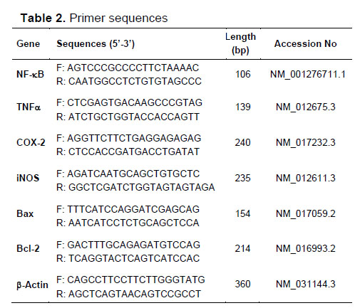
Real-Time PCR Analysis: Relative mRNA transcript levels of Bcl-2-associated X protein (Bax), B-cell lymphoma 2 (Bcl-2), cyclooxygenase-2 (COX-2), iNOS, NF-κB and TNFα genes in gastric tissues were analyzed by RT-PCR method. The first step was isolating total RNA from tissues using QIAzol Lysis Reagent (79306; Qiagen). NanoDrop (BioTek Epoch) was used to determine the amount of RNA obtained at the end of the processes. According to the results obtained, total RNA concentrations of all samples were equalized to 1000 ng/mL. At this stage, all procedures were performed in accordance with the manufacturer's instructions. After that, cDNA synthesis was performed using the iScript cDNA Synthesis Kit on the total RNAs collected (Bio-Rad). This procedure was carried out in accordance with the manufacturer's instructions, and the following reaction conditions were established:
1. Priming: 5 minutes at 25 °C
2. Reverse transcription: 20 minutes at 46 °C
3. RT inactivation: 1 minute at 95 °C
In the last step, cDNAs from all groups were mixed with iTaq Universal SYBR Green Supermix (BIORAD) and reaction conditions were set up in a Rotor-Gene Q (Qiagen) device according to the manufacturer's instructions. Primer sequences have been presented in Table 2. Reaction conditions:
1. Polymerase activation and DNA denaturation: 95 °C for 30 seconds,
2. Denaturation: 95 °C for 5 seconds (40 cycles),
3. Annealing/Elongation and Plate Reading: 60 °C for 30 seconds (40 cycles),
4. Melt Curve Analysis: 65-95 °C 2-5 seconds per increase of 0.5 °C
CT values were obtained from the device after the cycle was over. These data were standardized in accordance with β-Actin. According to the 2-ΔΔCT method developed by Livak and Schmittgen [39], normalization was performed.
Statistical Analysis: Shapiro-Wilk test was done to evaluate the samples as the sample number was under 50. Because of the distribution of the data is normal and the number of groups is more than 2, comparison of differences between groups were performed using one-way analysis variance (ANOVA) and Tukey’s multiple comparison test via SPSS software (version 20.0; SPSS, Chicago, IL). Results were shown as mean ± standard deviation. Statistical significance was defined as a P value <0.05.
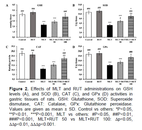
Effect of RUT on Enzymatic and Non-Enzymatic Antioxidant Markers in MLT-Induced Gastric Toxicity: In the study, it was determined that MLT reduced the activity of antioxidant enzymes relative to the control group (P<0.001). Furthermore, the levels of GSH, an essential antioxidant in the body, decreased by 46% in response to MLT therapy (P<0.001). This indicates that MLT causes in oxidative stress in gastric tissue. In contrary, GSH levels increased and SOD, CAT, and GPx activities approached those of the control group in the MLT- and RUT-treated groups compared to the MLT group (P<0.001). In addition, there was a statistically significant difference between the 50 mg/kg and 100 mg/kg doses of RUT for GSH, SOD, CAT, and GPx (P<0.05, P<0.01, P<0.01, P<0.05, respectively). Figure 2 presents the data for enzymatic and non-enzymatic indicators in gastric tissue.
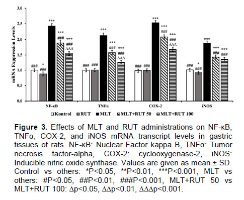
Effect of RUT on Inflammation in MLT-Induced Gastric Toxicity: Real-time PCR was used to determine the NF-κB, TNFα, COX-2, and iNOS mRNA transcript levels in gastric tissue to determine the inflammation. Analysis of mRNA transcripts revealed increased NF-κB, TNFα, COX-2 and iNOS (2.4-, 2.1-, 2.5-, and 1.9-fold, respectively) in the MLT group compared to the control group (P<0.001 for all). The levels of NF-κB, TNFα, COX-2, and iNOS were decreased in the RUT-treated group compared to the MLT-treated group (P<0.001 for both dosages). Figure 3 shows the mRNA transcript levels of NF-κB, TNFα, COX-2, and iNOS.
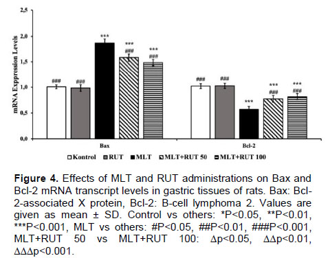
Effect of RUT on Apoptosis in MLT-Induced Gastric Toxicity: The degree of apoptosis in gastric tissue was determined by analyzing mRNA transcript levels of Bax and Bcl-2. Proapoptotic factor Bax levels were 1.9-fold higher in the MLT group than in the control group, according to the results of mRNA transcripts (P<0.001), whereas the levels of the anti-apoptotic factor Bcl-2 were lower (P<0.001) (Figure 4). With RUT co-administration, Bax transcript levels decreased compared to MLT alone, while Bcl-2 transcript levels increased slightly (P<0.001).
Enzymes such as SOD, CAT, and GPx constitute the first line of defense in preventing damage to reactive oxygen species. Whereas SOD catalyzes the conversion of superoxide radical to extremely deadly hydrogen peroxide and normal molecular oxygen, which may cause permanent cell damage, CAT and GPx break down hydrogen peroxide into water and molecular oxygen [43]. GSH is a nonenzymatic antioxidant that protects cells from the damaging effects of ROS such as free radicals, peroxides, and heavy metals. By binding free radicals, GSH may become oxidized (GSSG), and the GSSG/GSH ratio is a significant indicator of oxidative stress [44]. While MDA is a significant signal of oxidative stress, it is also an indicator of oxidative damage to membrane lipids, which corresponds strongly with the degree of lipid peroxidation [45]. Previous study shown that MLT inhibits antioxidant enzyme activity and induces lipid peroxidation and oxidative stress [46]. This research was conducted to explore the protective and beneficial effects of RUT on MLT-induced gastric toxicity. The research found that MLT induced an increase in oxidative stress and a reduction in the antioxidant system in the stomach tissue. On the other hand, it was revealed that RUT decreased stomach damage by decreasing the efficacy of free radicals by activating the antioxidant system. Similar to our findings, Celik et al. [47] showed that RUT protects cells from oxidative damage by enhancing the activity of antioxidant enzymes. It was revealed that MLT generated oxidative damage by causing an inverse association between SOD, CAT, and GPx enzyme activity and MDA levels in gastric tissues. On the other hand, it was discovered that RUT inhibits the worsening of the oxidant-antioxidant equilibrium by scavenging free radicals, resulting in a decline in MDA levels by restoring antioxidant enzyme activity and replenishing GSH reserves.
NF-κB, a transcription factor, is essential for the activation of proinflammatory cytokines and the initiation of the immune response in several organs [48,49]. In a previous study, it was discovered that MLT promotes inflammation in tissues via triggering NF-κB activation [50]. Experimental research indicated that OPs compounds can directly stimulate the release of pro-inflammatory cytokines. Rats treated with MAL demonstrated an increase in pro-inflammatory cytokines such as NF-κB and TNFα [51,52]. In the current research, it was established that MLT elevated NF-κB transcript levels in gastric tissues, causing oxidative stress and stomach damage; RUT therapy administered in conjunction with MLT reduced NF-κB transcript levels and gastric toxicity. NF-κB activates the TNFα, a key pro-inflammatory cytokine implicated in inflammation [53]. Preventing the translocation of NF-κB to the nucleus reduces the production of the COX-2 proinflammatory mediator. In the current research, an increase in TNFα and COX-2 transcript levels was found, and it was hypothesized that this was owing to an increase in NF-κB gene expressions caused by MLT-induced ROS generation. Arowoogun et al. [54] achieved comparable results to ours and found that RUT decreased COX-2 release.
Apoptosis is a natural mechanism of cell death [55]. Yet, when it develops uncontrollably, tissue function is lost. Apoptotic factor Bax and anti-apoptotic factor Bcl-2 play crucial roles in the apoptotic process and are commonly used biomarkers for determining apoptotic state [56]. Previous research has demonstrated that MLT activates apoptosis and enhances cell death [57,58]. MLT treatment resulted in increased Bax and reduced Bcl-2 expression in our research. On the other hand, it has been discovered that RUT may reduce apoptosis by inhibiting Bax expression and enhancing Bcl-2 expression. Similarly, a recent research found that Bax expression reduced after MLT therapy and considerably increased following RUT administration [6].
Thus, it was concluded that the use of RUT as a supportive treatment in MLT toxicity would be beneficial due to its anti-oxidant, anti-inflammatory, and anti-apoptotic effects in gastric toxicity caused by MLT, thereby reducing ROS formation and healing tissue and organ damage.
Material and Method
Research and Publication Ethics: All animal experiments were carried out at the Ataturk University Medical Experimental Research Center. The Ataturk University Animal Experiments Local Ethics Committee authorized this work (approval number: 2022–11/224).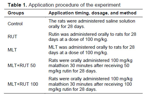
Results
Effect of RUT on Lipid Peroxidation in Malathion-Induced Gastric Toxicity: Figure 1 shows the effects of RUT against MLT-induced lipid peroxidation in gastric tissue. MLT increased MDA levels, an essential biomarker of lipid peroxidation by about 1.7-fold compared to the control group (P<0.001). MLT-induced lipid peroxidation levels decreased with RUT administration (15% in the MLT+RUT 50 group and 25% in the MLT+RUT 100 group) and approached control group levels (P<0.01).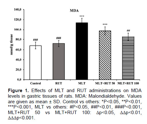
Discussion
Many studies have shown that organophosphate insecticides are among the most dangerous environmental contaminants that constitute a risk to human health. Organophosphates (OPs) are taken into the body through the respiratory system, the digestive system, and the skin. By blocking AChE in the neurological system, OPs mostly lead to respiratory and neuromuscular conduction problems [40]. MLT has entered these harmful substances as one of the environmental contaminants that represent a danger to human health. While several research have been conducted on MLT toxicity in the past, no viable therapy has yet been identified [41,42]. In this investigation, the effects of RUT against the gastric toxicity of MLT were studied, and the acquired results indicated that RUT was a potential agent against this toxicity.References
- RATLARDA BAZI NİTROSOAMİNLERİN DÜŞÜK MİKTARDA-UZUN SÜRELİ VERİLMESİNİN KAN, KARACİĞER VE BÖBREKLERDE OKSİDATİF STRES ÜZERİNE ETKİLERİ
- Paraquat İle Oluşturulmuş Oksidatif Stresin HepG2 Hücrelerinde Apoptozis Üzerine Etkisinin Araştırılması
- Geçiş Dönemindeki İneklerde Stres Parametreleri Üzerine Mineral Uygulamasının Etkileri
- Yumurtlama Öncesi Dönemde Aydınlatma Programında Yapılan Değişikliğin Etlik Piliç Ebeveynlerinde Bazı Stres Parametrelerine Etkisi
- Oral Olarak Verilen Panax ginseng ve Panax ginseng + Ganoderma lucidum Ekstraktlarının Hamsterlardaki Oksidatif Stres, Lipit Profili ve Karaciğer Protein Ekspresyonları Üzerine Etkileri