
Ratlarda Yüzeyel Venlerin Yakın Kızılötesi Görüntüleme Teknolojisi ve Lateks Görüntülenme Metodları Kullanılarak İncelenmesi
Cilt 38, Sayı 1
Ocak 2024 · Sayfalar 014-020
Ratlarda Yüzeyel Venlerin Yakın Kızılötesi Görüntüleme Teknolojisi ve Lateks Görüntülenme Metodları Kullanılarak İncelenmesi
Near Infrared Imaging Technology and Latex Imaging of Superficial Veins in Rats Investigation using Methods
Funda AKSÜNGER KARAAVCI1, Meryem KARAN2
2Firat University, Faculty of Veterinary Medicine, Department of Anatomy, Elazig, TÜRKİYE
Yakın kızılötesi ışın teknolojisne sahip cihazlar, derinin 8–15 milimetre (mm) altında periferik venlerin, gerçek zamanlı bir görüntüsünü oluşturur. Bu çalışmada 15 adet Wistar Albino cinsi rat kullanıldı. Çalışmanın amacı, damar görüntüleme cihazının (Accuvein av 400) ratlarda torasik ve pelvik ekstremite yüzeysel venlerinin görüntülenmesinde kullanılabilirliğini ortaya çıkarmaktır. İlk olarak materyallerin ekstremitelerinin ven görüntülemesi yapılmış ve bulgular kayıt altına alınmıştır. Daha sonra aynı materyallere lateks enjeksiyon yöntemi uygulanarak materyaller diseke edildi ve sonuçlar karşılaştırmalı olarak değerlendirilmiştir. Ratlarda v. cephalica'nın iki dal halinde başladığı, birinci dalın boyun uzunluğunun orta 1/3'ünde, ikinci dalın ise göğüs boşluğu girişinde v. jugularis externa’dan çıktığı saptandı. Regio colli'de v. cephalica'nın seyri ven görüntüleme cihazı ile izlenemezken, regio brachii'den itibaren görüntüleme mümkün oldu. Pelvik extremite yüzeysel venleri olan v. saphena medialis et lateralis'in seyirleri boyunca cihaz ile görüntülendiği görüldü. Ratlar üzerinde deneysel olarak yürütülen bu araştırmanın bulguları göz önüne alındığında, cihazın hayvanlarda yüzeysel venleri görüntülemede alternatif bir yöntem olarak kullanılabiliniceği düşünülmektedir. Ek morfolojik ve klinik çalışmalar, tekniğin hayvanlar üzerindeki etkinliğini gösterebilir.
Near-infrared imaging technology devices create a real-time image of the 8–15 millimeters (mm) peripheral veins under the skin. In this study, 15 Wistar Albino rats were used. Aim of this study was to reveal whether the vein-viewer device (Accuvein av 400) can visualize thoracic and pelvic limb superficial veins in rats. Firstly, vein viewer imaging of the limbs of the materials was performed and the findings were recorded. Afterwards, the materials were dissected by applying the latex injection method to the same animals and the results were evaluated comparatively. In rats, it was found that vena cephalica starts as two branches, the first branch is in the middle 1/3 of the neck length and the second branch originates from v. jugularis externa at the entrance to the thoracic cavity. While the run of vena cephalica in regio colli could not be viewed with the vein viewer device, it was possible to view it down from the regio brachii. It was observed that v. saphena medialis, and v. saphena lateralis which are the superficial veins of the pelvic limb, viewed with the vein viewer device throughout their run. Given the findings of this research this device may be considered as an alternative method for imaging superficial veins in animals. Additional morphological and clinical studies could show the techniques efficacy in animals.
Giriş
Rats are used in the sceintifi studies of various diseases, as well as in many vaccine-drug trials, anatomical research, and operative studies in both medicine and veterinary medicine. Although its anatomical features should be among the most well-known species, the literature review revealed a dearth of information regarding the circulatory system[1-3].Extremity veins are superficial and profound and blood flow is from superficial veins to profound veins. Superficial veins usually lie subcutaneously just under the skin, and because of their location, they are often preferred for intravenous injections in clinical settings[3-6].
Vena (V.) cephalica is commonly originates from v. jugularis externa in domestic mammals[7-11]. The vein usually joins with v. brachialis through mediana cubiti and also with v. mediana in dogs and rabbits[11-13]. It gives v. cephalica accessoria on the flexor surface of articulation (art.) cubiti[12,14,15]. Vena cephalica accessoria originates from v. cephalica medial to musculus (m.) extensor carpi radialis and the skin[10,16-19].
Vena iliaca interna leaves cavum pelvis with nervus (n.) ischiadicus from incisura (inc.) ischiadica major, and the continuation vein is called v. ischiadica[20]. The continuation of vena ischiadica in the ventral direction is v. saphena lateralis[3,12,20].
Colored fillers are often preferred in vascular anatomy studies[10,20-24]. The fillers used should not dissolve in water, should be resistant to acids and alkalis, and should be mixed homogeneously with some paints. Moreover, their chemical and physical properties should be standard, they should not stain extravascular tissues, and they should be easy to obtain[25]. Latex is most preferred as a filler[26-28].
The branches of the v. iliaca externa and the v. iliaca interna provide venous drainage of the pelvic limb. Vena femoralis, canalis femoralis separate from v. iliaca externa. Vena saphena medialis, one of the superficial veins of the pelvic limb, is separated from the v. femoralis[3,5,12,19].
The vein viewer devices are new systems which uses reflective near-infrared imaging (NIR) technology to create a simultaneous image of veins 8 to 15 millimeters (mm) deep in the skin and then project the image back onto the skin[29,30]. The working mechanism of this system is based on showing veins by reflecting on the skin with the help of infrared rays[31]. The 700-1000 nanometers (nm) wavelength ray makes direct contact with the skin of living organisms. Although this wavelength exceeds the absorption capacity of the skin and subcutaneous tissues, it is absorbed by the veins, contingent upon the concentration of deoxyhemoglobin in the venous veins[32]. Thus, it can be observed that blood produces a comparatively darker image, whereas the skin and subcutaneous tissues exhibit a lighter appearance. The reflected image emanating from the living is transmitted to a video camera, and the device includes a filter to reduce extraneous light in the video camera. The computer system integrated into the device processes the infrared image, which is subsequently projected onto the skin of the living through the use of a green or red projector[30,33-35]. The utilization of this device does not involve the emission of ionizing radiation or the production of thermal energy that could potentially cause harm or injury to the skin. Furthermore, the emitted ray from the apparatus poses no threat to the eyesight of either the patient or the operator. The fact that the device has a movable apparatus with feet on which it can be fixed creates an important advantage because the person who will perform the operation can use both hands. The device is especially used for iv catheterization of venous veins located subcutaneously in human medicine[33-36].
With the reflective near-infrared imaging system with ray, it is possible to view veins with diameters less than 0.2 mm at a depth of 0.5-1 mm and to detect veins with relatively larger diameters (1-2 mm) to an average depth of 3-4 mm. However, there is a 0.06 mm margin of error in determining the localization of veins up to 8-15 mm deep in the vein viewer device. The smallest vein diameter that is detected in this system is 0.2 mm, and the largest vein diameter is 5.08 mm[37,38]. There is a negative correlation between vein depth and contrast[39,40].
This study aims to investigate the branches of the superficial veins of the thoracic and pelvic extremities in rats by the latex injection method andthe vein imaging device.
Gereç ve Yöntem
The ethics committee decision of the study was taken from Firat University Animal Experiments Local Ethics Committee with the protocol number 2019/60 on 10.04.2019. 15 Wistar Albino rats (male) were used in this study.Anesthesia was conducted by applying xylazine hydrochloride 10 mg/kg intraperitoneally (ip) + ketamine hydrochloride 100 mg/kg ip, and after shaving, all limbs were viewed with a vein viewer device (Accuvein AV 400)[41]. In order to prevent coagulation during latex application, 450 IU/20 g Na Heparin was given through v. coccygea mediana to the materials whose imaging was completed, and the animals laid on their backs on the table. Vena cava caudalis and aorta abdominalis were uncovered in the materials whose abdominal cavity was opened. A 26 G (purple color) catheter was placed in the aorta abdominalis in the caudal direction, and a 22 G (blue color) catheter was placed in v. cava caudalis in the cranial direction and exsanguin was performed. The veins were washed by injecting 0.9% NaCl through the catheter in the aorta abdominalis. This process was continued until clear colored liquid came from v. cava caudalis[16]. After this stage, latex injection was performed in accordance with the literature[19,20,42]. Cotton pads soaked with 5% acetic acid were used to stop the latex leaking from the ruptured veins during the procedure[43]. Dissection procedure, skin and vein diameter measurements and statistical analyzes were performed in accordance with the literature[44].
Nomina Anatomica 2017 was used to name the veins[45].
Bulgular
Due of the venous flow's path towards the heart, some researchers have described the veins as moving from the periphery to the center. According to some studies, the arteries connect the center to the periphery. The second method was used for this study because it was more convenient.
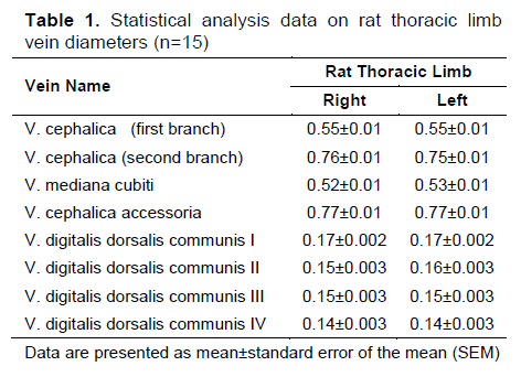
In rats (Figure 1-A/1, 1-A/2) v. cephalica began as two branches. The first branch emerged from v. jugularis externa in the middle 1/3 of the neck length and continued in the distal direction in the caudal of m. cleidobrachialis. The second branch, on the other hand, came out of v. jugularis externa at the entrance to the thoracic cavity and run first in m. pectoralis transversus and then in the cranial of m. cleidobrachialis, and its diameter was larger than the first branch (Table 1), (Figure 1-A/3). It was observed that these two branches merged at the border of the insertios of m.
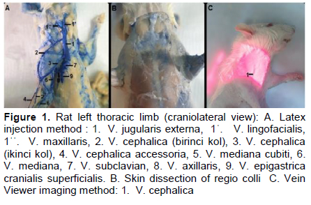
It was observed that vena cephalica gave v. cephalica accessoria in the flexor of art. cubiti (Figure 1-A/4) and that it run in the mediodistal direction and formed 2-3 connecting branches with v. mediana in the middle 1/3 of the antebrachium and that they run parallel to each other in the distal direction. The continuation of the vein was involved in the formation of the rete carpi dorsale.
Due to the adipose tissue and salivary glands located in the regio colli in both thoracic limb in rats, v. cephalica could not be viewed with the vein viewer device (Figure 1-B). In its run in the regio brachii, the vein was viewed under the skin thickness of 0.37±0.01 mm (right thoracic limb) and 0.36±0.01 (left thoracic limb) mm (Table 2) (Figure 1-C/1).
Vena cephalica accessoria originated from v. cephalica on the flexor surface of the elbow joint and run subcutaneously towards the distal. During this run, it was observed that it gave 1-2 branches to the rete carpi dorsale. While v. cephalica accessoria could not be viewed with the device at the point where it separated from v. cephalica in rats, it was clearly viewed under the skin thickness of 0.24±0.006 in the right thoracic limb and 0.27±0.014 (Table 2) in the left thoracic limb starting from the distal 1/3 level of the antebrachium. However, this image, which can be seen with the naked eye, could not be photographed with a camera or mobile phone to create a clear image.
It was determined that vena mediana cubiti originated from the second branch of v. cephalica in ten rats, and the vena mediana cubiti originated from the first branch of v. cephalica on the flexor surface of the art. cubiti in three rats and terminated in v. brachialis.
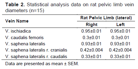
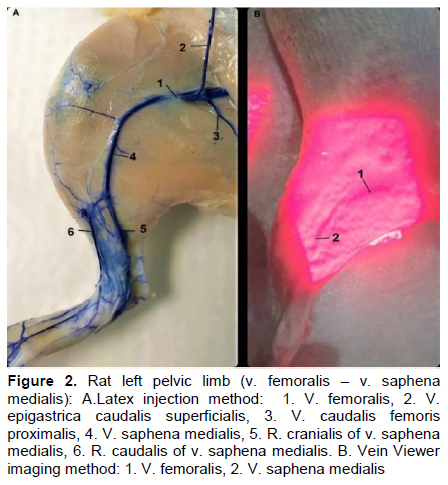
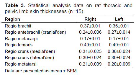
Vena femoralis was the distal continuation of v. iliaca externa after the ligamentum inguinale. Vena saphena medialis, which separated from the medial wall of the vena femoralis, was running in the distal direction together with the arteria saphena and n. saphenus (Figure 2-A/1, 2-A/4). Vena saphena medialis was viewed with the vein viewer device under the skin thickness of 0.31±0.025 mm in the right pelvic limb and 0.30±0.024 mm in the pelvic limb (Table 2).
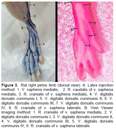
Ramus cranialis of vena saphena medialis gave many branches to the rete calcaneum. In rats, r. cranialis was involved in the formation of v. digitalis dorsalis communis I (Figure 3-A/4) by passing on the dorsal part of the foot at the level of the insertio of m. tibialis cranialis (Figure 3-A/2). Vena digitalis dorsalis communis I was on mm. extensor digitorum breves, just under the skin. Venae digitales dorsales propria running axially and abaxially was separating from this vein. The vein was viewed with a vein viewer along its run to distal starting from the level of separation from v. saphena medialis (Figure 3-B). However, it was determined that the branches going to the phalanxes could not be viewed.
Ramus caudalis of v. saphena medialis (Figure 2-A/5, 3-A/2) was running through the distal in the caudal of a. saphena, and just in front of the tendon calcaneus communis. The vein ended by dividing into v. plantaris lateralis and v. plantaris medialis at the level of art. tarsocralis. At the same time, it was observed that it gave many branches to the rete calcaneum during its course.
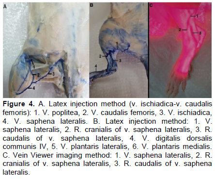
In rats, v. saphena lateralis (Figure 4-A/4, 4-B/1) had an average diameter of 0.93±0.02 mm (Table 2) and was formed by the union of v. ischiadica and v. caudalis femoris in the caudal of m. gastrocnemius. In five of the rats (Figure 4-B/1), it was determined that the vein showed a highly curved run. The presence of ramus anastomoticus cum v. saphena mediali extending between vena saphena lateralis and v. saphena medialis was not found. Vena saphena lateralis gave 1-3 branches to the fascia cruris during its run on m. gastocnemius lateralis. It was determined that immediately after these branches, the vein ended in the middle 1/3 of the crus by dividing into r. cranialis and r. caudalis (Figure
Ramus cranialis of v. saphena lateralis originated at the level of the middle of the crus and was located subcutaneously. It was observed that this vein run towards the distal at the border of the musculus tibialis cranialis and m. fibularis longus. It was observed that the continuation of the vein passed along the lateral border of the retinaculum extensorium proximale and passed to the dorsal part of the foot and continued as v. digitalis dorsalis communis IV (Figure 3-A/7, 4-A/4). The vein run was viewed with the vein viewer device from the point of separation from v. saphena lateralis (Figure 3-B/6, 4-B/2). The run of v. digitalis dorsalis communis IV (Figure 4-B/5), which is the continuation of the vein and given on the metatarsal bones, was visualized with the vein viewer device.
It was determined that ramus caudalis also separated from v. saphane lateralis at the middle level of the crus. This branch run at the border of m. soleus with the caput laterale of the musculus gastrocnemius, then proceeded to the distal in the craniolateral of tendo calcaneus communis and anastomosed with v. tarsea lateralis (Figure 4-B/2). The exit point of the ramus caudalis from v. saphena lateralis and its distal run were viewed with the vein viewer device (Figure 4-C/3).
No statistically significant difference was found between thoracic and pelvic limbs vein diameters in rats (Table 3).
It is stated that v. mediana cubiti, originating from v. cephalica at the level of articulatio cubiti, terminates in v. brachialis superficialis in carnivores, in the caudal vein of v. brachialis pair in pigs, and in v. brachialis in ruminants in mammals[3,7,12,14,42,46,47]. Bozkurt[22] states that v. cephalica joins with v. mediana through v. mediana cubiti in rabbits, while Özüdoğru[16] states that they did not find the presence of this vein in rabbits. In this study, the presence of v. mediana cubiti was detected in rats in accordance with the literature data.
Some researchers stated that v. cephalica merged with v. mediana in the palmar of the antebrachium in rats. In this study, it was observed that v. cephalica run in the mediodistal direction after giving v. cephalica accessoria in the flexor of art. cubiti, formed 2-3 connecting branches with v. mediana, and the two veins showed a parallel run towards the distal. In rats, there are differences in the literature data since v. cephalica does not merge with another vein on the palmar surface of the antebrachium[5,48].
It was stated that v. saphena medialis, which is one of the superficial veins of the pelvic limb, separated from the medial of v. femoralis in the distal direction and run together with a. saphena and n. saphenous in domestic mammals such as cattle, horses, cats and dogs[4,7,47,49], rats[5,50,51], and rabbits[17,18,52]. In the present study, the origin and run of v. saphena medialis were in parallel with the literature data[5,17,18,50,53].
It was reported that v. saphena medialis can be found in pairs in pigs[7,49], sheep and goats[54] and rabbits[18]. In this study, it was determined that all the rats had a single v. saphena medialis. Some researchers[18,50,53,55] reported that r. cranialis of v. saphena medialis gives many branches to the rete calcaneum in rats and rabbits. Greene[5] and Popesko et al[53] stated that in rats, the vein passed over the dorsal part of the foot and participated in the formation of v. digitalis dorsalis communis I. Some researchers[5,17,48,50,53,56] also stated that r. caudalis of vena saphena medialis ended by dividing into v. plantaris lateralis and v. plantaris medialis at the distal end of the crus in rats and rabbits, and that it gave many branches to rete calcaneum during its run. In the present study, the run of r. cranialis and r. caudalis of v. saphena medialis and the branches it gave are in parallel with the literature data[5,18,22,50,53].
Hebel and Stromberg[57] and Greene[5] stated that v. saphena lateralis originates from v. poplitea in rats. Popesko et al. [50] reported that the vein was formed by the union of v. ischiadica and v. caudalis femoris. In the present study, the data obtained on v. saphena lateralis in rats are in parallel with Popesko et al.’s finding.
It was reported that vena saphena lateralis divided into r. cranialis and r. caudalis at the mid-crus level in rats, r. cranialis gave vv. digitales dorsales II, III, IV on the dorsal part of the foot, and ramus caudalis divided into r. lateralis and r. medialis again[5]. The data obtained in our study about v. saphena lateralis and its branches are in parallel with the literature.
Vein viewer imaging method could not show that v. cephalica originated from v. jugularis externa in regio colli in rats. In the dissection, it was observed that the glandula mandibularis in this region was quite large and there was a dense fatty tissue around it. This is thought to be an obstacle to viewing with the vein viewer device. Imaging of vena cephalica with the vein viewer device showed parallelism with the latex findings down from regio brachii. While v. cephalica accessoria was detected in the distal 1/3 of the antebrachium with the device in rats, the entire run of the vein and the branches it gave to the dorsal foot could not be clearly viewed with the device. This is thought to be due to the fact that adipose tissue mass starting in the neck region progresses to the middle of the antebrachium.
One of the superficial veins of the pelvic limb, v. saphena medialis and its branches were in parallel with the latex findings of the vein with the device. Vena saphena lateralis, which is often preferred as an injection site, was also viewed with vein viewer, and the thin branches leading to the fascia cruris were also viewed. The viewing of vv. digitales dorsales communes, which is the continuation of the vein, was clearer than the thoracic limb.
It is stated that the vein viewer device is preferred in humans, especially during peripheral intravenous catheterization, in order to make the veins visible[58,59]. In this study, it was determined that especially the pelvic limb superficial veins in rats were successfully viewed and could be used for catheter applications.
The findings obtained in this study show that the superficial veins especially of the pelvic limbs in rats are clearly viewed with the vein viewer device. In the thoracic limb, v. cephalica accesoria, which is one of the preferred veins for intravenous interventions, was viewed with the device. The viewed vein runs show parallelism with the data obtained by the latex injection method. In the light of the data obtained, the use of the vein viewer device as an alternative method in viewing of superficial veins in animals. Due to this fact, it would be advantageous to carry out various studies by applying the vein viewer device in veterinary applications with additional research being planned after this point.
Tartışma
After originating from v. jugularis externa at the entrance of the thoracic cavity in domestic mammals other than cats, vena cephalica runs caudoventrally in the sulcus pectoralis lateralis between m. pectoralis descendens and m. brachiocephalicus[5,7,9,10,17,22]. In the present study, however, it was observed that v. cephalica was formed by the fusion of the middle 1/3 of v. jugularis externa and the two veins separating at the level of the thoracic cavity entrance at the level of the insertio of m. brachiocephalicus.References
- Determination of Arterial Oxygen Saturation in Newborn Lambs by Pulse Oximetry
- Comparative Isolation of Serum ß + Pre- ß Lipoproteins of Human and Various Domestic Animal Species By Heparin/MnCl2 Precipitation Method
- The Effects of Levamisole on the Pregnancy Rate, Avarage Litter Size, Sexuality, Living Rate and Lactation Period in Rats
- RATLARDA LEVAMİZOL’ÜN GEBE KALMA ORANI, YAVRU SAYISI, CİNSİYETİ, YAŞAMA ORANI VE LAKTASYON SÜRESİNE ETKİSİ
- Separation of Human and Various Animal Lipoproteins by Vertical Agarose Gel Electrophoresis