
Su Mandalarında (Bubalus Bubalis) Rickettsia aeschlimannii'nin varlığına dair İlk Moleküler Raporu; Ompa ve Ompb Genlerinin Filogenetik Analizlerine İlişkin Bir Not
Cilt 39, Sayı 1
Mart 2025 · Sayfalar 054-060
Su Mandalarında (Bubalus Bubalis) Rickettsia aeschlimannii'nin varlığına dair İlk Moleküler Raporu; Ompa ve Ompb Genlerinin Filogenetik Analizlerine İlişkin Bir Not
First Molecular Report of Rickettsia aeschlimannii in Water Buffalo (Bubalus Bubalis); with a Note of Phylogenetic Analyses of Ompa and Ompb Genes
Ömer Faruk ŞAHİN1, Ufuk EROL1, Kürşat ALTAY1
Son yıllarda vektör kaynaklı patojenlerin önemi artmış olup, moleküler çalışmalar dünyanın farklı bölgelerindeki yeni konaklarda bu patojenlerin varlığını ortaya koymuştur. Türkiye, vektör kaynaklı patojenlerin farklı konaklarda bildirildiği önemli ülkelerden biridir. Sığır, koyun ve keçilerde vektör kaynaklı patojenleri araştıran birçok çalışma olmasına rağmen, bu patojenlerin mandalardaki varlığı ve dağılımı hakkındaki veriler yetersizdir. Bu çalışmada, Türkiye'de ilk kez mandalarda Rickettsia türlerinin moleküler yöntemlerle araştırılması amaçlanmıştır. Bu amaçla, Sivas ilinde yedi ayrı bölgeden alınan 364 manda kan örneği, soy-spesifik PCR yöntemi kullanılarak Rickettsia türleri açısından araştırılmıştır. Ayrıca pozitif örnekteki ompA ve ompB genlerinin kısmi parçaları tür teşhisi ve filogenetik analizler yapmak amacıyla sekanslanmıştır. Çalışmada tespit edilen örneğin DNA dizi analizi sonuçları Rickettsia aeschlimannii ile enfekte olduğunu göstermiştir. Elde edilen ompA ve ompB genlerine ait DNA dizileri GenBank'a yüklenmiş olup sırasıyla PQ197208 ve PQ197209 erişim numaraları alınmıştır. Her iki genin BLASTn analizleri, Ri. aeschlimannii izolatımız ile GenBank'ta bulunan Ri. aeschlimannii izolatları arasında yüksek nükleotid benzerliklerinin (ompA için %98,93-100, ompB için %98,39-100) bulunduğunu ortaya koymuştur. Rickettsia aeschlimannii, insanlarda enfeksiyona neden olan en önemli vektör kaynaklı patojenlerden biridir. Bu, Türkiye ve dünyada su mandalarında Ri. aeschlimannii'nin ilk moleküler tespitidir. Bu sonuç, etkenin mandalarda sirküle olduğunu ve konak görevi gösterdiğini ortaya koymuştur. Ancak, mandaların Ri. aeschlimannii epidemiyolojisine katkısını daha iyi anlamak için hala büyük ölçekli moleküler çalışmalara ihtiyaç vardır.
The importance of vector-borne pathogens has increased in recent years and molecular studies have revealed the presence of these pathogens in new hosts in different parts of the world. Türkiye is one of the important countries where vector-borne pathogens are reported in different hosts. Although there are many studies investigating vector-borne pathogens in cattle, sheep, and goats, there is paucity of data on the presence and distribution of these pathogens in water buffaloes. In this study, it was aimed to investigate Rickettsia species in buffaloes by molecular methods for the first time in Türkiye. In this study, 364 water buffalo blood samples obtained from seven distinct in Sivas province were researched for Rickettsia species using genus-specific PCR assay. Moreover, the partial parts of the ompA and ompB genes of the positive sample were sequenced for species identification and to perform phylogenetic analyses. DNA sequence analysis results showed that the sample identified in the study was infected with Rickettsia aeschlimannii. The obtained DNA sequence belonging to ompA and ompB genes were submitted to GenBank and accession numbers were for ompA gene PQ197208 and ompB gene PQ197209. The BLASTn analyses of both genes revealed that high nucleotide similarities (98.93-100% for ompA and 98.39-100% for ompB) were present between our Ri. aeschlimannii isolate and Ri. aeschlimannii isolates present in the GenBank. Rickettsia aeschlimannii is one of the most important vector-borne pathogens causing infection in humans. This is the first molecular report of Ri. aeschlimannii in water buffalo in Türkiye and the world. This result indicated that the agent circulates in buffaloes. However, large-scale molecular studies are still needed to better understand the contribution of buffaloes to the epidemiology of Ri. aeschlimannii.
Giriş
Rickettsia species are obligate intracellular bacterial pathogens, and these are classified in the Rickettsia genus placed Rickettsiaceae family. Rickettsia species are grouped into four major branches typhus group rickettsiae, spotted fever group (SFG) rickettsiae, Rickettsia canadensis, and Rickettsia bellii group by researchers according to biological and clinical symptoms [1]. Rickettsiosis, caused by Rickettsia species, is recognized as one of the oldest vector-borne diseases, and the disease has been reported in different parts of the world[1-4].The spotted fever group Rickettsia (SFGR) species are mostly transmitted by hard ticks (Ixodidae), besides hard tick species, these are also spread by soft ticks (Argasidae) and other blood-feeding arthropods[1,5,6]. The spotted fever group Rickettsia species have worldwide distributions, and these species may lead to infection in domestic and wild animals and humans[1,6]. The spotted fever group Rickettsia species often cause non-specific clinical symptoms in hosts, such as eschar, local lymphadenopathy on the tick-bite site, headache, fatigue, pyrexia, muscle pain (localized or generalized), anorexia, and nausea. The clinical symptoms show up in five to seven days after tick-bite. The course of the disease may change according to the species that caused the infection, the time of diagnosis, and the immune status of the patient[1,2,7-9].
Different identification methods (staining, culture, serological, and molecular) are used for the detection of Rickettsia species in the hosts[5]. Staining (Giemsa, Diff-Quik, and acridine orange stains) and culture (cell, embryonated eggs, and lab animals) can be used for the detection of pathogens, but these methods have low sensitivity and specificity, moreover, culture methods take a long time and are laborious [5]. Serological methods (Weil-Felix, indirect immunoperoxidase (IIP), complement fixation, immunofluorescent assay (IFA), and enzyme-linked immunosorbent assay (ELISA), etc.) are preferred by researchers for the detection of Rickettsia species in hosts. However, these methods have several disadvantages, such as less sensitivity and specificity, laborious to perform, and cross-reaction with other Rickettsia species[5,10]. Compared to other techniques, molecular techniques have many advantages over other identification techniques, like high sensitivity and specificity and identification from different clinical samples[10]. Molecular techniques provide contributions to understanding the epidemiology of Rickettsia species, identifying different Rickettsia species in various hosts that never reported them, and reporting novel Rickettsia genotypes/species. Therefore, researchers have recommended the use of these techniques in studies on Rickettsia species[1,5,10].
Türkiye is a suitable country for many vector arthropods thanks to its climate and vegetation diversity. Many vectors and vector-borne pathogens have been detected in the country so far[11-19]. Rickettsia species have been also researched in the country and Rickettsia aeschlimannii (Ri. aeschlimannii), Ri. raoultii, Ri. slovaca, Ri. africae, Ri. helvetica, Ri. massiliae, Ri. felis, Ri. conorii subsp. conorii, Ri. sibirica mongolitimonae, Ri. hoogstraalii, Ri. monacensis, Candidatus Rickettsia barbariae Candidatus Rickettsia goldwasserii, and Candidatus Rickettsia vini have been reported in different hosts[20-28]. However, literature research showed that no data on Rickettsia species among water buffalo herds in Türkiye. This study aimed to investigate the Rickettsia species in buffaloes in Sivas province using molecular methods and to reveal the phylogenetic characteristics of the identified species by DNA sequence analysis.
Gereç ve Yöntem
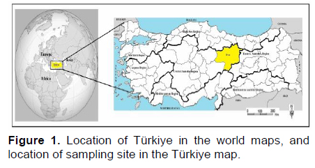
Research and Publication Ethics: All procedures done in this work involving animals were in accordance with the ethical standards approved by the Sivas Cumhuriyet University Animal Experiments Local Ethics Committee (Approval number: 09.02.2021–65202830–050.04.04–495).Study Area and Materials: Türkiye consists of seven geographical regions: Marmara, Aegean, Mediterranean, Central Anatolia, Black Sea, Eastern Anatolia, and South Eastern Anatolia. Sivas province is located in the Central Anatolia region and the intersection between Central Anatolia, Black Sea, and Eastern Anatolia regions (Figure 1). The city is the second-largest province in the country with a geographical area of approximately 28,400 km2. Sivas is an important region for water buffalo breeding due to its large pasture lands.
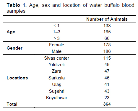
In this study, DNA belonging to 364 water buffalo blood samples was used. These materials were collected by Sahin et al.[18] for the other study, and stored in appropriate conditions in the research laboratory. Detailed information about the samples can be found in the study by Sahin et al. (18). Briefly, these blood samples were obtained from 119 buffalo herds in seven districts (Sivas city center, Koyulhisar, Zara, Yildizeli, Sarkisla, Susehri, and Ulas) of Sivas province (Table 1).
Molecular Research of Rickettsia spp. in Water Buffalo Blood Samples: The gDNAs were screened in terms of Rickettsia species with PCR assay using 190-70 (5’-ATGGCGAATATTTCTCCAAAA-3’) and 190.701 (5’-GTTCCGTTAATGGCAGCATCT-3’) amplified to ompA gene[29].
PCR assay was performed to a total volume 25 μL, including 10× PCR buffer (Invitrogen™, Carlsbad, USA), MgCl2 (50 mM) (Invitrogen™, Carlsbad, USA), 200 μM of each dNTP (Cat.No.: R0181, Thermo Scientific™, Lithuania), 1 μL (10 pmol/μL) of each of the primers, 1.25 U of Taq DNA polymerase (5U/μL) (Ref.No.: 100021276, Invitrogen™, Carlsbad, USA), 2.5 μL template DNA, and DNase-RNase-free sterile water.
The thermal cycling protocol used for PCR was 94°C for 5 min, followed by 35 cycles of 94°C for 1 min, 55°C for 1 min, 72°C for 1 min, and a final extension of 5 min at 72°C. The resulting PCR products were loaded onto a 1% agarose gel and then electrophoresed at 90 volts for 60 minutes. After electrophoresis, the agarose gel was stained by ethidium bromide for 20 min and visualized with a UV transilluminator for specific amplicons.
Preparation of the Samples for DNA Sequence Analyses: The positive sample detected in this work was sequenced for species identification and phylogenetic analyses. For these purposes, PCR assays were done to amplify ompA using primers 190-70 and 190-701 and ompB gene with the primers 120-2788 and 120-3599[29,30]. In these PCR assays, the EZ-FD PCR High Fidelity DNA polymerase kit (Cat.No.: 9K-005-0019, BioBasic) was used, and the PCR was done with reagents supplied by manufacturers and in a total volume of 25μL, including 10× EZ-FD Reaction buffer with 25mM MgCl2 (Lot.No.: O807R0K, BioBasic), 200 μM of each dNTP (Lot.No.: R22256RoX, BioBasic), 1 μL (10 pmol/μL) of each of the primers, 1.25 U of EZ-FD PCR High Fidelity DNA Taq polymerase (2.5U/μL) (Lot.No.: Q6A00120KF, BioBasic), 2.5 μL template DNA, and DNase-RNase-free sterile water. After PCR assays, PCR products were loaded into the agarose gel and checked in terms of appropriate amplicon sizes, and then, these products were sent to DNA sequence analyses.
DNA sequence analyses were performed in the commercial company (BM Labosis, Ankara). Before DNA sequence analyses, all PCR products were purified with the HighPrepTM PCR Clean-up System (Cat. No.: AC-60005, MagBio) following the manufacturer’s instructions. The sequence data were checked with FinchTV (version 1.4.0) software (Geospiza Inc., Seattle, Washington, USA) for chromatogram qualities, and nucleotides that had poor chromatogram qualities were trimmed. The consensus sequences were determined using MEGA-11 software[31]. The identified consensus sequences were deposited to GenBank, and accession numbers were determined.
The maximum likelihood method was used to construct phylogenetic trees using MEGA-11 software[31] to reveal the genetic variation between the Rickettsia species identified in this study and the Rickettsia species in GenBank. Before constructing the phylogenetic tree, it was determined that the best algorithm to be used in the phylogenetic tree of related pathogens was the Tamura-3 parameter model[32] using the Find Best-Fit Substitution Model in MEGA-11 and this algorithm was used in the phylogenetic tree. Bootstrap analysis (1,000 repetitions) was performed.
Bulgular
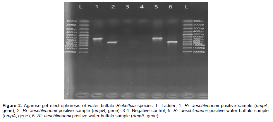
In this study, 364 water buffalo blood samples were screened for Rickettsia species using PCR assay, and one (0.27%) water buffalo was found to be infected with Rickettsia sp. (Figure 2). Species identification of Rickettsia species in the study was performed with DNA sequence analyses, and Ri. aeschlimannii was identified. These nucleotide sequences were uploaded to the GenBank and accession numbers were taken for ompA (PQ197208) and ompB (PQ197209) genes.
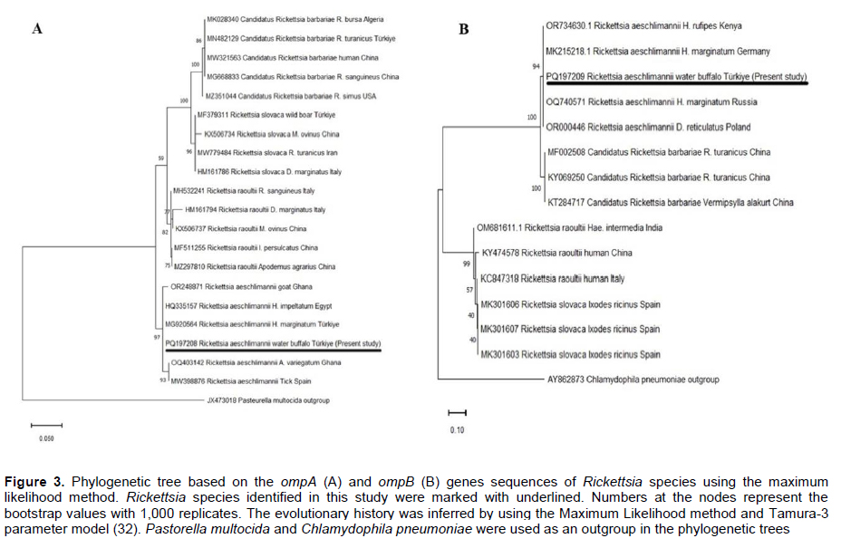
The ompA gene sequence analyses of the positive sample revealed 98.93-100% nucleotide similarities between Ri. aeschlimannii isolate identified in this study and Ri. aeschlimannii isolate present in the GenBank. In addition, our Ri. aeschlimannii isolates had 100% nucleotide identities with Ri. aeschlimannii identified in H. marginatum larvae (MG920564) from Türkiye, in H. lusitanicum (MH532238) from Italy, in H. marginatum (MT793816) from Lithuania, in H. impeltatum (HQ335157) from Egypt, and in goat (OR248871) from Ghana.
The BLASTn analyses of the consensus sequence of the ompB gene showed that there were 98.39-100% nucleotide resemblance between our Ri. aeschlimannii isolate and Ri. aeschlimannii isolate identified in various parts of the world. Moreover, 100% nucleotide identities were seen Ri. aeschlimannii isolate determined in the study and Ri. aeschlimannii isolate detected in H. marginatum (MK215218) from Germany, D. reticulatus (OR000446) from Poland, and H. rufipes (OR734630) from Kenya.
The phylogenetic trees based on ompA (Figure 3A) and ompB (Figure 3B) genes revealed our Ri. aeschlimannii isolate placed with the same clade with Ri. aeschlimannii isolates reported in different parts of the world and positioned in different branch from other Rickettsia species.
Tartışma
The importance of vector-borne pathogens increases year by year due to different reasons, such as climate change, deforestation, and expanded human settlement towards nature. Studies show that millions of people and animals live in areas at risk of vector-borne diseases[33,34]. Türkiye has a unique location, as the country is situated between the continents of Europe and Asia. This location makes the country a bridge for diseases to cross from one continent to another. In addition, Türkiye has a variety of climates suitable for the survival of many vector species[17]. Therefore, many studies have been conducted to understand the epidemiology, prevalence, and distribution of vector-borne pathogens in Türkiye and various vector-borne pathogens have been identified in the country, especially among domestic animals[17]. Water buffaloes are important domestic ruminants in Türkiye, but the number of studies conducted to investigate vector-borne pathogens in buffaloes in the country is quite low. Only four molecular studies were performed on these animals, in these studies, Anaplasma capra[18], Anaplasma phagocytophilum-like 1[19], Mycoplasma wenyonii, Candidatus Mycoplasma haemobos[36], and Theileria buffeli[35] were identified, to date. According to the literature review, no study presents on Rickettsia species in water buffalo in Türkiye. In this study, Rickettsia species were investigated in water buffalo herds for the first time and phylogenetic analyses of the detected Rickettsia species were performed.In the last two decades, many Rickettsia species, some of them novel species, have been identified among various hosts and their vectors in the different parts of the world using molecular-based techniques[1,4]. These studies also showed that Rickettsia species have been circulated in domestic ruminants, and DNA of these pathogens was detected in blood samples of cattle, sheep, and goats[37-39]. Serological studies were performed in water buffalo, and antibodies against Rickettsia species were detected in these animals[40]. In this study, 364 water buffalo blood samples were researched and one sample was found to be infected with Rickettsia species. This is the first molecular detection of Rickettsia DNA in a water buffalo blood sample. Our work and study performed by Pangjai et al.[40] demonstrated that water buffaloes may be exposed to Rickettsia species. The prevalence and distribution of vector-borne pathogens such as Rickettsia may vary according to the climatic characteristics of the study regions, the common vector species in the sampling regions, the specificity and sensitivity of the diagnostic methods used in the study, and the number of animals included in the study and their ages. In addition, since new Rickettsia species and strains have been detected in different hosts in recent years[1,4], it is thought that studies should be carried out to determine the epidemiological risk factors of these pathogens.
DNA sequence analyses have been done for different purposes such as the correction of PCR results, revealing phylogenetic analyses and genetic diversity of species, identification of novel species or genotypes of pathogens, and discovery of new host or epidemiological areas of pathogens in the studies[1,14,18]. The DNA sequence of one Rickettsia species identified in the work was done by targeting ompA and ompB genes. These sequence results revealed that Ri. aeschlimannii was circulated in a water buffalo herd in Sivas province. The ompA (98.93-100%) and ompB (98.39-100%) gene sequences showed that our Ri. aeschlimannii isolate had high nucleotide similarities with Ri. aeschlimannii isolates identified in different hosts from various parts of the world. With this study Ri. aeschlimannii was detected for the first time in water buffalo using PCR and DNA sequence analyses. Although Ri. aeschlimannii was detected in a water buffalo in the work, large-scale molecular studies involving buffaloes and their ectoparasites in different countries are needed to understand the contribution of these animals to the epidemiology of Ri. aeschlimannii.
Rickettsia aeschlimannii was identified firstly in ticks (H. marginatum) from Morocco[41]. Five years later, this pathogen was reported in humans returning to France from Morocco using serological and molecular techniques[42]. In the same and following years, Ri. aeschlimannii was detected in humans from South Africa[43], and Greece[8]. In these studies, clinical manifestations such as necrotic vesicular symptoms on the ankle, high fever, and maculopapular skin rash around the tick-bite sites were reported in patients, and these patients were treated with doxycycline for one week[8,42,43]. To date, several studies have been performed, and Ri. aeschlimannii was reported in different hosts (Meriones shawi, camel, human, Hyalomma marginatum, Rhipicephalus sanguineus etc.) from various parts of the world[4]. In this study, the DNA of Ri. aeschlimannii was detected for the first time in a water buffalo blood sample both in Türkiye and world. In Türkiye, Ri. aeschlimannii was identified in different cities from multiple hosts like Hyalomma spp. collected from cattle[27], hares[26], and humans[23,25], H. marginatum from wild boars[26], cattle[23], goats[28], sheep[28], and humans[20,22,23,25], H. aegyptium from human [20,23,25], and hedgehogs[24], H. excavatum from humans[25], Rhipicephalus spp. from goats[27], Rhipicephalus bursa from humans[20,25], R. turanicus from humans[25], cattle[28], and sheep[23], Haemaphysalis parva from human[25], Hae. punctata from human[25], Hae. sulcata from humans[25], Dermacentor marginatus from humans[25], and Ixodes ricinus from humans[25]. Above-mentioned studies revealed Ri. aeschlimannii is carried by multiple tick species in Türkiye. Sivas is the second biggest city in Türkiye, and different tick species, some of them vectors of Ri. aeschlimannii, circulate among domestic and wild animals in the city[44-46]. In studies conducted by Altay et al.[45,46] several tick-borne pathogens were reported in the ticks collected from cattle and sheep. Considering that Ri. aeschlimannii is a zoonotic pathogen transmitted by ticks, it is thought that buffalo breeders and people living in the region should take the necessary precautions against the pathogen.
In conclusion, Ri. aeschlimannii was reported for the first time with this study in a water buffalo. This molecular report showed that buffaloes, like other domestic ruminants, are hosts/reservoirs for vector-borne zoonotic pathogens. Vector-borne pathogens have a wide range of pathological effects on human and animal health. In addition, the economic loss due to these pathogens can reach billions of dollars, and studies showing the current prevalence and distribution of vector-borne pathogens among hosts are needed to reduce this economic loss.
References
- Parola P, Paddock CD, Socolovschi C, et al. Update on tick-borne rickettsioses around the world: A geographic approach. Clin Microbiol Rev 2013; 26: 657-702.
- Merhej V, Raoult D. Rickettsial evolution in the light of comparative genomics. Biol Rev 2011; 86: 379-405.
- Portillo A, Santibáñez S, García-Álvarez L, et al. Rickettsioses in Europe. Microbes Infect 2015; 17: 834-838.
- Seidi S, Omidi AH, Esmaeili S. Distribution of different Rickettsia species in countries of the WHO Eastern Mediterranean (WHO-EMRO) region: An overview. Travel Med Infecti Dis 2024; 102695.
- Luce-Fedrow A, Mullins K, Kostik AP, et al. Strategies for detecting rickettsiae and diagnosing rickettsial diseases. Future Microbiol 2015; 10: 537-564.
- Zhang YY, Sun YQ, Chen JJ, et al. Mapping the global distribution of spotted fever group rickettsiae: A systematic review with modelling analysis. Lancet Digit Health 2023; 5: e5-e15.
- Oteo JA, Portillo A. Tick-borne rickettsioses in Europe. Ticks Tick Borne Dis 2012; 3: 271-278.
- Germanakis A, Chochlakis D, Angelakis E, et al. Rickettsia aeschlimannii infection in a man, Greece. Emerg Infect Dis 2013; 19: 1176.
- Li H, Zhang PH, Huang Y, et al. Isolation and identification of Rickettsia raoultii in human cases: A surveillance study in 3 medical centers in China. Clin Infect Dis 2018; 66: 1109-1115.
- Husin NA, AbuBakar S, Khoo JJ. Current tools for the diagnosis and detection of spotted fever group Rickettsia. Acta Trop 2021; 218: 105887.
- Altay K, Dumanli N, Aktas M. Molecular identification, genetic diversity and distribution of Theileria and Babesia species infecting small ruminants. Vet Parasitol 2007; 147: 161-165.
- Altay K, Dumanli N, Aktas M. A study on ovine tick-borne hemoprotozoan parasites (Theileria and Babesia) in the East Black Sea Region of Turkey. Parasitol Res 2012; 111: 149-153.
- Altay K, Dumanli N, Aktaş M, et al. Survey of Anaplasma infections in small ruminants from East part of Turkey. Kafkas Üniv Vet Fak Derg 2014; 20: 1-4.
- Altay K, Erol U, Sahin OF. The first molecular detection of Anaplasma capra in domestic ruminants in the central part of Turkey, with genetic diversity and genotyping of Anaplasma capra. Trop Anim Health Prod 2022; 54: 129.
- Aktas M, Altay K, Dumanli N, et al. Molecular detection and identification of Ehrlichia and Anaplasma species in ixodid ticks. Parasitol Res 2009; 104: 1243-1248.
- Dumanlı N, Altay K, Aydin MF. Tick species of cattle, sheep and goats in Turkey. Turkiye Klinikleri J Vet Sci 2012; 3: 67-72.
- Inci A, Yildirim A, Duzlu O, et al. Tick-borne diseases in Turkey: A review based on one health perspective. PLoS Negl Trop Dis 2016; 10: e0005021.
- Sahin OF, Erol U, Altay K. Buffaloes as new hosts for Anaplasma capra: molecular prevalence and phylogeny based on gtlA, groEL, and 16S rRNA genes. Res Vet Sci 2022; 152: 458-464.
- Sahin OF, Erol U, Duzlu O, et al. Molecular survey of Anaplasma phagocytophilum and related variants in water buffaloes: the first detection of Anaplasma phagocytophilum-like 1. Comp Immunol Microbiol Infect Dis 2023a; 98: 102004.
- Gargili A, Palomar AM, Midilli K, et al. Rickettsia species in ticks removed from humans in Istanbul, Turkey. Vector Borne Zoonotic Dis 2012; 12: 938-941.
- Keskin A, Koprulu TK, Bursali A, et al. First record of Ixodes arboricola (Ixodida: Ixodidae) from Turkey with presence of Candidatus Rickettsia vini (Rickettsiales: Rickettsiaceae). J Med Entomol 2014; 51: 864-867.
- Keskin A, Bursali A, Keskin A, et al. Molecular detection of spotted fever group rickettsiae in ticks removed from humans in Turkey. Ticks Tick Borne Dis 2016; 7: 951-953.
- Orkun Ö, Karaer Z, Çakmak A, et al. Identification of tick-borne pathogens in ticks feeding on humans in Turkey. PLoS Negl Trop Dis 2014; 8: e3067.
- Orkun Ö, Çakmak A, Nalbantoğlu S, et al. Molecular detection of a novel Babesia sp. and pathogenic spotted fever group rickettsiae in ticks collected from hedgehogs in Turkey: Haemaphysalis erinacei, a novel candidate vector for the genus Babesia. Infect Genet Evol 2019; 69: 190-198.
- Karasartova D, Gureser AS, Gokce T, et al. Bacterial and protozoal pathogens found in ticks collected from humans in Corum province of Turkey. PLoS Negl Trop Dis 2018; 12: e0006395.
- Orkun Ö, Çakmak A. Molecular identification of tick-borne bacteria in wild animals and their ticks in Central Anatolia, Turkey. Comp Immunol Microbiol Infect Dis 2019; 63: 58-65
- Demir S, Erkunt Alak S, Köseoğlu AE, et al. Molecular investigation of Rickettsia spp. and Francisella tularensis in ticks from three provinces of Turkey. Exp Appl Acarol 2020; 81: 239-253.
- Orkun Ö. Comprehensive screening of tick-borne microorganisms indicates that a great variety of pathogens are circulating between hard ticks (Ixodoidea: Ixodidae) and domestic ruminants in natural foci of Anatolia. Ticks Tick Borne Dis 2022; 13: 102027.
- Fournier PE, Roux V, Raoult D. Phylogenetic analysis of spotted fever group Rickettsiae by study of the outer surface protein rOmpA. Int J Syst Bacteriol 1998; 48: 839-849.
- Roux V, Raoult D. Phylogenetic analysis of members of the genus Rickettsia using the gene encoding the outer-membrane protein rOmpB (ompB). Int J Syst Evol Microbiol 2000; 50: 1449-1455.
- Tamura K, Stecher G, Kumar S. MEGA11: molecular evolutionary genetics analysis version 11. Mol Biol Evol 2021; 38: 3022-3027.
- Tamura K. Estimation of the number of nucleotide substitutions when there are strong transition-transversion and G + C-content biases. Mol Biol Evol 1992; 9: 678-687.
- Wilson AL, Courtenay O, Kelly-Hope LA, et al. The importance of vector control for the control and elimination of vector-borne diseases. PLoS Negl Trop Dis 2020; 14: e0007831.
- Rocklöv J, Dubrow R. Climate change: an enduring challenge for vector-borne disease prevention and control. Nat Immunol 2020; 21: 479-483.
- Sahin OF, Erol U, Altay K. Sivas Yöresinde Mandalarda Theileria Türlerinin Moleküler Yöntemlerle Araştırılması. 23. Parazitoloji Kongresi, 95-104, 2023.
- Erol U, Sahin OF, Altay K. Molecular prevalence of bovine hemoplasmosis in Turkey with first detection of Mycoplasma wenyonii and Candidatus Mycoplasma haemobos in cattle and water buffalo. Vet Res Commun 2023; 47: 207-215.
- Liang CW, Zhao JB, Li J, et al. Spotted fever group Rickettsia in Yunnan Province, China. Vector Borne Zoonotic Dis 2012; 12: 281-286.
- Ortuño A, Pons I, Quesada M, et al. Evaluation of the presence of Rickettsia slovaca infection in domestic ruminants in Catalonia, Northeastern Spain. Vector Borne Zoonotic Dis 2012; 12: 1019-1022.
- Addo SO, Bentil, RE, Yartey KN, et al. First molecular identification of multiple tick-borne pathogens in livestock within Kassena-Nankana, Ghana. Animal Diseases 2023; 3: 1.
- Pangjai D, Saengsawang P, Kidsin K, et al. Seroprevalence of Orientia tsutsugamushi and Rickettsia typhi in water buffaloes (Bubalus bubalis) from Southern Thailand. Vet World 2023; 16: 1600.
- Beati L, Meskini M, Thiers B, et al. Rickettsia aeschlimannii sp. nov., a new spotted fever group rickettsia associated with Hyalomma marginatum ticks. Int J Syst Bacteriol 1997; 47: 548-554.
- Raoult D, Fournier PE, Abboud P, et al. First documented human Rickettsia aeschlimannii infection. Emerg Infect Dis 2002: 8; 748.
- Pretorius AM, Birtles RJ. Rickettsia aeschlimannii: a new pathogenic spotted fever group rickettsia, South Africa. Emerg Infect Dis 2002; 8: 874.
- Bursali A, Keskin A, Şimşek E, et al. A survey of ticks (Acari: Ixodida) infesting some wild animals from Sivas, Turkey. Exp Appl Acarol 2015; 66: 293-299.
- Altay K, Ataş AD, Özkan E. Molecular survey of Theileria and Babesia species in small ruminants and ticks from Sivas region of Turkey. Manas J Agric Vet Life Sci 2017; 7: 30-39.
- Altay K, Atas AD, Ograk YZ, et al. Survey of Theileria, Babesia and Anaplasma infections of cattle and ticks from Sivas region of Turkey. Erciyes Üniv Vet Fak Derg 2020; 17: 32-38.
- TÜRKİYE'DE SIĞIR HYPODERMOSİS'İ ÜZERİNE EPİDEMİYOLOJİK ARAŞTIRMALAR
- TABANIDAE SPECIES (DIPTERA) IN THE ELAZIĞ PROVINCE OF TURKEY
- ELAZIĞ BÖLGESİNDE TABANİDAE( DİPTERA) TÜRLERİ
- Türkiye'nin Doğu ve Güneydoğu Anadolu Bölgelerinde Hayvan Kuduzunun Epidemiyolojisi
- Türkiye'nin Bitlis Yöresinde İnsanlara Tutunan Kene Türlerinin Belirlenmesine Yönelik Ön Bir Çalışma