
Mycobacterium avium Subsp. paratuberculosis Açısından Seropozitif Sığırlarda Dışkı Karakteristiklerinin İncelenmesi
Volume 40, Issue 2
June 2026 · Pages 113-120
Mycobacterium avium Subsp. paratuberculosis Açısından Seropozitif Sığırlarda Dışkı Karakteristiklerinin İncelenmesi
Evaluation of Fecal Characteristics in Cattle Seropositive for Mycobacterium avium Subsp. paratuberculosis
Yiğit KAÇAR1, Zafer MECİTOĞLU1, İ. Taci CANGÜL2, Thomas WITTEK3, Ezgi YUMUŞAK2, Hasan BATMAZ1
2Uludag University, Faculty of Veterinary Medicine, Department of Medical Pathology, Bursa, TÜRKİYE
3University Clinics for Ruminants, University of Veterinary Medicine, Vienna, AUSTRİA
Paratüberküloz, Mycobacterium avium subsp. paratuberculosis (MAP) tarafından oluşturulan, sığırlarda ilerleyici ishal ve zayıflama ile karakterize, kronik, tedavi edilemeyen ve ekonomik açıdan önemli bir hastalıktır. Bu çalışmanın amacı, MAP-seropozitif sığırlarda dışkı kıvamı ve dışkı yapışkanlığını değerlendirmek ve bu parametrelerin klinik hastalık şiddeti ile ilişkisini incelemektir. Çalışmaya, ELISA ile doğrulanmış klinik paratüberküloz tanısı bulunan 20 Holştayn sığır (n=20) ile ELISA-negatif ve klinik olarak sağlıklı 10 Holştayn sığırdan oluşan kontrol grubu (n=10) dahil edildi. Başlangıçta, dışkı kıvamı ve dışkı yapışkanlığı aynı gözlemci tarafından basitleştirilmiş üç puanlı bir skorlama sistemi kullanılarak değerlendirildi. Kombine dışkı kıvamı ve yapışkanlık skorlarına göre MAP-seropozitif sığırlar hafif (n=10) ve orta–şiddetli (n=10) klinik gruplara ayrıldı. Daha sonra, hafif gruptan dört ve orta–şiddetli gruptan dört olmak üzere, kesime giden MAP-seropozitif sığırlardan elde edilen ileal doku örneklerinde histopatolojik inceleme gerçekleştirildi. Dışkı yapışkanlık skorları orta–şiddetli grupta hafif gruba kıyasla anlamlı derecede daha yüksek bulundu (2.10 vs. 1.00; p<0.001), buna karşın dışkı kıvamı skorları arasında istatistiksel farklılık gözlenmedi (2.00 vs. 1.40). Histopatolojik değerlendirmede klinik gruplar arasında villus uzunluğu, kript derinliği, goblet hücre sayısı ve inflamasyon skorları açısından değişkenlik saptanmış olmakla birlikte, bu bulgular klinik dışkı değişikliklerinin şiddeti ile paralellik göstermedi. Sonuç olarak, dışkı yapışkanlığı sığırlarda daha önce tanımlanmamış bir dışkı özelliği olup, bu çalışmada MAP-seropozitif hayvanlarda ilk kez değerlendirilmiştir. Artmış dışkı yapışkanlığının paratuberkülozlu sığırlarda klinik olarak fark edilebilir olabileceği ve rutin klinik değerlendirmede dışkı kıvamı değerlendirmesini tamamlayıcı bir parametre olarak kullanılabileceği düşünülmektedir.
Paratuberculosis, caused by Mycobacterium avium subsp. paratuberculosis (MAP), is a chronic, incurable, and economically important disease of cattle characterized by progressive diarrhea and weight loss. This study aimed to evaluate fecal consistency and fecal stickiness in MAP-seropositive cattle and to investigate their relationship with clinical disease severity. Twenty Holstein cattle with ELISA-confirmed clinical paratuberculosis (n=20) and ten ELISA-negative, clinically healthy control cattle (n=10) were included. Initially, fecal consistency and fecal stickiness were assessed using a simplified three-point scoring system by the same observer. Based on combined fecal consistency and stickiness scores, MAP-seropositive cattle were classified into mild (n=10) and moderate-to-severe (n=10) clinical groups. Subsequently, ileal histopathological examination was performed on tissue samples obtained at slaughter from MAP-seropositive cattle, including four animals from the mild group and four from the moderate–severe group. Fecal stickiness scores were significantly higher in the moderate-to-severe group than in the mild group (2.10 vs. 1.00; p<0.001), whereas fecal consistency scores showed only limited differences (2.00 vs. 1.40). Histopathological evaluation revealed variations in villus length, crypt depth, goblet cell count, and inflammatory scores between clinical groups; however, these findings did not demonstrate a clear correlation with the severity of clinical fecal alterations. Overall, fecal stickiness represents a previously undescribed fecal characteristic in cattle and is evaluated here for the first time in MAP-seropositive animals. Increased fecal stickiness may be clinically noticeable in cattle with paratuberculosis and could complement fecal consistency assessment during routine clinical evaluation.
Introduction
Paratuberculosis (Johne's disease) is a chronic infectious disease caused by the acid-fast bacterium Mycobacterium avium subsp. paratuberculosis (MAP), primarily affecting the intestinal tract of ruminants. Although cattle are the principal host, MAP infections have also been reported in other domestic and wild ruminant species, and the disease remains major clinical and economic importance in cattle production systems [1-3].Transmission occurs predominantly via the fecal–oral route, with infection risk being highest early in life through contact with contaminated environments and, potentially, milk or colostrum [3-5]. The disease progresses slowly and insidiously, with infected animals often remaining asymptomatic for prolonged periods. Paratuberculosis is classically described as a progressive condition with four stages: silent infection, subclinical disease, clinical disease, and advanced clinical disease [6]. Following ingestion, MAP establishes a persistent infection in the ileum, leading to chronic granulomatous enteritis and progressive impairment of intestinal structure and function [5,6,8].
Clinical manifestations are more commonly observed in cattle older than two years, with prevalence increasing with age, and typically include chronic diarrhea, progressive weight loss, reduced milk production, and decreased appetite; in advanced stages, cachexia and death may occur [3,8-10]. Importantly, the clinical stage represents only a small proportion of infected animals, as epidemiological studies suggest that for each clinically affected cow in MAP-infected herds, approximately 20–25 subclinically infected but asymptomatic animals may be present [11,12].
Paratuberculosis is a widespread and economically significant disease of dairy cattle, both globally and in Türkiye [3,13,14]. The infection is endemic in many regions and is characterized by a high within-herd prevalence, largely driven by a prolonged subclinical phase that facilitates silent transmission. Beyond its impact on animal health and welfare, paratuberculosis leads to substantial economic losses due to reduced milk production and quality, impaired fertility, decreased carcass value, and increased culling rates [3,15-18]. As no curative treatment is currently available, practical clinical tools that support early recognition and assessment of disease severity under field conditions are of particular importance [3,8].
The diagnosis of paratuberculosis is based on a range of laboratory methods, applied either individually or in various combinations. Although fecal culture is considered the gold standard, its prolonged incubation period limits routine use under field conditions. Consequently, faster diagnostic approaches are commonly applied, including serological assays—particularly ELISA—Ziehl–Neelsen staining, interferon-gamma tests, molecular techniques such as IS900 PCR, and rapid lateral flow assays [3,6,10,19,20].
In contemporary farm animal practice, various clinical scoring systems are routinely used to support rapid decision-making and disease monitoring under field conditions. These systems provide practical, low-cost tools for the phenotypic assessment of animal health and are widely applied in areas such as respiratory disease evaluation [21,22], lameness assessment [23,24], rumen fill scoring [25,26] and fecal consistency [25,27,28] scoring, particularly in neonatal calves. Standardized scoring systems improve objectivity, facilitate communication between clinicians, and allow more consistent monitoring of disease progression and treatment response [29].
Although altered faecal consistency (including diarrhoea) is commonly associated with paratuberculosis [3,6,30], its assessment often remains subjective. Moreover, the potential diagnostic or clinical relevance of other physical faecal properties, such as stickiness—which may reflect alterations in intestinal function, inflammatory processes, or malabsorption—has not been systematically evaluated in MAP-infected cattle. Therefore, this study aimed to evaluate and compare faecal consistency and faecal stickiness between MAP-seropositive cattle and seronegative controls, with the objective of exploring faecal stickiness as a potential auxiliary clinical indicator of paratuberculosis.
Material and Method
Research and Publication Ethics: This study was conducted after approval from the Bursa Uludağ University Animal Experiments Local Ethics Committee (Approval No: 2024-01/05).Animals and Study Design: A convenience sample of 30 Holstein cows in their 2nd and 3rd lactation stages that were housed in the same farm and managed under identical feeding, housing, and herd health programs to minimize management-related confounding factors, was selected from a herd undergoing routine screening for paratuberculosis. Serum samples from all animals were tested for antibodies against Mycobacterium avium subsp. paratuberculosis (MAP) using a commercial ELISA kit (IDEXX MAP Ab Test, IDEXX Laboratories Inc., USA) according to the manufacturer's instructions. Based on ELISA results and clinical evaluation, animals were allocated into two groups: a MAP-seropositive group (n=20) and a MAP-seronegative control group (n=10). The seropositive cattle exhibited clinical signs consistent with paratuberculosis, including diarrhea of varying severity and weight loss, whereas the control group consisted of clinically healthy animals with negative ELISA results. Following group allocation, fecal samples were collected and evaluated from each animal. Fecal evaluations were performed on fresh fecal material, either immediately after spontaneous defecation or from samples obtained by rectal palpation when spontaneous defecation was not observed.
Fecal consistency and stickiness scoring were performed throughout the study by the same researcher who was blinded to the MAP serostatus of the animals at the time of evaluation. The fecal consistency scoring was adapted from previously published bovine clinical scoring systems [29,31-33]. For fecal stickiness—a parameter that has not been previously systematically assessed in cattle—a novel scoring system was developed in the present study. This was achieved by modifying and simplifying adhesiveness- or stickiness-based scoring approaches originally described for other species, such as poultry, mice, and dogs [34-36]. In both scoring systems, the number of score categories was reduced to improve practicality and reproducibility for routine clinical use.
In clinical cases of paratuberculosis, fecal consistency was scored using a three-level scale: normal (1), soft (2), and slightly watery (3), based on modifications of previously published reports [29,31-33]. Additionally, fecal stickiness was categorized into three levels: mild (1), moderate (2), and severe (3) (Figure 1).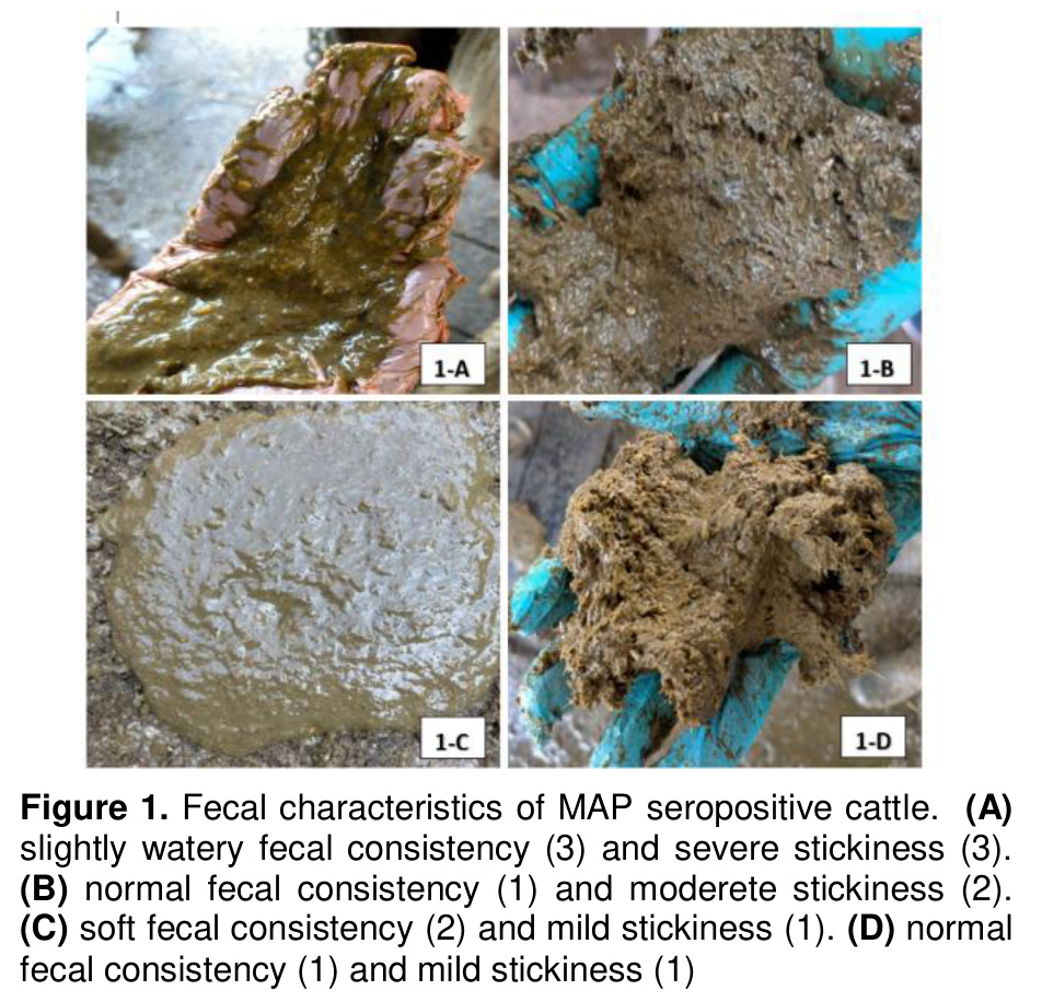
Stickiness was visually assessed by using gloved fingers to handle the feces and evaluating the degree to which the fecal matter adhered to the gloves. Mild Stickiness (Score 1): Feces do not adhere to surfaces or gloves, or only show minimal adherence. Moderate Stickiness (Score 2): Feces exhibit moderate adherence to surfaces and gloves, making them more difficult to separate. Severe Stickiness (Score 3): Feces strongly adhere to surfaces and gloves, making them difficult to completely remove (Figure 1).
Fecal consistency and stickiness scores were compared between MAP-seropositive cattle (n=20) and MAP-seronegative controls (n=10). Subsequently, to assess the relationship between fecal characteristics and clinical disease severity, MAP-seropositive cattle were further classified into mild (n=10) and moderate-to-severe (n=10) categories based on combined fecal consistency and stickiness scores. Classification criteria were defined as follows: fecal samples with a consistency score of 1–2 and a stickiness score of 1 were classified into the mild group, whereas samples with a fecal stickiness score of 2–3, as well as those with a consistency score of 3 but a stickiness score of 1, were classified into the moderate-to-severe group.
Histopathological Analysis: Additionally, intestinal tissue samples were collected from eight MAP-seropositive cattle at slaughter. Gross and histopathological examinations were performed. For histopathological evaluation, samples were obtained from ileal regions showing the most severe macroscopic lesions, characterized by marked wall thickening and brain-like appearance. After routine processing, sections were stained with hematoxylin and eosin, with Alcian blue used additionally for goblet cell evaluation, and were examined in a blinded manner. Ten randomly selected microscopic fields per sample were evaluated at ×400 magnification, and mean values were calculated for villus length, crypt depth, and goblet cell count. Inflammatory cell infiltration was scored semi-quantitatively on a four-point scale: 1 (0–25 cells), 2 (26–50 cells), 3 (51–75 cells), and 4 (>75 cells), with higher scores indicating more severe inflammation [37,38].
Statistical Analysis: The data were processed and analyzed using SigmaPlot 15 (Version 15; Systat Software Inc., San Jose, CA, USA) statistical software. The normality of the data was assessed using the Shapiro–Wilk test. As most variables did not meet the assumption of normal distribution, non-parametric statistical methods were applied. Descriptive statistics are presented as mean ± standard deviation (SD), median with interquartile range (Q1–Q3), and minimum–maximum values for all variables, to provide a comprehensive description of data distribution. Comparisons between groups were performed using the Mann–Whitney U test. Spearman's rank correlation analysis was used to assess relationships between parameters. A p-value <0.05 was considered statistically significant for all analyses.
Results
The main clinical signs observed in the paratuberculosis-positive cattle included diarrhea of varying severity, softening of the feces, and weight loss. Notably, the feces were soft and sticky in the MAP-positive cattle. The gross appearance of the feces from MAP-seropositive cattle is presented in Figure 1. When the fecal consistency and fecal stickiness scores of 20 MAP-seropositive cattle were evaluated, 10 cattle were classified into the moderate-severe clinical group, while 10 were placed in the mild clinical group. In the mild cases, fecal consistency scores were closer to normal (1), with an average score of 1.40, while the moderate-severe group had an average score of 2.0. In paratuberculosis cases, changes of fecal stickiness were more pronounced than consistency. In the mild group, the stickiness score remained within normal limits (score 1), while in the severe group, a significant increase was observed, with a score of 2.10 (p<0.001). The fecal consistency and fecal stickiness scores for cattle in the mild and moderate-severe clinical categories are presented in Table 1 and Table 2. In the feces of MAP-seropositive animals, a particularly sticky appearance and altered consistency were noted.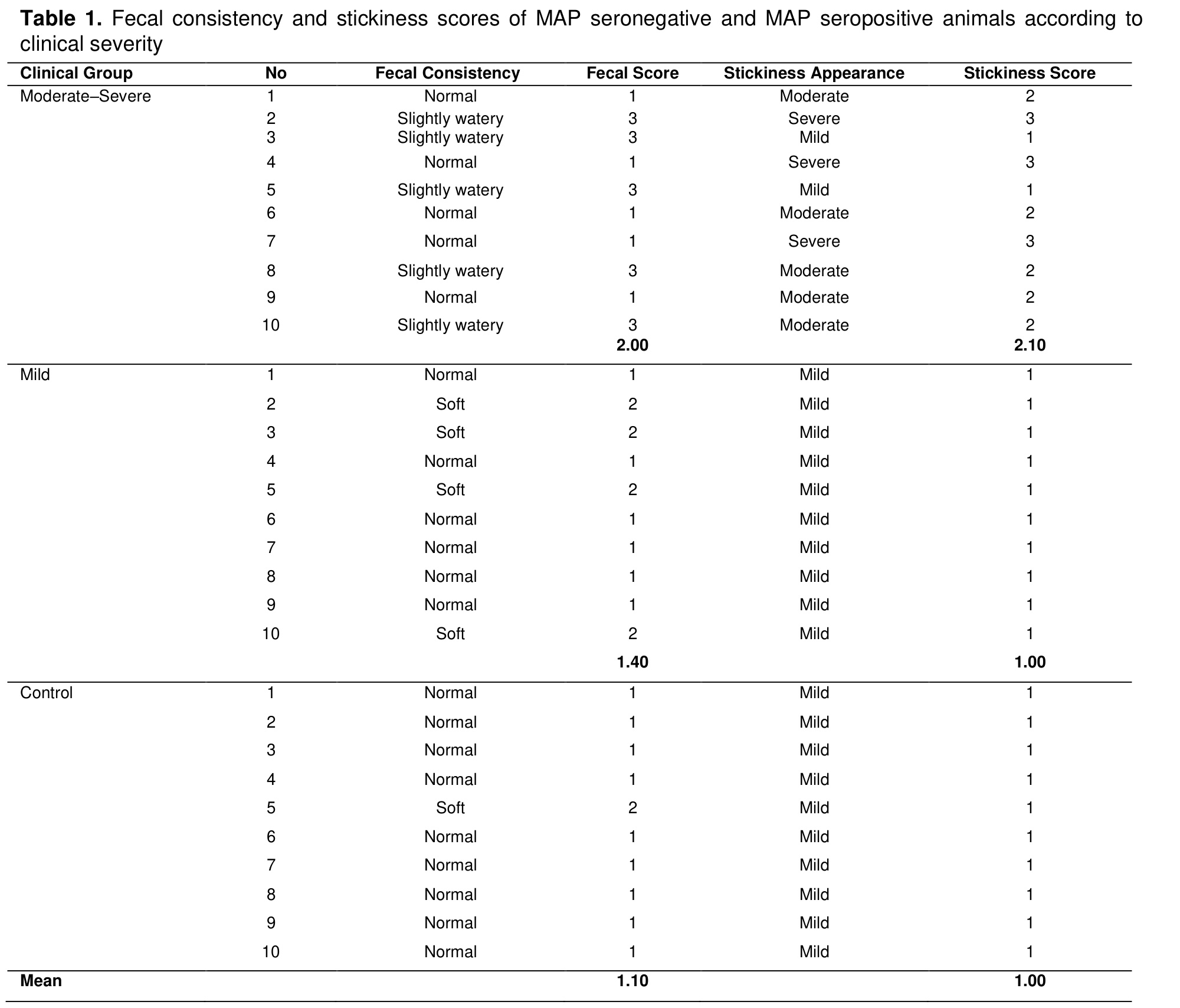
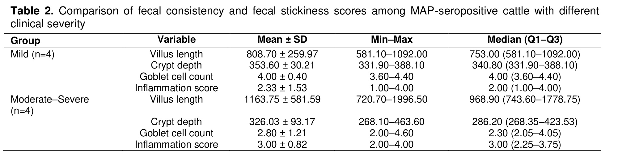
No significant correlation was observed between fecal consistency and fecal stickiness scores when evaluated across all animals (n=30; r=0.12, p=0.514) or within MAP-positive animals only (n=20; r=−0.01, p=0.957). When clinical subgroups were analyzed separately, the correlation between fecal consistency and stickiness in the moderate–severe group was weak and not statistically significant (n=10; r=−0.42, p=0.213). Correlation analysis could not be reliably computed in the mild (n=10) and control (n=10) groups due to the absence of variability in stickiness scores. Histopathological evaluation revealed descriptively higher values of villus length and inflammation scores and lower goblet cell counts in the moderate–severe group compared with the mild group, whereas crypt depth values showed considerable overlap between groups (Table 3).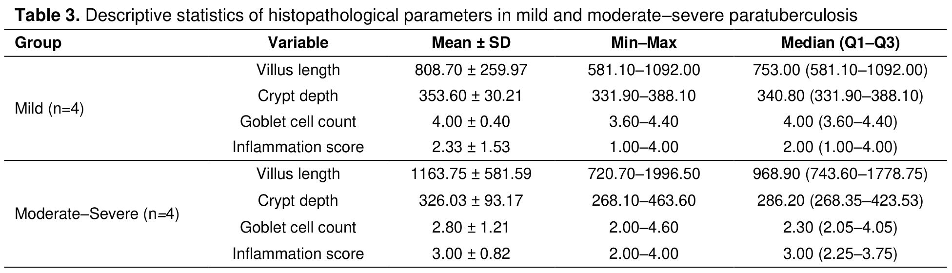
Discussion
Feces are increasingly used as a non-invasive biological material in both human [39,40] and veterinary medicine [41,42] and has therefore become the focus of numerous clinical and experimental studies. Changes in fecal characteristics may reflect underlying alterations in gastrointestinal physiology, inflammation, or nutrient absorption, making fecal evaluation clinically relevant not only through biochemical or molecular biomarkers but also through practical assessment of physical properties.In both human medicine [43,44] and veterinary practice—including dogs [45,46], pigs [47], cats [48], calves [41,49], and horses [50]—fecal scoring systems are widely used to support diagnosis, disease monitoring, and assessment of treatment response. Across the majority of studies in both veterinary and human medicine, fecal scoring systems primarily rely on fecal consistency or fluidity as the main quantitative parameter [43,48,50,51]. In some studies, additional fecal characteristics—including shape, color, odor, or the presence of mucus—have also been evaluated; however, these parameters are generally recorded descriptively or considered supportive observations rather than being incorporated as independent scoring criteria [28,41]. Similarly, in human medicine, stool scoring systems are predominantly consistency-based [43], while features such as stool color or amount are occasionally documented as complementary parameters rather than central components of the scoring framework [44].
In farm animals, fecal scoring is commonly applied in the assessment of diarrhea and gastrointestinal disorders in both adult cattle [51,52] and calves [28,41]. However, the majority of scoring systems in farm animal practice have been developed for neonatal calf diarrhea, where three-point, four-point [28,41,52], or five-point [25,51] scales based mainly on fecal consistency are widely used, and in some cases color or odor is also considered. These systems have proven useful for monitoring calf health and disease progression under field conditions.
In adult cattle, fecal scoring is applied more selectively than in neonatal animals and has primarily been used in the evaluation of digestive disturbances and feeding-related disorders [28,41]. In particular, fecal consistency scoring has been incorporated into the assessment of subacute ruminal acidosis and related nutritional imbalances in lactating dairy cows [53]. In addition, fecal scoring has been used as a practical tool for the general assessment of digestive function and ration adequacy during lactation, especially in relation to dietary fiber content and feeding management [25,52]. Within the multi-component scoring system proposed by Zaaijer and Noordhuizen [25]—which integrates rumen fill, fecal consistency, the undigested fecal fraction, and rectal reproductive assessment—fecal consistency and the undigested fecal fraction were identified as rapid, field-based indicators of energy balance and feeding efficiency in dairy cows. Deviations in these fecal parameters were shown to be associated with an increased risk of fertility disorders, highlighting their value as supportive markers of nutritional inadequacy rather than standalone diagnostic tools. Similarly, Ireland-Perry and Stallings [52] demonstrated that fecal consistency in lactating Holstein cows is responsive to dietary composition, particularly fiber level and protein source, with lower dietary fiber being associated with more liquid feces and lower fecal scores. However, these authors also reported that fecal consistency could not be accurately predicted based on dietary and cow-level parameters alone, indicating that fecal scoring in adult cattle is best interpreted as a practical and rapid indicator of dietary effects, rather than a precise quantitative measure.
Despite the well-recognized occurrence of altered fecal consistency in adult cattle affected by paratuberculosis [1,10], the literature addressing fecal scoring in MAP-seropositive cattle remains limited [30], and fecal assessment in this disease has largely been descriptive rather than systematic. In one of the few longitudinal studies addressing this issue, Vass-Bognár et al. [30] evaluated fecal consistency in MAP-infected dairy cows across the lactation period and demonstrated that fecal scores remained largely stable in subclinically infected animals and did not show a progressive decline. In contrast, cows progressing to clinical paratuberculosis exhibited a pronounced and continuous deterioration in fecal consistency, accompanied by body condition loss and increased bacterial shedding. Collectively, these findings indicate that fecal scoring reflects disease progression and clinical severity rather than serving as an early or stand-alone marker of subclinical MAP infection.
In the present study, fecal consistency and fecal stickiness were evaluated for the first time in MAP-seropositive adult cattle. Our findings demonstrate that, with increasing clinical severity of paratuberculosis—as defined by the presence of chronic weight loss, poor body condition, reduced milk yield, and persistent diarrhea consistent with clinical classification criteria—fecal stickiness increased more consistently and more markedly than fecal consistency. Although fecal consistency scores tended to be higher in moderate-to-severe cases, this difference did not reach statistical significance, whereas fecal stickiness scores showed a clear and statistically significant increase. This observation suggests that, particularly in chronically affected animals classified based on clinical examination findings, fecal stickiness may represent a more sensitive indicator of disease severity than fecal consistency alone.
To the best of our knowledge, fecal stickiness has not previously been evaluated in cattle. In other animal species, however, fecal stickiness or adhesiveness has been assessed using different visual scoring approaches. Ray et al. [34] evaluated fecal stickiness in poultry by visually scoring the amount of feces adhering to wire floors on a six-point scale ranging from 0 to 5, with higher scores indicating greater fecal adhesion. Takemori et al. [35] assessed fecal characteristics in mice using a four-point scoring system, in which fecal stickiness was defined by adherence of feces to forceps during handling. Yamka et al. [36] evaluated fecal quality in dogs using separate visual scoring systems for volume, stickiness, and adhesiveness, with stickiness assessed by insertion of a metal spatula and graded on a five-point scale from non-sticky to very sticky. These studies indicate that fecal stickiness represents a distinct physical property reflecting alterations in intestinal function beyond simple changes in moisture or consistency. Building on these approaches, we adapted and simplified previously described methods to develop a practical three-point fecal stickiness scoring system for routine clinical evaluation in adult cattle with paratuberculosis.
In this research, fecal stickiness exhibited a pronounced and statistically significant increase in the moderate-to-severe paratuberculosis group compared with mildly affected animals (2.10 vs. 1.00; p<0.001), indicating that this parameter may reflect disease severity more sensitively than fecal consistency alone. The increased fecal stickiness observed in moderate-to-severe cases may be related to the chronic granulomatous inflammation of the intestinal mucosa characteristic of MAP infection, supported in our cases by descriptively higher inflammation scores, lower goblet cell counts, and variations in villus length and crypt depth in the moderate-to-severe group compared with the mild group.
Classical descriptions of bovine paratuberculosis emphasize that diarrhea is typically not accompanied by grossly visible blood or mucus [5,6,11]; therefore, increased fecal stickiness observed in the present study is more likely to be associated with the intensity of inflammatory cell infiltration and villus–crypt degeneration, leading to impaired intestinal absorption and altered fecal physical properties that are not fully captured by conventional fecal consistency scoring [5]. This interpretation is further supported by previous observations indicating that the occurrence of clinical signs is not necessarily related to the severity of histological lesions in paratuberculosis [7]. From a clinical perspective, fecal stickiness is easily assessed during routine examination, does not require additional equipment, and may serve as a practical adjunct indicator of disease severity under field conditions.
The absence of a significant correlation between fecal stickiness and fecal consistency indicates that these parameters reflect distinct aspects of gastrointestinal alteration in paratuberculosis. While fecal consistency primarily relates to changes in water content and motility, fecal stickiness may be more closely associated with inflammatory processes, mucosal alterations, or changes in microbial composition. Thus, fecal stickiness may provide complementary clinical information beyond conventional fecal consistency scoring, particularly in animals with moderate-to-severe disease.
An important strength of this study is the standardized assessment of fecal characteristics by a single, blinded observer, which minimized inter-observer variability and reduced potential bias related to knowledge of MAP serostatus. Furthermore, the use of a simplified scoring system enhances the feasibility of applying fecal stickiness evaluation in routine herd health monitoring and clinical practice.
Several limitations should be acknowledged. The study relied on serological classification without fecal culture or PCR confirmation, which precluded precise staging of MAP infection and assessment of fecal shedding status. Additionally, the sample size was limited, particularly for subgroup analyses based on clinical severity. Despite these limitations, the present study demonstrates that fecal stickiness is a distinct and clinically relevant feature of paratuberculosis in adult cattle and may serve as a practical adjunct to fecal consistency scoring. The significant increase in fecal stickiness in moderate-to-severe cases highlights its potential utility in assessing clinical severity and disease progression. Incorporating fecal stickiness into routine clinical evaluation may improve the field diagnosis and monitoring of paratuberculosis, particularly in chronic cases where conventional fecal consistency scoring alone may be insufficient. Future studies incorporating combined diagnostic approaches, larger populations, and longitudinal follow-up—particularly focusing on earlier or subclinical stages of infection—are warranted to further validate fecal stickiness as a marker of disease severity and progression and to better define its clinical relevance across different stages of MAP infection.
References
- Okuni JB, Hansen S, Eltom KH, et al. Paratuberculosis: A potential zoonosis and a neglected disease in Africa. Microorganisms 2020; 8: 1007.rn
- Stanitznig A, Khol JL, Lambacher B, et al. Prevalence of Mycobacterium avium subspecies paratuberculosis and hepatitis E in New World camelids in Austria. Vet Rec 2017; 181: 46.rn
- Whittington R, Donat K, Weber MF, et al. Control of paratuberculosis: Who, why and how. A review of 48 countries. BMC Vet Res 2019; 15: 198.rn
- Khol JL, Braun AL, Slana I, et al. Testing of milk replacers for Mycobacterium avium subsp. paratuberculosis by PCR and bacterial culture as a possible source for Johne's disease (paratuberculosis) in calves. Prev Vet Med 2017; 144: 53-56.rn
- Sweeney RW. Pathogenesis of paratuberculosis. Vet Clin North Am Food Anim Pract 2011; 27: 537-v.rn
- Fecteau ME. Paratuberculosis in cattle. Vet Clin North Am Food Anim Pract 2018; 34: 209-222.rn
- Clarke CJ. The pathology and pathogenesis of paratuberculosis in ruminants and other species. J Comp Pathol 1997; 116: 217-261.rn
- Behr MA, Collins DM. Paratuberculosis: Organism, disease, control. Wallingford (UK): CABI; 2010.rn
- Otter A, Cranwell M. Differential diagnosis of diarrhoea in adult cattle. In Pract 2007; 29: 9-19.rn
- Belknap EB, Navarre CB. Differentiation of gastrointestinal diseases in adult cattle. Vet Clin North Am Food Anim Pract 2000; 16: 59-86.rn
- Whitlock RH, Buergelt C. Preclinical and clinical manifestations of paratuberculosis (including pathology). Vet Clin North Am Food Anim Pract 1996; 12: 345-356.rn
- Magombedze G, Ngonghala CN, Lanzas C. Evaluation of the "iceberg phenomenon" in Johne's disease through mathematical modelling. PLoS One 2013; 8: e76636.rn
- Ozturk D, Pehlivanoğlu F, Tok AA, et al. Seroprevalence of paratuberculosis in the Burdur province (Turkey), in dairy cattle using the enzyme linked immunosorbent assay (ELISA). Isr J Vet Med 2010; 65: 53-57.rn
- Gümüşsoy KS, Ica T, Abay S, et al. Serological and molecular diagnosis of paratuberculosis in dairy cattle. Turk J Vet Anim Sci 2015; 39:147-153.rn
- Griss S, Knific T, Buzzell A, et al. A scoping review on associations between paratuberculosis and productivity in cattle. Front Vet Sci 2024;11: 1352623.rn
- Gonda MG, Chang YM, Shook GE, Collins MT, Kirkpatrick BW. Effect of Mycobacterium paratuberculosis infection on production, reproduction, and health traits in US Holsteins. Prev Vet Med 2007; 80: 103-119.rn
- Hendrick SH, Kelton DF, Leslie KE, et al. Effect of paratuberculosis on culling, milk production, and milk quality in dairy herds. J Am Vet Med Assoc 2005; 227: 1302-1308.rn
- Lombard JE, Garry FB, McCluskey BJ, Wagner BA. Risk of removal and effects on milk production associated with paratuberculosis status in dairy cows. J Am Vet Med Assoc 2005; 227: 1975-1981.rn
- Britton LE, Cassidy JP, O'Donovan J, et al. Potential application of emerging diagnostic techniques to the diagnosis of bovine Johne's disease (paratuberculosis). Vet J 2016; 209: 32-39.rn
- Agrawal A, Varshney R, Gattani A, et al. Gold nanoparticle based immunochromatographic biosensor for rapid diagnosis of Mycobacterium avium subspecies paratuberculosis infection using recombinant protein. J Microbiol Methods 2020; 177: 106024.rn
- McGuirk SM, Peek SF. Timely diagnosis of dairy calf respiratory disease using a standardized scoring system. Anim Health Res Rev 2014; 15: 145-147.rn
- Love WJ, Lehenbauer TW, Kass PH, et al. Development of a novel clinical scoring system for on-farm diagnosis of bovine respiratory disease in pre-weaned dairy calves. PeerJ 2014; 4: e2380rn
- Sprecher DJ, Hostetler DE, Kaneene JB. A lameness scoring system that uses posture and gait to predict dairy cattle reproductive performance. Theriogenology 1997; 47:1179-1187.rn
- Flower FC, Weary DM. Effect of hoof pathologies on subjective assessments of dairy cow gait. J Dairy Sci 2006; 89: 139-146.rn
- Zaaijer D, Noordhuizen JPTM. A novel scoring system for monitoring the relationship between nutritional efficiency and fertility in dairy cows. Ir Vet J 2003; 56: 145-151.rn
- Burfeind O, Sepúlveda P, von Keyserlingk MA, et al. Technical note: Evaluation of a scoring system for rumen fill in dairy cows. J Dairy Sci 2010; 93: 3635-3640.rn
- Constable PD, Hinchcliff KW, Done SH, Grünberg W. Veterinary Medicine: A Textbook of the Diseases of Cattle, Horses, Sheep, Pigs, and Goats. 11th Edition, Elsevier; 2017.rn
- Larson LL, Owen FG, Albright JL, et al. Guidelines toward more uniformity in measuring and reporting calf experimental data. J Dairy Sci 1977; 60: 989-991.rn
- Batmaz H. Buzağı sağlığı. In: Batmaz H (Editor). Sığırlarda Sürü Sağlığı ve Yönetimi. Genişletilmiş 2. Baskı. Ankara: Nobel Tıp Kitabevleri; 2021: 105.rn
- Vass-Bognár B, Bakony M, Fornyos K, et al. When is the best time to test paratuberculosis positivity? Observations from a follow-up study in Hungarian dairy herds. Front Vet Sci 2025; 12: 1570915.rn
- Noordhuizen J. Dairy Herd Health and Management – A Guide for Veterinarians and Dairy Professionals. Leicestershire (UK): Context; 2012: 99.rn
- Petrovski KR, Cusack P, Malmo J, et al. The value of 'cow signs' in the assessment of the quality of nutrition on dairy farms. Animals (Basel) 2022; 12: 1352.rn
- Soren S, Roy B, Gore JA, et al. Influence of NutriSigma® on lactational performance of Sahiwal cows in the declining milk production stage. Indian J Anim Health 2019; 58: 101-110.rn
- Ray S, Pubols MH, McGinnis J. The effect of a purified guar degrading enzyme on chick growth. Poult Sci 1982; 61: 488-494.rn
- Takemori H, Hamamoto A, Isogawa K, et al. Mouse model of metformin-induced diarrhea. BMJ Open Diabetes Res Care 2020; 8: e000898.rn
- Yamka RM, Harmon DL, Schoenherr WD, et al. In vivo measurement of flatulence and nutrient digestibility in dogs fed poultry by-product meal, conventional soybean meal, and low-oligosaccharide low-phytate soybean meal. Am J Vet Res 2006; 67: 88-94.rn
- Cangul IT, Gul NY, Topal A, et al. Evaluation of topical tripeptide–copper complex and zinc oxide on open-wound healing in rabbits. Vet Dermatol 2006; 17: 417-423.rn
- Gul NY, Topal A, Cangul IT, Yanik K. Effects of tripeptide–copper complex and He–Ne laser on wound healing in rabbits. Vet Dermatol 2008; 19: 7-14.rn
- Callaway TR, Dowd SE, Edrington TS, et al. Evaluation of bacterial diversity in the rumen and feces of cattle fed different levels of dried distillers grains plus solubles using bacterial tag-encoded FLX amplicon pyrosequencing. J Anim Sci 2010; 88: 3977-3983.rn
- Zhgun ES, Ilina EN. Fecal metabolites as non-invasive biomarkers of gut diseases. Acta Naturae 2020; 12: 4-14.rn
- McGuirk SM. Disease management of dairy calves and heifers. Vet Clin North Am Food Anim Pract 2008; 24: 139-153.rn
- Suchodolski JS, Markel ME, Garcia-Mazcorro JF, et al. The fecal microbiome in dogs with acute diarrhea and idiopathic inflammatory bowel disease. PLoS One 2012; 7: e51907.rn
- Lewis SJ, Heaton KW. Stool form scale as a useful guide to intestinal transit time. Scand J Gastroenterol 1997; 32: 920-924.rn
- Bekkali N, Hamers SL, Reitsma JB, van Toledo L, Benninga MA. Infant stool form scale: Development and results. J Pediatr 2009; 154: 521-526.rn
- Cavett CL, Tonero M, Marks SL, et al. Consistency of faecal scoring using two canine faecal scoring systems. J Small Anim Pract 2021; 62: 167-173.rn
- Grellet A, Feugier A, Chastant-Maillard S, et al. Validation of a fecal scoring scale in puppies during the weaning period. Prev Vet Med 2012; 106: 315-323.rn
- Pons AM, González B, Pujols J, et al. A proposal of a fecal scoring system based on physicochemical analyses of suckling pig feces. Porc Health Manag 2025; 11: 5.rn
- German AC, Cunliffe NA, Morgan KL. Faecal consistency and risk factors for diarrhoea and constipation in cats in UK rehoming shelters. J Feline Med Surg 2017;19: 57-65.rn
- Renaud DL, Buss L, Wilms JN, Steele MA. Is fecal consistency scoring an accurate measure of fecal dry matter in dairy calves? J Dairy Sci 2020; 103: 10709-10714.rn
- Kaya Karasu G, Gümüş H. Evaluating the impact of starch intake on fecal consistency scores in equines. Turk J Vet Anim Sci 2025; 49: 199-206.rn
- Muhsen RK, Al-Autaish HHN. Fecal pH and fecal score in local cattle. J Kerbala Agric Sci 2017; 4: 44-47.rn
- Ireland-Perry RL, Stallings CC. Fecal consistency as related to dietary composition in lactating Holstein cows. J Dairy Sci 1993; 76: 1074-1082.rn
- Kleen JL, Hooijer GA, Rehage J, Noordhuizen JPTM. Subacute ruminal acidosis in Dutch dairy herds. Vet Rec 2009; 164: 681-684.
- YEMLERLE ALINAN YÜKSEK DÜZEYDE İYOTUN SIĞIRLARDA BAZI KLİNİK HEMATOLOJİK VE BİYOKİMYASAL PARAMETRELER ÜZERİNE ETKİSİ
- SIĞIR THEİLERİOSİSİ
- ŞAP AŞISI UYGULANAN BESİ SIĞIRLARINDA ANTİOKSİDAN VİTAMİNLERİN KLİNİK VE BAZI HEMATOLOJİK PARAMETRELER İLE ANTİOKSİDAN ENZİM VE LİPİT PEROKSİDASYON DÜZEYLERİNE ETKİLERİ
- SIĞIRDA FARİNGEAL PENETRAN BİR YORGAN İĞNESİ OLGUSU
- KIŞ MEVSİMİNDE KARS YÖRESİNDE ZAVOT SIĞIRLARININ GENEL, YAŞ VE CİNSİYETE GÖRE VENÖZ KAN GAZI DEĞERLERİ