
Nigella sativa ve Aloe vera’nın Yanık İyileşmesi Üzerine Etkileri: Klinik ve Histopatolojik Değerlendirmeler
Cilt 40, Sayı 3
Ekim 2026 · Sayfalar 209-215
Nigella sativa ve Aloe vera’nın Yanık İyileşmesi Üzerine Etkileri: Klinik ve Histopatolojik Değerlendirmeler
Effects of Nigella sativa and Aloe vera on Burn Healing: Clinical and Histopathological Evaluations
Şeyda Hilal KAYA1, Ali Said DURMUŞ1
Bu çalışmada, Nigella sativa (NS) ve Aloe vera’nın (AV) deneysel deri yanıklarının iyileşmesi üzerindeki klinik ve histopatolojik etkileri değerlendirildi. Otuz altı rat kontrol (Cold krem, C), NS ve AV olmak üzere üç eşit gruba ayrıldı. Dorsal torasik bölgede 1.5 cm çapında 2. derece yanık oluşturuldu ve günde iki kez cold krem, NS ve AV topikal olarak uygulandı. Yanık ve epitelizasyon alanları 0., 4., 7., 10. ve 14. günlerde ölçüldü. Yedinci ve 14. günlerde histopatolojik inceleme için her gruptan 6 rat ötenazi edildi.
Makroskopik incelemelerde, iyileşmenin 4., 7. ve 10. günlerde AV grubunda daha iyi olduğu; 14. günde ise AV ve NS gruplarında kontrol grubuna göre daha iyi olduğu gözlendi (p<0.0001).
Histopatolojik muayenelerde, kontrol grubunda 7. günde şiddetli nekroz ve 7.-14. günlerde şiddetli düzeyde mononükleer (Mn) hücre infiltrasyonu saptandı. Ödem, 7. günde AV ve NS gruplarında şiddetli düzeydeydi. Epitelizasyon 7. günde kontrol grubunda tam olarak şekillenmemişken, NS grubunda orta, AV grubunda ileri düzeydeydi. Ondördüncü günde AV ve NS gruplarında epitelizasyon tamamlanmıştı. Granülasyon dokusu oluşumu AV grubunda ileri düzeydeydi.
Klinik ve histopatolojik bulgular, AV’nin nekroz oluşumunu önleyerek ve epitelizasyonu destekleyerek deri yanıklarının iyileşmesini artırdığını göstermiştir. Sonuç olarak, elde edilen veriler NS ve AV’nin termal deri yanıklarının tedavisinde güvenle kullanılabileceğini ortaya koymaktadır.
In this study, clinical and histopathological effects of Nigella sativa (NS) and Aloe vera (AV) on the healing of experimental skin burns were evaluated. Thirty-six rats were divided into three equal groups: control (cold cream, C), NS, and AV. A 1.5 cm diameter second-degree burn was created on the dorsal thoracic region and cold cream, NS, and AV were applied topically twice daily. The burn and epithelialization areas were measured on days 0, 4, 7, 10, and 14. On days 7 and 14, six rats from each group were euthanized for histopathological examination.
Macroscopic examinations revealed that the healing was better in the AV group on days 4, 7, and 10; on day 14, healing was better in both the AV and NS groups compared to the control group (p<0.0001).
Histopathological examinations showed severe necrosis on day 7 and severe mononuclear (Mn) cell infiltration between days 7 and 14 in the control group. Oedema was severe in the AV and NS groups on day 7. Epithelialization was incomplete in the control group on day 7, moderate in the NS group, and advanced in the AV group. Epithelialization was complete in the AV and NS groups on day 14. Granulation tissue formation was advanced in the AV group.
Clinical and histopathological findings demonstrated that AV enhances the healing of skin burns by preventing necrosis and promoting epithelialization. In conclusion, the obtained data indicate that NS and AV can be safely used in the treatment of thermal skin burns.
Giriş
In the treatment of thermal burns, ointments containing antibiotics, antimicrobials, and antiseptics, as well as those that promote epithelialization and possess antioxidant properties, are used with the aim of achieving rapid tissue repair [1-3]. Research is still ongoing to identify the most effective treatment method for burns [4].Nigella sativa (NS) is composed of approximately 36-38% fixed oils along with proteins, alkaloids, and saponins, and it also contains 0.4-2.5% essential oils. In addition, it includes several bioactive constituents such as thymoquinone, dithymoquinone [5], and thymol [6]. Through these compounds, it exhibits antineoplastic, antibacterial, antifungal, anthelmintic, antidiabetic, and antioxidant effects [7]. NS has been reported to show therapeutic efficacy in conditions including inflammation, bronchitis, asthma, and eczema [8]. It is also reported to support wound healing by stimulating angiogenesis, increasing fibroblast proliferation and collagen synthesis, and reducing leukocyte count, tissue damage, and bacterial contamination [9].
Aloe vera (AV) possesses topical anesthetic, tissue penetration, antimicrobial, anti-inflammatory, and immunomodulatory properties [10]. AV, containing vitamins C and E, essential amino acids, and polysaccharides, is a powerful moisturizer that protects the skin barrier [11]. It has been reported that AV shortens the treatment duration by supporting vascularization and accelerating healing and epithelialization [12]. Additionally, it has been reported to be effective against various bacterial pathogens that cause infection in burn wounds, as well as certain Candida species [3].
This study aimed to systematically assess the therapeutic effects of NS and AV on burn wound healing in an experimental rat model and to clinically and histopathologically compare the efficacy of these two herbal treatment agents.
Gereç ve Yöntem
Research and Publication Ethics: The present study was performed following approval from the Firat University Animal Experiments Local Ethics Committee (FU-HADYEK) (approval date: 30.06.2021; decision no: 2011/11) and was financially supported by the Scientific Research Projects Coordination Unit of Fırat University (FÜBAP) (Project No: VF.21.20).Animals and Experimental Groups: This study utilized 36 female Wistar albino rats aged 2–3 months. The animals were randomly assigned to three main groups (n=12 each), and each group was further divided into two subgroups based on experimental duration (7 and 14 days), with six rats per subgroup. Before the experimental procedures, all animals were acclimatized for at least one week in plastic cages under controlled environmental conditions (temperature and humidity), with unrestricted access to standard laboratory chow and water.
Anesthesia: General anesthesia was induced by intramuscular administration of 6 mg/kg Xylazine hydrochloride (Rompun, Bayer, 23.32 mg/mL), followed 10 minutes later by 85 mg/kg Ketamine hydrochloride (Ketalar, Parke-Davis, 50 mg/mL).
Creation of the Burn Model: After shaving the dorsal thoracic regions of the rats, the areas were disinfected with a 10% povidone-iodine solution. A standardized full-thickness second-degree burn lesion with a 1.5 cm diameter was induced in each animal. For this purpose, a brass probe of the same diameter was heated in boiling water (100°C) until thermal equilibrium was achieved, then gently applied to the dorsal thoracic region without pressure and maintained in contact for 15 seconds. Following the procedure, all rats received intraperitoneal resuscitation with Lactated Ringer’s solution at a dose of 2 mL/100 g body weight [6,9,13-15]. A total of 36 rats were then allocated equally into three experimental groups. Cold cream (control, K) was applied topically twice daily to the 12 rats in the first group, NS to the 12 rats in the second group, and AV to the 12 rats in the third group.
Clinical Examinations: Digital photographs of the burn areas were taken from a distance of 8 cm under standard conditions on days 0, 4, 7, 10, and 14 following burn induction. The wound healing process was evaluated using these images. A quantitative assessment of wound healing was carried out using ImageJ software (National Institutes of Health, Bethesda, MD, USA) [16]. In this process, wound areas were quantified from digital images acquired at each time point using ImageJ, and the wound closure rate was calculated over time.
Histopathological Examinations: After burn induction, six animals from each experimental group were euthanized on days 7 and 14. Skin specimens, including the burn area and adjacent normal tissue, were harvested for histopathological evaluation.
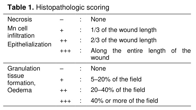
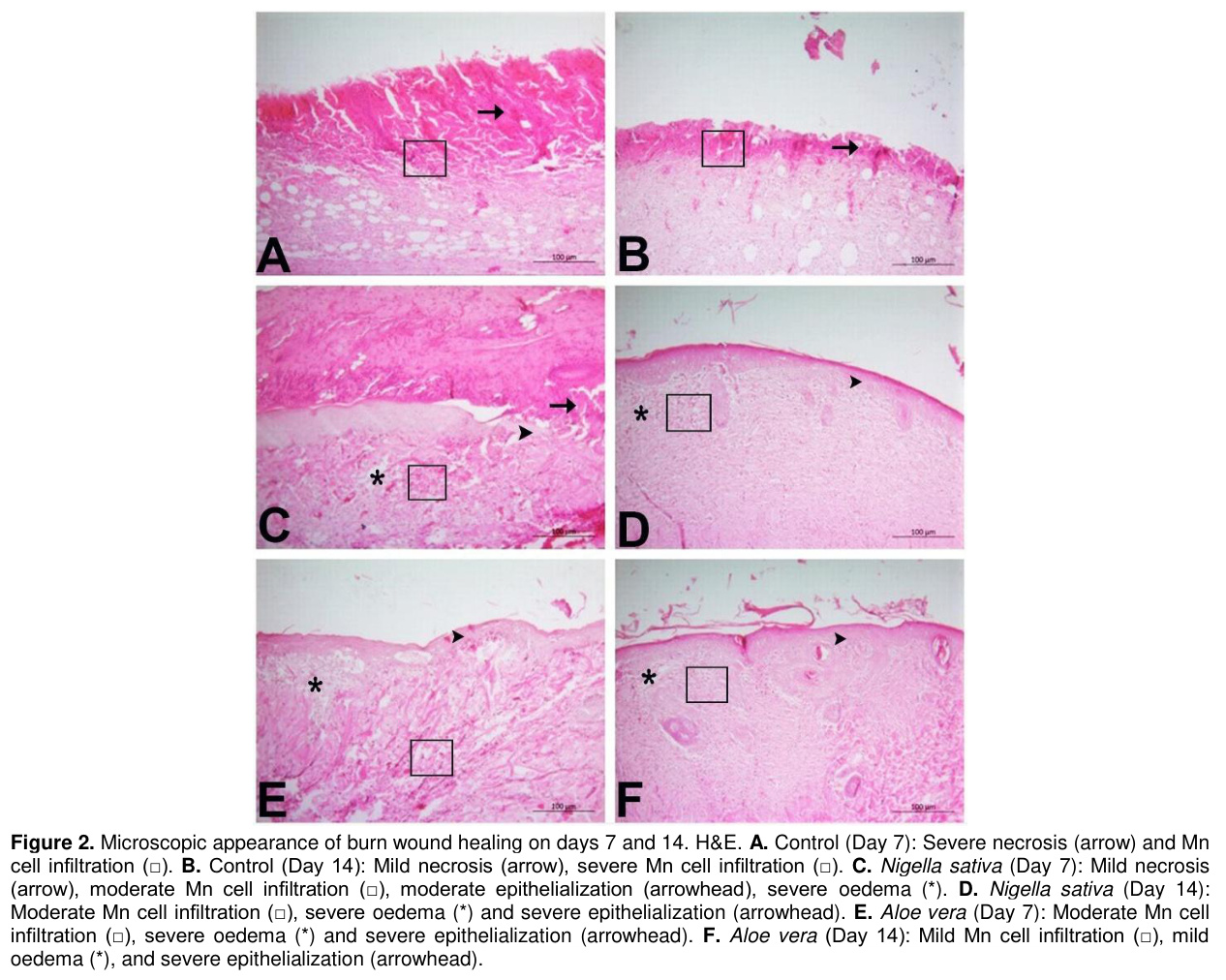
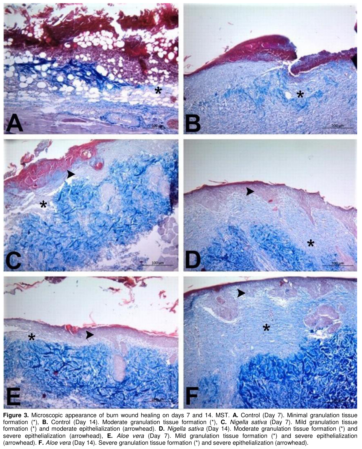
The skin specimens were fixed in 10% neutral buffered formalin. After fixation, routine tissue processing was performed through graded alcohols and xylene, followed by paraffin embedding. Sections of 4 µm thickness were cut and placed on poly-lysine-coated slides. Hematoxylin and eosin staining was applied for routine histological evaluation, while Masson’s trichrome staining was used to assess granulation tissue formation. All slides were examined under a light microscope for necrosis, mononuclear (Mn) cell infiltration, oedema, and epithelialization. Histopathological findings were semi-quantitatively graded as absent (−), mild (+), moderate (++), or severe (+++) (Table 1).
Statistical Analysis: All collected data were analyzed using IBM SPSS Statistics software (version 22). The Shapiro-Wilk normality test was performed to determine whether the raw values of all measured parameters followed a normal distribution. For parametric data showing a normal distribution, a one-way analysis of variance (ANOVA) was performed to determine differences between groups, and the post hoc Tukey test was used for pairwise comparisons. Differences in histopathological findings between groups were determined using the Kruskal-Wallis test, a nonparametric test, and the group showing the difference was identified using the Mann-Whitney U test. A p-value less than 0.05 was considered statistically significant.
Bulgular
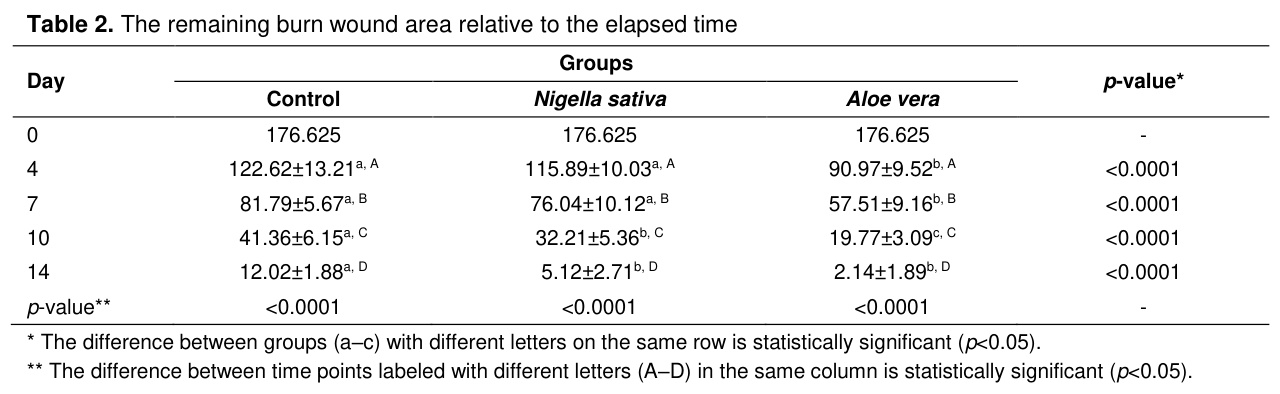
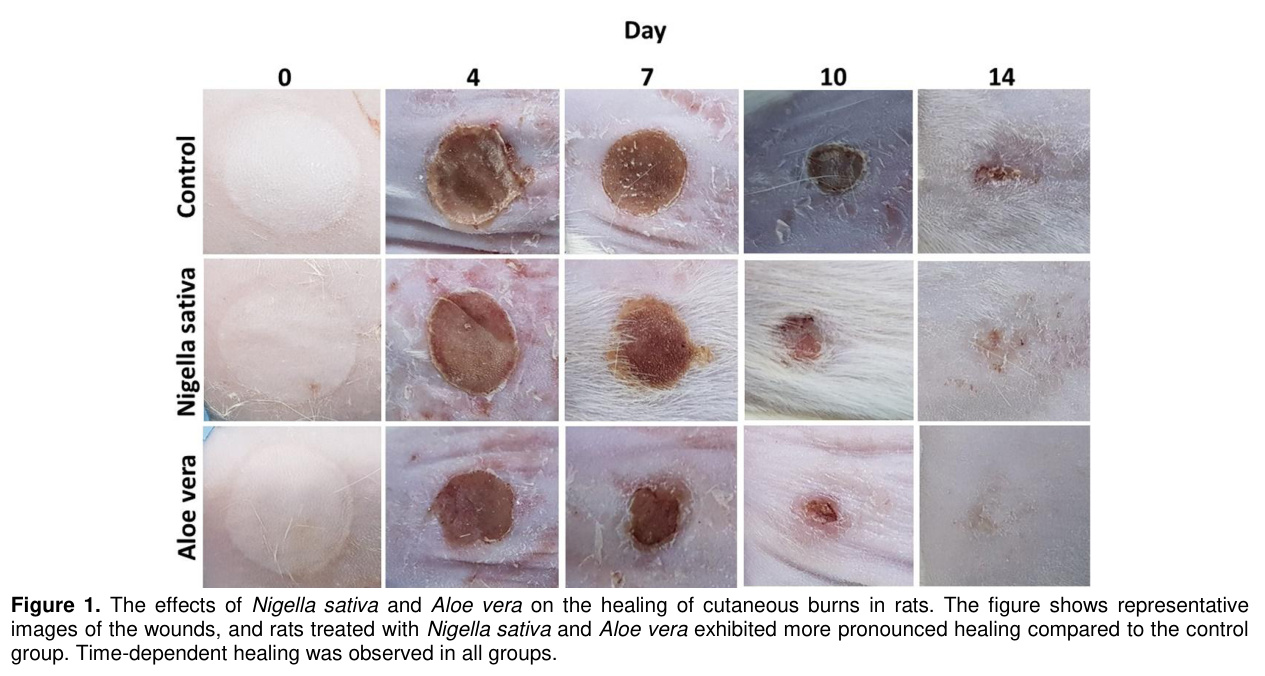
Clinical Findings: Macroscopic examinations revealed marked inflammation and erythema in the burn area on day 1. On days 4 and 7, healing in the AV group was observed to be better than in the other groups (p<0.0001). While scab detachment occurred earlier in the AV group compared to the NS group, no scab detachment was observed in the control group for 14 days. On the 10th day, it was determined that healing in the NS and AV groups was better than in the control group, with the best healing observed in the AV group (p<0.0001). By the fourteenth day, healing in the treatment groups was better than in the control group (p<0.0001); although there was no statistical difference between the NS and AV groups, it was determined that healing in the AV group was numerically better than in the NS group.Macroscopic images illustrating the effects of NS and AV on cutaneous burn healing in rats are presented in Figure 1, while data on the remaining wound area according to the post-burn period are presented in Table 2.
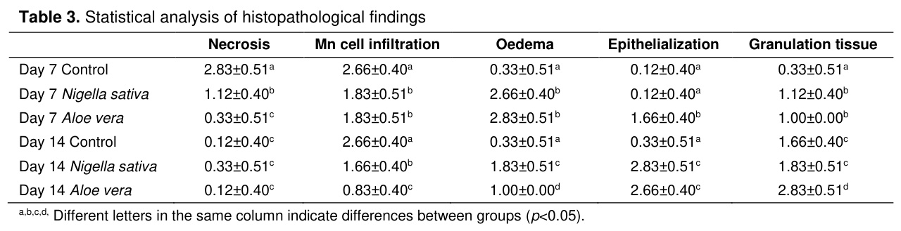
Histopathological Findings: On days 7 and 14, significant differences were detected among the experimental groups regarding necrosis, Mn cell infiltration, oedema, epithelialization, and granulation tissue formation (Table 3).
On day 7, severe necrosis was observed in the control group, while mild necrosis was observed in the NS group. On day 14, mild necrosis was observed in the NS group, while no necrosis was detected in the control and AV groups.
On day 7, Mn cell infiltration was assessed as severe in the control group and moderate in the NS and AV groups. In the AV group, Mn cell infiltration decreased to a mild level on day 14. Oedema was evaluated as severe in both NS and AV groups on day 7, while it decreased to a moderate level in the NS group and a mild level in the AV group on day 14. Epithelialization was not yet fully established in the control group. On day 7, epithelialization was moderate in the NS group and more advanced in the AV group. By day 14, complete epithelialization was observed in both treatment groups.
Although granulation tissue had not yet fully begun to form in the control group by day 7,mild formation was observed in the NS and AV groups. By day 14, granulation tissue formation was graded as moderate in both the control and NS groups, whereas the AV group exhibited advanced granulation tissue development (Figure 3).
Tartışma
Burn healing is a dynamic, multi-stage biological process that begins following tissue damage and involves phases of inflammation, proliferation, and remodeling. The primary clinical goal in this process is to ensure that the wound heals as quickly as possible with minimal scar formation by preventing the development of complications, particularly infection [6,9,13,15,17]. The effects of plant-derived agents on wound healing have long been studied in both traditional and modern medicine [17]. The aim of this study was to investigate the clinical and histopathological effects of NS and AV on an experimental burn model established in rats and to determine any differences in efficacy between the groups.In this study, the burn model was created by applying a 1.5 cm diameter brass probe heated to 100°C to the dorsal region of rats for 15 seconds, in accordance with similar methodologies reported in the literature [6,9,13-15]. The full-thickness second-degree skin burn created by this method allowed for the establishment of a standard tissue injury and the objective monitoring of the healing process. Wound area measurement is of critical importance for the reliability of results in experimental studies. It has been reported that individual variations and measurement errors in drawings made on live animals can lead to deviations in area calculations [6,9,13,14,17]. In this study, digital measurements were performed using ImageJ software instead of traditional acetate paper and millimeter-based calculation methods, with the aim of minimizing measurement errors and enhancing the objectivity of the results.
In this study, macroscopic evaluations revealed that wound area reduction in the AV group on days 4, 7, and 10 was statistically significantly greater than in all other groups (p<0.0001). By day 14, significant improvement was observed in both the NS and AV groups compared to the control group (p<0.0001). However, the data indicate that the AV group’s rate of healing is higher than that of the NS group, suggesting that AV may be a more effective alternative in burn treatment.
In the first few days following a burn injury, the tissues surrounding the central necrotic area may progress to necrosis due to inflammation and oxidative stress. This can lead to an expansion of the area requiring surgical intervention and increased scar formation. In this study, the fact that necrosis remained at a lower level in the treatment groups compared to the control group demonstrates that NS and AV have the capacity to limit tissue loss in the stasis area.
It has been reported that NS possesses antimicrobial, antioxidant, anti-inflammatory, and immunomodulatory properties and accelerates the histopathological healing process in burn models [6]. It is stated that the positive effects of NS on wound healing are associated with the induction of angiogenesis, increased fibroblast proliferation, and stimulation of collagen synthesis. It is also noted that it contributes to reducing tissue damage and bacterial infection by regulating leukocyte activity [18].
AV is widely used due to its antibacterial, antiviral, and anti-inflammatory properties [19,20]. It has been reported that AV is more effective in chronic wounds than in acute wounds and accelerates the healing process of burn wounds due to its anti-inflammatory effects, enhanced immune activity, antibacterial and antiviral effects, and reduced histamine activity [21].
In the histopathological evaluation, collagen deposition and granulation tissue formation during the proliferative phase (key indicators of wound healing) were taken into account [22,23]. By day 7, a significant increase in granulation tissue was observed in the NS and AV groups compared to the control group. By the fourteenth day, it was notable that granulation tissue levels were higher in the AV group compared to the other groups.
When evaluated in terms of epithelialization, the fact that the epithelialization rate in the AV group was significantly higher than in all other groups on day 7 demonstrates the significant positive effect of AV on early-stage epithelialization. The higher levels of necrosis observed in the control group on day 7 and of mononuclear cell infiltration on days 7 and 14; in contrast, the more advanced epithelialization observed in the NS and AV groups on day 14, indicate that NS and AV positively influence the healing process.
One limitation of the present study is that repeated measurements obtained from the same animals were evaluated primarily through time-point-based group comparisons. In addition, the relatively limited sample size in histopathological evaluations should be considered when interpreting the findings. Future studies using longitudinal statistical models and larger sample sizes may provide additional insight into healing dynamics over time.
Sonuç
In conclusion, the findings of this study demonstrate that both AV and NS significantly accelerate burn healing at both the clinical and histopathological levels. However, AV exhibited more pronounced efficacy compared to NS in terms of limiting necrosis, initiating early-stage epithelialization, and achieving faster wound bed reduction. While this study supports the potential of herbal agents in burn treatment, it also highlights the need for comprehensive research evaluating different dosages, application forms, and long-term outcomes to fully integrate these findings into clinical practice and veterinary medicine.References
- Merev HF. İkinci Derece Yanık Yarasında % 1’lik Gümüş Sulfadiazin ile Chitosan, Aloe Vera, Panthenol Kombinasyonu Lokal Etkilerinin Karşılaştırılması. Tıpta Uzmanlık Tezi, Edirne: Trakya Üniversitesi Tıp Fakültesi Plastik Rekonstrüktif ve Estetik Cerrahi Anabilim Dalı, 2016.
- Sancak T. İkinci Derece Yanık Yaralarının Sağaltımında Enjekte Edilebilen Trombositten Zengin Fibrin Kullanımının Termografik ve Histopatolojik Değerlendirilmesi: Tavşan Modeli. Doktora Tezi, Van: Van Yüzüncü Yıl Üniversitesi Sağlık Bilimleri Enstitüsü, 2018.
- Soykan B. Sıçanlarda Yanık Yarasının Murt Ağacı Ekstresi, Amnion Sıvısı ve Gümüş Sülfadiazin İle Sağaltımının Karşılaştırılması. Yüksek Lisans Tezi, Afyonkarahisar: Afyon Kocatepe Üniversitesi Sağlık Bilimleri Enstitüsü, 2020.
- Kaymaz MB. Yanık Yarası Üzerine Cotinus Coggygria (Duman Ağacı) Yaprak Ekstresi ve Fenitoinin Etkileri. Doktora Tezi, Malatya: İnönü Üniversitesi Sağlık Bilimleri Enstitüsü, 2018.
- Aljabre SHM, Randhawa MA, Akhtar N, et al. Antidermatophyte activity of ether extract of Nigella sativa and its active principle, thymoquinone. J Ethnopharmacol 2005; 101(1-3): 116-119.
- Yaman İ, Durmuş AS, Çeribaşı S, Yaman M. Effects of Nigella sativa and silver sulfadiazine on burn wound healing in rats. Vet Med-Czech 2010; 55(12): 619-624.
- Han MC, Durmuş AS, Sağlıyan A, et al. Effects of Nigella sativa and Hypericum perforatum on wound healing. Turk J Vet Anim Sci 2017; 41(1): 99-105.
- El Gazzar M, El Mezayen R, Marecki JC, et al. Anti-inflammatory effect of thymoquinone in a mouse model of allergic lung inflammation. Int Immunopharmacol 2006; 6(7): 1135-1142.
- Durmuş AS, Han MC, Yaman İ. Comperative evaluation of collagenase and silver sulfadiazine on burned wound healing in rats. FÜ Sağ Bil Vet Derg 2009; 23(3): 135-139.
- Kılıç N. The effect of Aloe vera gel on experimentally induced peritoneal adhesions in rats. Revue Med Vet 2005; 156(7): 409-413.
- Majumder R, Das C K, Mandal M. Lead bioactive compounds of Aloe vera as potential anticancer agent. Pharmacol Res 2019; 148: 104416.
- Faydaoğlu E, Sürücüoğlu M. Tıbbi ve aromatik bitkilerin antimikrobiyal, antioksidan aktiviteleri ve kullanım olanakları. Erzincan Üniversitesi Fen Bilimleri Enstitüsü Dergisi 2013; 6(2): 233-265.
- Han MC, Durmuş AS, Karabulut E, Yaman İ. Effects of Turkish propolis and silver sulfadiazine on burn wound healing in rats. Revue Med Vet 2005; 156(12): 624-627.
- Hosnuter M, Gurel A, Babuccu O, et al. The effect of CAPE on lipid peroxidation and nitric oxide levels in the plasma of rats following thermal injury. Burns 2004; 30: 121-125.
- Uckun FM, Orhan C, Tuzcu M, et al. RJX improves wound healing in diabetic rats. Front Endocrinol 2022; 13: 874291.
- Cheng KY, Lin ZH, Cheng YP, et al. Wound healing in streptozotocin-induced diabetic rats using atmospheric-pressure argon plasma jet. Sci Rep 2018; 8(1): 12214.
- Durmus AS, Yaman M, Can HN. Effects of extractum cepae, heparin, allantoin gel and silver sulfadiazine on burn wound healing: An experimental study in a rat model. Vet Med-Czech 2012; 57(6): 287-292.
- Shahani MY, Memon S, Shahani SB, Bano U, Arain SN. Effect of Nigella sativa extract oil on granulation tissue in cutaneous wound: An experimental study in a rabbit model. Med Forum Mon 2013; 24: 72-77.
- Reddy CU, Reddy KS, Reddy JJ. Aloe vera-A wound healer. Asian Journal of Oral Health and Allied Sciences 2011; 1: 91-92.
- Sahu PK, Giri DD, Singh R. et al. Therapeutic and medicinal uses of Aloe vera: A review. Pharmacol Pharm 2013; 4: 599-610.
- Dat AD, Poon F, Pham KBT, Doust J. Aloe vera for treating acute and chronic wounds. Cochrane Database Syst Rev 2012; 2: CD008762.
- Guo S, DiPietro LA. Factors affecting wound healing. J Dent Res 2010; 89: 219-229.
- Çiçek M. Deneysel Olarak Oluşturulan Yara Modelinde Akupunktur’un Yara İyileşmesi Üzerine Etkisinin Araştırılması. Yüksek Lisans Tezi, Tokat: Gaziosmanpaşa Üniversitesi, Sağlık Bilimler Enstitüsü, 2015.
- Determination of Arterial Oxygen Saturation in Newborn Lambs by Pulse Oximetry
- Comparative Isolation of Serum ß + Pre- ß Lipoproteins of Human and Various Domestic Animal Species By Heparin/MnCl2 Precipitation Method
- The Effects of Levamisole on the Pregnancy Rate, Avarage Litter Size, Sexuality, Living Rate and Lactation Period in Rats
- RATLARDA LEVAMİZOL’ÜN GEBE KALMA ORANI, YAVRU SAYISI, CİNSİYETİ, YAŞAMA ORANI VE LAKTASYON SÜRESİNE ETKİSİ
- Separation of Human and Various Animal Lipoproteins by Vertical Agarose Gel Electrophoresis