[ Ana Sayfa | Editörler | Danışma Kurulu | Dergi Hakkında | İçindekiler | Arşiv | Yayın Arama | Yazarlara Bilgi | E-Posta ]
Fırat Üniversitesi Sağlık Bilimleri Veteriner Dergisi
2023, Cilt 37, Sayı 3, Sayfa(lar) 230-236
[ Özet ] [ PDF ] [ Benzer Makaleler ] [ Yazara E-Posta ] [ Editöre E-Posta ]
Deneysel omurilik hasarı oluşturulan ratlarda shilajitin karaciğer ve böbrek üzerine etkisinin histopatolojik ve biyokimyasal incelenmesi
Tunahan SANCAK1, Çağlar OKULMUŞ2, Mehmet Edip AKYOL3, Ömer Faruk KELEŞ4, Eyüp ÇETİN 5
1Sivas Cumhuriyet University, Veterinary Faculty, Department of Surgery, Sivas, TÜRKİYE
2İzmir/Bornova Veterinary Control Institute, Department of Biochemistry, İzmir, TÜRKİYE
3Van Yuzuncu Yil University, Faculty of Medicine, Department of Neurosurgery, Van, TÜRKİYE
4Van Yuzuncu Yil University, Faculty of Veterinary, Department of Pathology, Van, TÜRKİYE
5Health Sciences University, Haydarpasa Training and Research Hospital, Department of Neurosurgery, İstanbul, TÜRKİYE
Anahtar Kelimeler: Böbrek, karaciğer, omurilik hasarı, shilajit
Özet
Amaç: Bu çalışmada deneysel omurilik hasarı oluşturulan ratlarda shilajitin karaciğer ve böbrek üzerine olan etkisinin belirlenmesi amaçlanmıştır.

Gereç ve Yöntem: Bu çalışmada omurilik hasarı oluşturulan ratlara 150 mg/kg ve 250 mg/kg dozlarda 1., 2., ve 3. günlerde shilajit tedavisi uygulanmıştır. Uygulama sonrasında ratlar 14. günde sakrifiye edilerek kan, karaciğer ve böbrek dokuları alınmıştır.

Bulgular: Karaciğer ve böbrek histopatolojik olarak incelendiğinde shilajitin yüksek dozlarının dokularda daha iyi koruyucu etkiye sahip olduğu düşük doz shilajitin ise kısmi düzeyde koruyucu etki gösterdiği belirlenmiştir. Biyokimyasal analizde ise shilajit uygulanan grupların kontrol grubuna istinaden serum serum aspartat aminotransferaz (AST), alanin aminotransferaz (ALT), laktat dehidrojenaz (LDH), kreatin kinaz (CK), kreatin ve üre konsantrasyonlarının daha düşük, albümin (ALB) ve total protein (TP) konsantrasyonlarının ise daha yüksek çıkmıştır (P aralığı: 0.05–0.001).

Öneriler: Sonuç olarak, bu çalışmada ratlarda spinal kord hasarı sonrasında Shilajit uygulamasının antiinflamatuar özelliği ile karaciğer ve böbrekte lezyon oluşumunu önemli ölçüde baskıladığı histopatolojik ve biyokimyasal olarak ortaya konmuştur.

  • Başa Dön
  • Özet
  • Giriş
  • Materyal ve Metot
  • Bulgular
  • Tartışma
  • Kaynaklar
  • Giriş
    It is well known that the spinal cord is prone to injuries causing damage to its neural structures, which can lead to the destruction of its locomotor and sensory functions. As one of the most significant traumas of the central nervous system (CNS), spinal cord injuries can have devastating neurological consequences. Primary spinal cord injury is caused by the destruction of neurons and axons due to mechanical causes such as traction injury and cerebral trauma, while histological and neurological damages, ischemia, apoptosis, necrosis, edema, inflammation, and formation of reactive oxygen species are the causes of secondary damage 1-5. Primary mechanical spinal cord injuries can rapidly trigger secondary injury and induce an inflammatory response and neuronal apoptosis in the damaged area6.

    Spinal cord injury disrupts the autonomic nervous system and impairs the coordination of organ functions in the body7. The resulting neurogenic damage can affect many systems of the body, such as the function of the lungs, liver, intestines, kidneys, and bladder, hormone release, and sexual function, due to biochemical, molecular, and cellular changes in the body. After the injury, the regeneration of spinal cord axons is very limited due to the low growth capacity of neurons, the presence of central nervous system myelin growth inhibitory factors, glial scar tissue formation, and lack of neurotrophic factors and nerve growth factors 8-10.

    The treatment of spinal cord injuries is still challenging in clinical practice due to the poor regeneration ability of neurons6. Despite recent advances in medicine, most spinal cord injury treatments are for temporary minimal functional recovery11. Common treatment modalities for spinal cord injuries include the use of anti-inflammatory drugs (ketorolac, minocycline, riluzole, magnesium, etc.), decompression surgery (decompression and instrumentation) to stabilize the spine, and supportive therapy to prevent secondary damage12. In spinal cord injuries, the circulation initiates a systemic inflammation in the tissues, defined by a rapid rise of chemokines and leukocytes in the lungs, liver, and kidneys, resulting in edema and hemorrhage. This systemic inflammation causes acute and chronic multiple organ dysfunction in patients with spinal cord injury13,14.

    Antioxidant and anti-inflammatory agents have been found to be clinically beneficial in dysfunctions that occur in organs because of spinal cord injury 10. Shilajit, also referred to as Salajit, Shilajatu, Mumie, or Mummiyo, is a semi-solid mineral pitch with a brown to black color that is mainly exuded from steep rocks in the Himalayas (especially in altitudes of 1000 to 5000 meters) and mountains in other nations like Afghanistan, Pakistan, Iran, Nepal, China, and Tibet15. Shilajit is one of the major Herbomineral remedies containing fulvic acid and humic acid with antioxidant properties. The Shilajit contains Shilajityl acetate, shilajitol, shilaceatatechol, shilaxanthone, shilanthranil and also consist major organic constituents included benzoic acid, hippuric acid, fatty acids, resin and waxy materials, gums, albuminoids and vegetable matter with benzoic acid being the active ingredient16. Shilajit has extensive medicinal value in various systems of traditional medicine and is widely used to treat a number of diseases worldwide. Numerous studies have shown that Shilajit has various bioactivity such as antioxidant, anti-inflammatory, antiviral, and immunomodulatory properties17-22.

    The aim of this study was to investigate histopathologically and biochemically the effect of shilajit on liver and kidney structure and function in spinal cord injury-induced rats.

  • Başa Dön
  • Özet
  • Giriş
  • Materyal ve Metot
  • Bulgular
  • Tartışma
  • Kaynaklar
  • Materyal ve Metot
    Research and Publication Ethics: This study is subject to the permission of Sivas Cumhuriyet University Animal Experiments Local Ethics Committee dated 12.05.2023 and numbered 65202830-050.04.04.

    Animal Material: In the study, 24 male Wistar albino rats, each weighing 200-250 g, were used. Adult and pathogen-free, male Wistar albino rats were procured from Van Yüzüncü Yıl University University Experimental Medicine Application and Research Center. The animals were fed ad libitum and kept in 12 h of light and 12 h of darkness per day. Their habitat had an average temperature of 26°C and 60% relative humidity.

    Xylazine hydrochloride (Rompun, Bayer Türk Kimya Sanayi, İstanbul) 10 mg/kg and Ketamine hydrochloride (Ketalar, Pfizer PFE İlaçları AŞ, İstanbul) 50 mg/kg were administered intraperitoneally to the rats to achieve general anesthesia before the procedure.

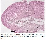
    Operation Method: Under general anesthesia, the operation areas of the rats were shaved and sterilized. Spinal cord injury was induced in all groups according to the modified Allens method23 (Figure 1). For this purpose, a 2 cm incision was made in the midline at the level of the 8th thoracic vertebra of the rats, and the spinous process and laminae of T8 were removed and the spinal cord was exposed. Subsequently, a 10-gram weight was dropped on the spinal cord at a distance of 5 cm and left on it for 3 min, and the layers were sutured in accordance with the anatomy after the operation. After the operation, a 5 mL intraperitoneal saline infusion was performed.


    Büyütmek İçin Tıklayın
    Figure 1: Spinal Injury Group: Showing the edema, degeneration and inflammatory reaction in the area of spinal injury.

    Trial Groups: Following the operation, 24 rats were randomly assigned to three groups (n: 8 rats). No medication was administered to the control group rats. In this study, rats were divided into two groups: those to receive a low dosage (150 mg/kg) and those receiving a high dosage (250 mg/kg)24.

    1. Control Group: It is the group in which spinal cord injury was induced but no medication was administered.

    2. Low-dose group: This group received intraperitoneal Shilajit at a dose of 150 mg/kg at the post-op 1st h, 1st day, 2nd day, and 3rd day after spinal cord injury was induced.

    3. High dose group: This is the group in which intraperitoneal Shilajit is given at a dose of 250 mg/kg at the post-op 1st h, 1st day, 2nd day, and 3rd day after spinal cord injury was induced.

    On the 14th day, under general anesthesia, the blood samples were collected from the heart of rats and they were euthanized. After euthanasia, blood samples were sent to the laboratory for biochemical analysis, and liver and kidney tissue samples were sent to the laboratory for histopathological examination.

    Histopathological Examination: After the experiment, the kidney and liver tissues of the necropsied rats were fixed in a 10% buffered formalin solution. The tissue samples were embedded in paraffin blocks after routine tissue follow-up procedures. Using a Rotary microtome, sections of 4 um were taken from paraffin blocks. Sections were stained with hematoxylin-eosin (HE), examined, and photographed under a light microscope (Nikon 80i, DS-RI2; Nikon, Japan).

    Biochemical Analysis: Blood samples taken during the experimental period were centrifuged at 3500 rpm for 10 min. Sera separated from blood were stored at -80°C until analyzed for biochemical parameters. Serum glucose, alanine aminotransferase (ALT), aspartate aminotransferase (AST), alkaline phosphatase (ALP), triglyceride (TG), total cholesterol (TC), creatine, BUN, lactate dehydrogenase (LDH), creatine kinase (CK), total protein (TP), albumin (ALB) and total bilirubin (T-Bil) levels were determined by the spectrophotometric method in an autoanalyzer (Mindray BS-200, China) with commercial test kits (Mindray, China).

    Statistical Analysis: The data were analyzed using the SPSS Windows 20.0 statistical software package. Mean values and standard deviations were calculated for each evaluated indicator. One-way analysis of variance (ANOVA) was conducted to analyze all parameters followed by a Bonferroni Post-hoc test for pairwise comparisons. The results were considered significant at P<0.05.

  • Başa Dön
  • Özet
  • Giriş
  • Materyal ve Metot
  • Bulgular
  • Tartışma
  • Kaynaklar
  • Bulgular
    Histopathological Findings
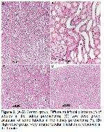
    1. Kidney: The control group: The most remarkable morphologic change in the histologic examination of the kidneys was the presence of multifocal dilatation (*) in the both the cortex and medullary tubules and lymphatics. The epithelium of the dilated tubules was flattened. Moreover, atrophy of the glomerular tangle and enlargement of Bowman's space was found in some glomeruli. Mild degenerative-necrotic changes in tubular epithelial cells were also observed in some tubules (Figure 2A-B).

    Low-dose group: Remarkably, the changes detected in the control group were found at milder levels (*) in this group (Figure 2C).

    The high-dose group: Although dilated tubules were observed very rarely in the renal parenchyma (*), an almost normal histologic appearance was observed in general (Figure 2D).


    Büyütmek İçin Tıklayın
    Figure 2: (A-B) Control group: Diffuse multifocal dilatation (*) of tubules in the kidney parenchyma; (C) Low dose group: Dilatation of some tubules in the kidney parenchyma (*); (D) High dose group: Very sparse tubular dilatation is observed in the tubules.

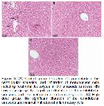
    2. Liver: The control group: The most striking morphologic change in the histologic examination of the liver was dilatation specifically in the centrilobular sinusoids and infiltration of inflammatory cells (hepatitis) as focal foci or scattered in the sinusoids (Figure 3A).

    Low-dose group: In the liver parenchyma, the sinusoids appeared almost normal and the infiltration of inflammatory cells was much less intense than in the Control Group (Figure 3B).


    Büyütmek İçin Tıklayın
    Figure 3: (A) Control group: Dilatation (*), particularly in the centrilobular sinusoids, and infiltration of mononuclear cells including neutrophil leukocytes in the sinusoids (arrows). (B) Low dose group: No significant dilatation of the centrilobular sinusoids and less infiltration of inflammatory cells. (C) High dose group: No significant dilatation of the centrilobular sinusoids and minimal infiltration of inflammatory cells.

    The high-dose group: It was found that the sinusoids were almost normal in the liver parenchyma and the infiltration of inflammatory cells in the sinusoids was minimal (sparse) (Figure 3C).

    In the histopathological examinations, some morphologic changes (tubular dilatation and hepatitis) were detected in the kidneys and livers. In groups treated with shilajit, these morphologic changes remarkably were, dose-dependently, much less intense than in the Control Group. It is considered that sinusoidal dilatation and infiltration of inflammatory cells were induced in the liver by the effect of inflammatory chemical mediators released due to experimentally induced spinal cord injury, and that the administration of the Shilajit significantly suppressed the formation of these lesions with its anti-inflammatory properties; in addition, tubular dilatation caused by impaired intrarenal urine flow as a result of spinal cord injury was restored by the phytochemical compounds of the Shilajit.

    Biochemical Analysis: The effects of the surgical procedure and Shilajit treatments on serum concentrations of glucose, ALT, AST, ALP, TG, TK, creatine, urea, BUN, LDH, TP, ALB, and T-Bil are shown in Table 1. In all biochemical parameters, there was no significant difference found between the low-dose (150 mg/kg) and high-dose (200 mg/kg) shilajit-treated groups (P>0.05). Glucose, ALP, TG, TK, and T-Bil concentration changes between the groups in the present study were insignificant (P>0.05). The group treated with low dose (150 mg/kg) shilajit had lower serum ALT, AST, LDH, Creatine, and CK (P<0.001) concentrations (P<0.05) and higher ALB concentrations (P<0.001) than the control group. In treatment with high dose (250 mg/kg) shilajit, meanwhile, this change was more pronounced and serum ALT (P<0.001), AST (P<0.001), CK (P<0.001), creatine, BUN, and LDH concentrations were lower (P<0.05), whereas TP and ALB (P<0.001) concentrations were higher (P<0.05).


    Büyütmek İçin Tıklayın
    Table 1: Mean serum glucose, ALT, AST, ALP, TG, TK, CK, creatine, BUN, LDH, TP, ALB, and T-Bil concentrations following the surgical procedure and different doses of Shilajit treatment in rats

  • Başa Dön
  • Özet
  • Giriş
  • Materyal ve Metot
  • Bulgular
  • Tartışma
  • Kaynaklar
  • Tartışma
    Experimental models have been developed in recent years to unravel the pathophysiology and effects of spinal cord injuries, the cellular and molecular mechanisms that underlie tissue regeneration and repair in the injured spinal cord, and to monitor the injury process25. Most injuries do not completely damage the spinal cord. Primary damage has been described by four main characteristic mechanisms. These are (a) impact and permanent compression, (b) impact and temporary compression, (c) irritation, and (d) laceration/ transection. Compression and compression types are the most common types in spinal cord injuries25,26. In order to evaluate treatment efficacy and associated mechanisms in spinal cord injuries, a weight-drop contusion method was developed. After performing laminectomy at level T8, Ko et al.27 and Lv et al.28 induced experimental spinal cord injury by dropping a 10g weight from a height of 5 cm onto the spinal cord surface. The spinal cord injury generation method used in the present study is similar to the method used in the studies of Ko et al.27 and Lv et al.28 (Figure 1).

    Spinal cord injury is a devastating event that results in significant physical disabilities for affected individuals. Aside from local damage to the spinal cord, patients with spinal cord injuries suffer from various complications including multiple organ dysfunction and failure as a result of systemic inflammatory responses. Injured patients are likely to develop neurogenic pain, depression, lung damage, cardiovascular disease, liver damage, renal dysfunction, urinary tract infection, and increased susceptibility to pathogen infections, all of which impede functional recovery and may even pose a life-threatening situation29-31. In the current study, lesions occurred in the kidneys, and liver of rats (Figure 2A-B, 3A). These conditions were histopathologically demonstrated. The lesions in the liver and kidneys observed in the study shows similar characteristics as described by Gris et al.29, Bao et al.30, and Sun et al.31.

    Spinal cord injury not only affects motor and sensory functions but also leads to serious and long-term complications due to impaired nerve conduction to vital organs. Autonomic innervation of the kidney is interrupted due to spinal cord injury. Consequently, renal arterial blood flow, sodium and potassium excretion, and renal plasma flow are decreased, leading to renal dysfunction32-34. In their study, Kandhare et al.35 found that spinal cord injury-induced rats displayed a change in glomerular permeability due to kidney damage. In the studies by Shunmugavel et al.36 and Akakin et al.37, degeneration of renal tubules was observed in spinal cord injury-induced rats. Besides, Sakarcan et al.38 observed enlargement in Bowman's cavity, atrophy in glomeruli, and degeneration in tubules in spinal cord injury-induced rats. The findings of the above-mentioned studies are align with the histopathology results (Figure 2) of our study in the kidney tissue.

    Liver function is regulated by autonomic parasympathetic innervation from the brain stem and sympathetic innervation from the thoracic spinal cord. Hence, spinal cord injury at or above the thoracic levels disrupts the main regulatory mechanisms for hepatic functions. Furthermore, the liver even has a major role in initiating and prolonging systemic inflammation after spinal cord injury39,40. In the study by Hundt et al.41, leukocyte infiltration was formed in the liver after the experimental spinal cord injury in rats, and in the study by Mohamed et al.40, dilatation and cellular infiltration in the sinusoids due to liver injury in spinal cord injury-induced rats were determined. Moreover, in the study by Goodus et al.42, leukocyte cell infiltrations were also observed in the liver in spinal cord injury-induced rats. In the present study, the histopathological changes in the liver (Figure 2) are similar to those observed by Mohamed et al.40, Hundt et al.41, and Goodus et al.42.

    The pathophysiology of spinal cord injury comprises a series of destructive events that occur acutely or chronically, including ischemia, oxidative stress, inflammatory changes, apoptosis, necrosis, and locomotor dysfunction. A variety of therapeutic strategies are being developed to overcome neurodegenerative events and reduce secondary neuronal damage. Despite continued research into neuroprotective and neurodegenerative therapies, no treatment has been demonstrated to provide full recovery in spinal cord injuries. Thus, researchers are still searching for new medical therapies for the treatment of spinal cord injuries43-45. For a successful outcome in spinal cord injuries, drugs such as vasopressors, steroids, anti-inflammatory agents, and rehabilitation therapies should be combined with a consideration of the complex pathophysiology of the disease46. Shilajit is a compound used in conventional medicine that exhibits anti-inflammatory, analgesic, antioxidant, and neuroprotective activities47,48. Likewise, in this study in which different doses of shilajit were used, its positive effects on the liver and kidney were also proven histopathologically and biochemically. Despite the minimal pathological changes observed in the liver and kidneys in the groups administered high-dose shilajit (250 mg/kg), the pathological changes were moderate when administered at a low dose (150 mg/kg), whereas the pathological changes were advanced in the untreated group (Figure 2-3). The results of serum biochemistry in the present study (Table 1) provide evidence of spinal cord injury-induced damage to the kidney and liver tissue in rats and histopathological findings showing the healing effect of shilajit on these tissues. Compared with the control group, the decrease in ALT and AST serum concentrations, and increase in TP (P<0.05) and ALB (P<0.001) levels in the groups treated with low (P<0.05) and high (P<0.001) dose shilajit reflect functional liver recovery and the healing role of shilajit. AST and ALT in particular are important liver enzymes that are found in higher concentrations in the cytoplasm. Elevated AST and ALT are directly proportional to acute injury40,49. Additionally, BUN and creatine enzymes35,39, which are among the parameters indicating renal function, were lower (P<0.05) in the shilajit-treated groups compared to the control group, which biochemically proves the histopathological changes occurring in the kidneys due to spinal cord injury in shilajit-treated rats. Furthermore, CK and LDH enzymes, which are markers of skeletal muscle injuries50, decreased more significantly (P<0.05) in the serum levels of the high shilajit-treated group in the present study, which biochemically reveals the tissue damage inhibitory property of this substance.

    Direk et al.10 demonstrated that atrophy of kidney glomeruli and degeneration of tubules occurred in spinal cord injury-induced rats. Cyclotrichium origanifolium (mountain mint) and Thymbra spicata L. var. spicata (zahter) they used in their studies partially prevented kidney damage. This damage caused degeneration in the kidneys, dilation in the Bowman space, and atrophy in the glomerulus in spinal cord injury-induced rats, as reported by Sakarcan et al.38 in their study. It was shown that riboflavin had a protective effect against these damages in the kidneys in the studies conducted. Similarly, in the present study, low-dose shilajit treatment offered a partial protective effect on kidney damage in spinal cord injury-induced rats compared with the control group, whereas this was more pronounced in high-dose shilajit treatment, and an effect with normal kidney histology was observed. This suggests that shilajit administration at a dose of 250 mg/kg may prevent kidney damage after spinal cord injury better than a 150 mg/kg dose.

    Mohamed et al.40 showed that liver damage occurred in spinal cord injury-induced rats and revealed that liver damage decreased with granulocyte colony-stimulating factor (G-CSF) used for treatment. In their study, Bao et al.51 used anti-CD11d to reduce the damage in the treatment of liver damage after experimental spinal cord injury. In the current study, it has been confirmed through both histopathology results and changes in liver enzyme levels that the use of a high dose of Shilajit (250 mg/kg) significantly reduces the liver damage resulting from spinal cord injury when compared to the control group. Low-dose shilajit administration (150 mg/kg) partially prevented liver damage following spinal cord injury. This suggests that shilajit at a dose of 250 mg/kg to be used after spinal cord injury may prevent liver damage better than the used protocol at a dose of 150 mg/kg.

    In conclusion, the protective effect of the shilajit compound against liver and kidney dysfunctions due to spinal cord injury in rats was investigated. In this study, it has been revealed histopathologically and biochemically that administration of the Shilajit significantly suppressed the formation of the lesions in liver and kidney with its anti-inflammatory properties after spinal cord injury in rats. In order to gain a better understanding of the mechanism of action of shilajit, further experimental studies need to be conducted.

  • Başa Dön
  • Özet
  • Giriş
  • Materyal ve Metot
  • Bulgular
  • Tartışma
  • Kaynaklar
  • Kaynaklar

    1) Ramer LM, Ramer MS, Bradbury EJ. Restoring function after spinal cord injury: Towards clinical translation of experimental strategies. Lancet Neurol 2014; 13: 1241-1256.

    2) Bi Y, Zhu Y, Zhang M, et al. Effect of shikonin on spinal cord injury in rats via regulation of HMGB1/TLR4/NF-kB Signaling Pathway. Cell Physiol Biochem 2017; 4: 481-49.

    3) Ruzicka J, Machova-Urdzikova L, Gillick J. A comparative study of three different types of stem cells for treatment of rat spinal cord injury. Cell Transplant 2017; 26: 585-603.

    4) Cizkova D, Cubinkova V, Smolek T, et al. Localized intrathecal delivery of mesenchymal stromal cells conditioned medium improves functional recovery in a rat model of spinal cord injury. Int J Mol Sci 2018; 19: 870-883.

    5) Liu W, Tang P, Wang J, et al. Extracellular vesicles derived from melatonin‐preconditioned mesenchymal stem cells containing USP29 repair traumatic spinal cord injury by stabilizing NRF2. J Pineal Res 2021; 71: e12769.

    6) Yu T, Zhao C, Hou S, et al. Exosomes secreted from miRNA-29b-modified mesenchymal stem cells repaired spinal cord injury in rats. BJMBR 2019; 52: 1-8.

    7) Kigerl KA, Mostacada K, Popovich PG. Gut microbiota are disease-modifying factors after traumatic spinal cord injury. Neurotherapeutics 2018; 15: 60-67.

    8) Liu J, Han D, Wang Z, et al. Clinical analysis of the treatment of spinal cord injury with umbilical cord mesenchymal stem cells. Cytotherapy 2013; 15: 185-191.

    9) Goodus MT, Sauerbeck AD, Popovich PG, Bruno RS, McTigue DM. Dietary green tea extract prior to spinal cord injury prevents hepatic iron overload but does not improve chronic hepatic and spinal cord pathology in rats. J Neurotrauma 2018; 35: 2872-2882.

    10) Direk FK, Tuncer MC, Deveci E, ve ark. Deneysel travmatik omurilik hasarında Thymbra Spicata L. var Spicata ve Cyclotrichium Origanifolium’un böbrek üzerine koruyucu etkisinin araştırılması. Dicle Tip Derg 2021; 48: 299-308.

    11) Song YH, Agrawal NK, Griffin JM, Schmidt CE. Recent advances in nanotherapeutic strategies for spinal cord injury repair. Adv Drug Deliv Rev 2019; 148: 38-59.

    12) Venkatesh K, Ghosh SK, Mullick M, Manivasagam G, Sen D. Spinal cord injury: Pathophysiology, treatment strategies, associated challenges, and future implications. Cell Tissue Res 2019; 377: 125-151.

    13) Sauerbeck AD, Laws JL, Bandaru VV, et al. Spinal cord injury causes chronic liver pathology in rats. J Neurotrauma 2015; 32: 159-169.

    14) Bloom O, Herman PE, Spungen AM. Systemic inflammation in traumatic spinal cord injury. Exp Neurol 2020; 325: 113143.

    15) Jafari M, Forootanfar H, Ameri A, et al. Antioxidant, cytotoxic and hyperalgesia-suppressing activity of a native Shilajit obtained from Bahr Aseman mountains. Pak J Pharm Sc 2019; 32: 2167-2173.

    16) Pandit SB, Pagar HJ, Patel TR, Darwade AP, Jadhav SA. In vivo study of nephroprotective potential of shilajit by using cisplatin induced nephrotoxicity model. AJPP 2018; 4: 680-685.

    17) Azizi S, Kheirandiah R, Azari O, Torabi N. Potential pharmaceutic effect of Shilajit (mumie) on experimental osteoarthritis in rat. Comp Clin Path 2018; 27: 755-764.

    18) Ojha R, Gupta AK, Pathak R, Pandey SK. Shilajit an elixir of ayurveda: A literary review of traditional usage as well as modern findings. IJAR 2021; 7(8): 323-332.

    19) Jambi EJ, Alshubaily FA. Shilajit potentiates the effect of chemotherapeutic drugs and mitigates metastasis induced liver and kidney damages in osteosarcoma rats. Saudi J Biol Sci 2022; 29: 103393.

    20) Derhami A, Rajabi S, Rad GH, Jafari F. The Effect of Shilajit on Carboplatin-induced Thrombocytopenia and Oxidative Stress in Rats. J Pharm Negat 2022; 621-625.

    21) Alshubaily FA, Jambi, EJ. Correlation between antioxidant and anti-osteoporotic activities of shilajit loaded into chitosan nanoparticles and their effects on osteoporosis in rats. Polymers 2022; 14: 3972.

    22) Jadhav P, Pagar H. Evaluation of Antidepressant activity of Shilajit in experimental animals. AJPT 2021; 9: 01-05.

    23) Allen AR . Surgery of experimental lesion of spinal cord equivalent to crush injury of fracture dislocation of spinal column a preliminary report. J Am Med Assoc 1911; 57: 878-880.

    24) Firozsalari FG, Shahrokhi N, Hadad MK, Asadikaram G, Atashbar J. Effect of shilajit on the levels of pro-inflammatory and anti-inflammation cytokines in hepatic injury in male rats. J Maz Univ Med Sci 2018; 28: 1-13.

    25) Alizadeh A, Dyck SM, Karimi-Abdolrezaee S. Traumatic spinal cord injury: An overview of pathophysiology, models and acute injury mechanisms. Front Neurol 2019; 10: 282.

    26) Hayta E, Elden H. Acute spinal cord injury: A review of pathophysiology and potential of non-steroidal anti-inflammatory drugs for pharmacological intervention. J Chem Neuroanat 2018; 87: 25-31.

    27) Ko WK, Kim SJ, Jo MJ, et al. Ursodeoxycholic acid inhibits inflammatory responses and promotes functional recovery after spinal cord injury in rats. Mol Neurobiol 2019; 56: 267-277.

    28) Lv R, Du L, Zhang L, Zhang Z. Polydatin attenuates spinal cord injury in rats by inhibiting oxidative stress and microglia apoptosis via Nrf2/HO-1 pathway. Life Sci 2019; 217: 119-127.

    29) Gris D, Hamilton EF, Weaver LC. The systemic inflammatory response after spinal cord injury damages lungs and kidneys. Exp Neurol 2008; 211: 259-270.

    30) Bao F, Omana V, Brown A, Weaver LC. The systemic inflammatory response after spinal cord injury in the rat is decreased by α4β1 integrin blockade. J Neurotrauma 2012; 29: 1626-1637.

    31) Sun X, Jones ZB, Chen XM, et al. Multiple organ dysfunction and systemic inflammation after spinal cord injury: A complex relationship. J Neuroinflammation 2016; 13: 1-11.

    32) Mestre H, Alkon T, Salazar S, Ibarra A. Spinal cord injury sequelae alter drug pharmacokinetics: An overview. Spinal Cord 2011; 49: 955-960.

    33) Rodríguez-Romero V, Guízar-Sahagún G, Castañeda-Hernández G, Reyes JL, Cruz-Antonio L. Early systemic alterations in severe spinal cord injury: An experimental study on the impact of injury level on renal function. Spine 2018; 43: E885-E890.

    34) Ghnenis AB, Jones C, Sefiani A, et al. Evaluation of the cardiometabolic disorders after spinal cord injury in mice. Biology 2022; 11: 495.

    35) Kandhare AD, Shivakumar V, Rajmane A, Ghosh P, Bodhankar SL. Evaluation of the neuroprotective effect of chrysin via modulation of endogenous biomarkers in a rat model of spinal cord injury. J Nat Med 2014; 68: 586-603.

    36) Shunmugavel A, Khan M, Te Chou PC, et al. Simvastatin protects bladder and renal functions following spinal cord injury in rats. J Inflamm 2010; 7: 1-10.

    37) Akakin D, Kiran D, Ozkan N, et al. Protective effects of melatonin against spinal cord injury induced oxidative damage in rat kidney: A morphological and biochemical study. Acta Histochemica 2013; 115: 827-834.

    38) Sakarcan S, Erşahin M, Eminoğlu ME, et al. Riboflavin treatment reduces apoptosis and oxidative DNA damage in a rat spinal cord injury model. Clin Exp Health Sci 2017; 7: 55-63.

    39) Goodus MT, McTigue DM. Hepatic dysfunction after spinal cord injury: A vicious cycle of central and peripheral pathology? Exp Neurol 2020; 325: 113160.

    40) Mohamed DA, Mohamed NM, Abdelrahaman S. Histological and biochemical changes in adult male rat liver after spinal cord injury with evaluation of the role of granulocyte-colony stimulating factor. Ultrastruct Pathol 2020; 44: 395-411.

    41) Hundt H, Fleming JC, Phillips JT, Lawendy A, et al. Assessment of hepatic inflammation after spinal cord injury using intravital microscopy. Injury 2011; 42: 691-696.

    42) Goodus MT, Carson KE, Sauerbeck AD, et al. Liver inflammation at the time of spinal cord injury enhances intraspinal pathology, liver injury, metabolic syndrome and locomotor deficits. Exp Neurol 2021; 342: 113725.

    43) Anjum A, Yazid MD, Fauzi Daud M, et al. Spinal cord injury: Pathophysiology, multimolecular interactions, and underlying recovery mechanisms. Int J Mol Sci 2020; 21: 7533.

    44) Öztürk G, Silav G, İncir S, ve ark. Ratlarda deneysel spinal kord hasar modelinde genisteinin nöroprotektif etkisinin araştırılması, diffüz tensor görüntüleme ile değerlendirilmesi. IGUSABDER. 2020; 11: 130-149.

    45) Yurtal Z, Kutlu T, Altuğ ME, ve ark. Investigation of neuroprotective effect of kefir in experimental spinal cord injury. Ankara Univ Vet Fak Derg 2023; 70: 9-19.

    46) Boyalı O, Civelek E, Kabataş S. Travmatik omurilik yaralanmasında konservatif tedavi (güncel farmakolojik tedavi yöntemleri). Türk Nöroşir Derg 2020; 30: 466-474.

    47) Das AS, El Masry M, Gnyawali SC, et al. Skin transcriptome of middle-aged women supplemented with natural herbo-mineral shilajit shows induction of microvascular and extracellular matrix mechanisms. J Am Coll Nutr 2019; 38: 526-536.

    48) Pandey, PS. Shilajit—A wonder drug of ayurveda: An overview. Int J Pharm Sci Rev Res 2019; 59: 140-143.

    49) Albayrak S, Atci IB, Kalayci M, et al. Effect of carnosine, methylprednisolone and their combined application on irisin levels in the plasma and brain of rats with acute spinal cord injury. Neuropeptides 2015; 52: 47-54.

    50) Barros NDA., Aidar FJ, Marçal AC, et al. Effects of resistance training on oxidative stress markers and muscle damage in spinal cord injured rats. Biology 2022; 11: 32.

    51) Bao F, Brown A, Dekaban GA, et al. CD11d integrin blockade reduces the systemic inflammatory response syndrome after spinal cord injury. Exp Neurol 2011; 231: 272-283.

  • Başa Dön
  • Özet
  • Giriş
  • Materyal ve Metot
  • Bulgular
  • Tartışma
  • Kaynaklar
  • [ Başa Dön ] [ Özet ] [ PDF ] [ Benzer Makaleler ] [ Yazara E-Posta ] [ Editöre E-Posta ]
    [ Ana Sayfa | Editörler | Danışma Kurulu | Dergi Hakkında | İçindekiler | Arşiv | Yayın Arama | Yazarlara Bilgi | E-Posta ]