[ Ana Sayfa | Editörler | Danışma Kurulu | Dergi Hakkında | İçindekiler | Arşiv | Yayın Arama | Yazarlara Bilgi | E-Posta ]
Fırat Üniversitesi Sağlık Bilimleri Veteriner Dergisi
2014, Cilt 28, Sayı 1, Sayfa(lar) 019-024
[ Özet ] [ PDF ] [ Benzer Makaleler ] [ Yazara E-Posta ] [ Editöre E-Posta ]
Türkiye'de Gökkuşağı Alabalığı (Oncorhynchus mykiss Walbaum, 1792) Ovarial Sıvılarından İzole Edilmiş İnfeksiyöz Pankreatik Nekrozis Virüsünün, VP2 Bölgesinin Kısmi Nükleotid Dizin Analizinin Belirlenmesi
Metin GÜRÇAY1, Ayşe PARMAKSIZ1, Ahmet SAİT2, Selehattin GÜRÇAY3
1Veteriner Kontrol Enstitüsü, Elazığ, TÜRKİYE
2Veteriner Kontrol Enstitüsü, İstanbul, TÜRKİYE
3Su Ürünleri İstasyonu, Elazığ, TÜRKİYE
Anahtar Kelimeler: Infectious pancreatic necrosis virus, ovarial sıvı, Gökkuşağı alabalığı, nükleotid sekansı, viral proteininin VP2 bölgesi
Özet
Bu çalışmada, gökkuşağı alabalığı (Oncorhynchus mykiss, Walbaum 1792) ovarial sıvılarından izole edilen, İnfeksiyöz Pankreatik Nekrozis Virüsünün (IPNV) viral proteininin VP2 bölgesinin kısmi nükleotid sekansı belirlendi. Nükleotid sekansının daha önceden dünyada izole edilmiş IPNV izolatları ile genetik yakınlığı araştırıldı. Bu amaç için, Elazığ bölgesindeki gökkuşağı alabalıklarının ovarial sıvılarından IPNV izole edildi, RT-PCR metoduyla identifiye edildi ve bu izolatın viral proteininin VP2 bölgesinin kısmi nükleotid sekans analizi yapıldı. Bu izolatın nükleotid ve bundan çıkarılan aminoasit sekans verisi GenBankda, KC525214 ELAZIĞ erişim numarası ile yayınlandı. Bu izolat için filogenetik analiz yapıldı. KC525214 ELAZIĞ isolatının, filogenetik analiz sonucunda Genogroup 5 içinde bulunduğu ve Fransa, Norveç, Danimarka, Tayvan, İspanya ve Amerika izolatları ile serotip A2 grubunda bulunan izolatlarla yakından ilişkili olduğunu ortaya çıkardı. Bu bulgular, ovarian sıvıların transferleri, IPNV'nin taşınma ve yayılmasına aracılık ettiğine işaret etmektedir. Bu çalışma ile, Türkiye'de IPNV viral proteininin, VP2 bölgesinin kısmi nükleotid dizinanalizi gerçekleştirilmiş oldu. Bu çalışma sonuçları gelecekte oluşabilecek IPNV salgınları ve epidemiyolojik çalışmalar için önem arz etmektedir.
  • Başa Dön
  • Özet
  • Giriş
  • Materyal ve Metot
  • Bulgular
  • Tartışma
  • Kaynaklar
  • Giriş
    IPNV belongs to the Birnaviridae family, whose members are characterized by a genome which consists of two segments of double stranded RNA, A and B, and a naked icosahedral single-shelled capsid. Segment A contains two open reading frames (ORF), the largest one is translated into a polyprotein which includes the major protein of the viral capsid (VP2). Segment B contains a single ORF which is translated as the RNA dependent RNA polymerase found as free VP1 or as a genome linked protein1.

    The production of rainbow trout (Oncorhynhus mykiss) using cage culture systems in the Eastern Anatolia Region of Turkey has been becoming widespread. It is expected that production levels will increase significantly in the next decade, due to government subsidies. The presence of the Infectious Pancreatic Necrosis Virus (IPNV) in the rainbow trout population does not always result in mortality or clinical disease and the virus is commonly associated with apparently healthy asymptomatic fry stocks and broodstock2 or with environmental reservoirs3,4. The trade in breeding materials are important in terms of transmitting infectious pancreatic necrosis virus in cage culture systems5.

    The aim of this study was to perform the partial nucleotide sequence VP2 region of IPNV isolated from ovarian fluid of rainbow trout breeding in cage culture system in Turkey in order to learn more about its orgin and genotype. The nucleotide sequence of VP2 region for IPNV isolate was firstly revealed by this study in Turkey.

  • Başa Dön
  • Özet
  • Giriş
  • Materyal ve Metot
  • Bulgular
  • Tartışma
  • Kaynaklar
  • Materyal ve Metot
    IPNV Isolate and Cell Line: An IPNV isolate E135/10 (Table 1), was genetically characterized and used for RT-PCR assay. Monolayers of BF-2 (Bluegill fry-2) were used for propagation of IPNV, they were cultured in DMEM (Dulbecco's Modified Eagle's Medium) supplemented with 10% fetal bovine serum and 50 mg L- 1 gentamicin in polystyrene bottles (area: 72 cm2) at15°C6.

    Virus Propagation and RNA Extraction: Homogenisated ovarian fluid was inoculated onto confluent monolayer of BF-2 cells. When an advanced cytopathic effect was observed, the cell culture supernatants were harvested as virus source and stored at -20°C until used.

    Viral RNA from supernatants of virus stocks was extracted according to the manufacturer's instructions with Total RNA Easy Mini Kit (Qiagen)7. The concentration and purity of the extracted total RNA was determined by measuring the absorbance ratio at 260 nm over 280 nm using a spectrophotometer (Nanodrop ND-1000 UV/VIS). RNA was diluated as 100ng/ μL

    Reverse Transcription Polymerase Chain Reaction (RT-PCR): The polymerase chain reaction amplification was performed as described by Williams et al.8 using WB1 (5'- CCGCAACTTACTTGAGATCCATTATGC-3') and WB2 (5´CGTCTGGTTCAGATTCCACCTGTAGTG-3´) primers flank a 206 bp DNA fragment for IPNV. The position of the primers for VP2 is based on the first nucleotide of the start codon of the viral mRNA encoding the IPNV polyprotein.

    The RT-PCR was made with Qiagen One- Step RTPCR kit (Qiagen, Germany)7,8. The 20 μL reaction mixture contained 7 μL Molecular Grade Water, 0.8 μL 10 pmol of forward and reverse primers (Table 1), 4.0 μL buffer, 0.8 μL dNTP mix, 0.8 μL enzyme mix, 4.0 μL 5x Q-Solution, 2.6 μL template RNA. The thermocycling profile was as follows: reverse transcription at 55ºC for 30 min, initial denaturation and activation of polymerase at 94ºC for 2 min, followed by 35 cycles of denaturation, annealing and extension at 94ºC for 45 sec, 50ºC for 1 min and 72ºC for 2 min, respectively, and final elongation at 72ºC for 7 min (Thermal Cycler, Techne Plus). The RT-PCR products were analysed by electrophoresis at 80V for 1 h on 2% agarose gel stained with ethidium bromide. PCR products with a molecular size of 206 bp were considered indicative for IPNV.


    Büyütmek İçin Tıklayın
    Table 1: An isolate of IPNV used in this study. E135/10 was isolated in Virology laboratory

    Sequence Analysis: PCR products were purified by a QIA_quick gel extraction kit (Qiagen) and sequenced by ABI 3130xl genetic analyzer (Applied Biosystem, USA).

    Controls:
    Positive control: Path-IPNV, Genesig, Primer design Ltd.United Kingdom.

    Negative control: Distilled water.

  • Başa Dön
  • Özet
  • Giriş
  • Materyal ve Metot
  • Bulgular
  • Tartışma
  • Kaynaklar
  • Bulgular
    A 206-bp fragment of the IPNV VP2 gene was amplified using RNA prepared from ovarian fluid isolate (E135/10) (Figure 1). Sequence obtained from the isolate was edited with the software BioEdit version 7.0.5.39 which was also used to define reading frames and to translate them into aminoacid sequence (Figure 2A,B).


    Büyütmek İçin Tıklayın
    Figure 1: IPNV amplification from E135/10 isolate in 2% agarose gel electrophoresis in 1x TBE at 80 volts by 60 min, PC: Positive control, E135/10 IPNV isolate, NC: Negative contol


    Büyütmek İçin Tıklayın
    Figure 2: A- IPNV segment A cDNA plus-strand sequence from 20 bases to the 225 (205 bases), B- IPNV segment A aminoacid sequence. The alignment between nucleotide and amino acid sequences of VP2 genes of 12 IPNV isolated Genogroup 5, A2 serotype group, nucleotid and aminoacid differences

    The nucleotide and deduced amino acid sequence data reported in this paper have been deposited in GenBank with the following accession number: KC525214 ELAZIĞ. The obtained sequence was compared and aligned with previously published VP2 IPNV sequences10 by using MEGA 511 MEGA 5 was also used to draw the amino acid based phylogenetic trees using the neighbor-joining method, 1000 bootstrap replicates were performed for analysis to assess the likelihood of the tree construction (Figure 3). The genogroup for our isolate were defined by direct sequence comparison. We observed low genetic diversity among isolates from the A2 Serotype (Figure 2A); this diversity is associated with point mutations, which could be introduced into the genome by replication errors, as a common property of virus RNA polymerase is the lack of exonuclease ability12. Point mutations at the nucleotides were resulted to range the aminoacid sequence (Figure 2B).


    Büyütmek İçin Tıklayın
    Figure 3: Phylogenetic relationship of IPNV isolate from Turkey, Norway, Denmark, Spain, Taiwan, France and United States based on alignments of amino acids of the VP2 gene in genogroup 5 and A2 serotype group

  • Başa Dön
  • Özet
  • Giriş
  • Materyal ve Metot
  • Bulgular
  • Tartışma
  • Kaynaklar
  • Tartışma
    Infectious pancreatic necrosis virus is highly prevalent in rainbow trout cultured in cage culture systems in the Eastern Anatolia region13. One of the main problems in epidemiological studies of IPNV is the difficulty of typing new isolates due to the large range of serotypes exist. A variety of IPNV isolates from around the world are currently known, and there is great interest in classifying them. Various techniques have been used to achieve this goal, the first being serology14 and restriction fragment length polymorphisms15,16. Presently, the most commonly used technique to determine single-nucleotide changes between sequences has been the sequencing of several IPNV fragments of genome segments A and B17,18. In this study, it was determined nucleotide sequence VP2 region of IPNV isolate (Figure 1). The result of this study 
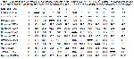
has shown that overall, the KC525214 ELAZIĞ isolate was identical to and clustered with the Norwegian, French, Danish, Taiwanese, Spanish and American isolates into one genogroup closely related to the VP2 reference sequence (95.1-96.6%). Norwegian IPNV isolates were generally the most similar to KC525214 ELAZIĞ (96.6%) (Table 2). In contrast, all the England, Canada and Japan reference sequences were more distant. Isolate used in the present study belong to one genogroup equivalent to genogroup 5 proposed by Blake et al.10 and are therefore consistent with the A2 serotype (Figure 2A,B; Figure 3).


    Büyütmek İçin Tıklayın
    Table 2: Comparison of infectious pancreatic necrosis virus (IPNV) KC525214 ELAZIĞ isolate, showing percentage nucleotid sequence identity matrix based on sequences of the VP2* gene in previously reported isolates in A2 serotype

    Currently, the most common system for analyzing the genetic diversity of a virus and classifying virus genogroups is through genomic sequencing of genes that generally code for the virion surface proteins or that encode the virus polymerase. For IPNV, several authors have used the sequences of nucleotides and amino acids to make such classifications based on the VP2 region or the entire ORF of segment A15,16,19,20 some of authors are also used for genogrouping IPNV based on VP118,21 or the VP3 and VP5 genes21.

    In this study, we used the sequence of nucleotides 20- 225 of genome segment A, which constitute a fragment of VP2 (Figure 3). The genogroups in the phylogenetic tree are in concordance with the classification proposed by Blake et al.10.

    Infectious pancreatic necrosis virus was first reported from rainbow trout by Candan22 in Turkey and after that was investigated by Ogut and Altuntas24, Albayrak and Ozan23, Gürçay et al.13 IPNV is now endemic and factors supporting endemicity are not fully explored at present in Turkey. It was reported that the severity of IPNV in rainbow trout depends on species, strain and age of the fish25,26 along with the two key disposing factors, stress and temperature24.

    The result of sequence analysis in this study implied that the origin of IPNV isolated from trout farms in the Eastern Anatolia was the hatcheries where Oncorhynchus mykiss eggs transported from Norwegian, French, Danish, Taiwanese, Spanish and American isolates (Figure 3). The findings strongly indicate that trout become infected in the hatcheries in the broodstock, and then the infection becomes latent. This result also suggests that probably IPNV, highly contagious leading to rapid spread in the region, became endemic in the region due to the transfer of latently infected fish and reproductive fluids. American serotype that was transferred to Spain by the importation of rainbow trout eggs from North American farms17. Similarly, as occurred with the importation of IPNV Eastern Anatolia region, study has shown that the KC525214 ELAZIĞ isolate is closely related to the Norwegian, French, Danish, Taiwanese, Spanish and American isolates.

    KC525214 ELAZIĞ isolate used in the present study belong to one genogroup equivalent to genogroup 5 proposed by Blake et al.10 and are therefore consistent with the A2 serotype. In the other study, nucleotide sequences of the VP2/NS region of IPNV showed that all isolates collected in the Black Sea region and surrounding areas were determined belonged to the genogroup III Ogut and Altıntas27.

    In conclusion, the findings presented here support the view that the transfer of breeding materials is associated with the transmission of pathogens. To prevent the introduction of the infectious diseases into a recirculating system, the best recommendation is to hatch eggs at the facility or buy fingerlings from a certified disease-free source. Additionally, newly arrived fingerlings should be quarantined before introduction into the system and reared in a pathogen-protected environment.

    Acknowledgments
    The authors would like to thank Tarımsal Araştırmalar ve Politikalar Genel Müdürlüğü (TAGEM) for its financial support.

  • Başa Dön
  • Özet
  • Giriş
  • Materyal ve Metot
  • Bulgular
  • Tartışma
  • Kaynaklar
  • Kaynaklar

    1) Dobos P. The molecular biology of infectious pancreatic necrosis virüs (IPNV). Ann Rev Fish Dis 1995; 5: 25-54.

    2) Sadasiv EC. Immunological and pathological responses of salmonids to infectious pancreatic necrosis virus (IPNV). Ann Rev Fish Dis 1995; 5: 209-223.

    3) McAllister PE, Bebak J. Infectious pancreatic necrosis virus in the environment: Relationship to effluent from aquaculture facilities. J Fish Dis 1997; 20: 201-207.

    4) Rivas CC, Cepeda CP, Dopazo B, Novoa MN, Barja JL. Marine environment as reservoir of birnaviruses from poikilothermic animals. Aquaculture 1993; 115: 83-194.

    5) Espinoza E, Farı´as G, Soler M, Kuznar J. Identity between infectious pancreatic necrosis virus VR-299 and a Chilean isolate. Intervirology 1985; 24: 58-60.

    6) Anonymous. Infectious haematopoietic necrosis. Chapter 2.3.4, Manual of Diagnostic Tests for Aquatic Animals, 2012: 300-313.

    7) Dopoza CP, Barja JL. Diagnosis and identification of IPNV in salmonids by molecular methods. In, Cunningham CO (Editor). Molecular Diagnosis of Salmonid Disease. Netherlands: Kluwer Academic Publishers, 2002: 23-48.

    8) Williams K, Blake S, Sweeney A, Singer JT, Nicholson BL. Multiplex reverse transcriptase PCR assay for simultaneous detection of three fish viruses. J Clin Microbiol 1999; 37: 4139-4141.

    9) Hall TA. BioEdit: A user-friendly biological sequence alignment editor and analysis program for Windows. Nucleic Acids Symposium Series 1999; 41: 95-98.

    10) Blake SL, Ma JY, Caporale DA, Jairath S, Nicholson BL. Phylogenetic relationships of aquatic birnaviruses based on deduced aminoacid sequences of genome segment A cDNA. Dis Aqua Org 200; 145: 89-102.

    11) Tamura K, Dudley J, Nei M, Kumar S. MEGA4. Molecular Evolutionary Genetics Analysis (MEGA) software version 4.0. Mol Biol Evol 2007; 24: 1596-1599.

    12) Sanju´an R, Nebot MR, Chirico N, Mansky LM, Belshaw R. Viral mutation rates. J Virol 2010; 84: 9733-9748.

    13) Gürçay M, Turan T, Parmaksız A. A study on the presence of Infectious Pancreatic Necrosis Virus infections in farmed rainbow trout (Oncorhynchus mykiss Walbaum, 1792) in Turkey. Kafkas Univ Vet Fak Derg 2013; 19: 141-146.

    14) Hill B J, Way K. Serological classification of infectious pancreatic necrosis (IPN) virus and other aquatic birnaviruses. Ann Rev Fish Dis 1995; 5: 55-77.

    15) Heppell J, Berthiaume L, Tarrab E, Lecomte J, Arella M. Evidence of genomic variations between infectious pancreatic necrosis virus strains determined by restriction fragment profiles. J Gen Virol 1992; 73: 2863-2870.

    16) Cutrin JM, Barja JL, Nicholson BL,et al. Restriction fragment length polymorphism and sequence analysis: Anapproach for genotyping infectious pancreatic necrosis virus reference strainsand other aquabirnaviruses isolated from northwestern Spain. Appl Envi-ron Microbiol 2004; 70: 1059-1067.

    17) Zhang CX, Suzuki S. Comparison of the RNA polymerase genes of marine birnavirus strains and other birnaviruses. Arch Virol 2003;148: 745-758.

    18) Nishizawa T, Kinoshita S, Yoshimizu M. An approach for genogrouping of Japanese isolates of aquabirnaviruses in a new genogroup, VII, based on the VP2/NS junction region. J Gen Virol 2005; 86: 1973-1978.

    19) Havarstein LS, Kalland KH, Christie KE, Endresen C: Sequence of the large double-stranded RNA segment of the N1 strain of infectious pancreatic necrosis virus: A comparison with other Birnaviridae J Gen Virol, 1990; 71: 299-308.

    20) Barrera-Mej´ıa M, Sim´on-Mart´ınez J, Ulloa-Arvizu R. Salgado-Miranda C, Soriano-Vargas E. Molecular characterization of the VP1 gene of a Mexican isolate of infectious pancreatic necrosis virus. Can J fish Aqua Sci 2010; 74: 218-222.

    21) Zhang CX, Suzuki S: Aquabirnaviruses isolated from marine organisms form a distinct genogroup from other aquabirnaviruses. J Fish Dis 2004; 27: 633-643.

    22) Candan A. First report on the diagnosis of infectious pancreatic necrosis (IPN) based on reverse transcription polymerase chain reaction (RT-PCR) in Turkey, Bull Eur Ass Fish Pathol 2002; 22: 45-48.

    23) Albayrak H, Özan E. Gökkuşağı alabalıklarında (Oncorhynchus mykiss Walbaum, 1792) infeksiyöz pankreatik nekrozis ve infeksiyöz hematopoietik nekrozis virus enfeksiyonlarının varlığının araştırılması. Ankara Üniv Vet Fak Derg 2010; 57: 125-129.

    24) Ogut H, Altuntas C. Occurrence and prevalence of infectious pancreatic necrosis virus in rainbow trout (Oncorhynchus mykiss) cultured in cages in the Black Sea. Aqua Res 2012; 43: 1550-1556.

    25) Silim A, Elazhary MASY, Lagace A. Susceptibility of trouts of different species and origins to various isolates of infectious pancreatic necrosis virus. Can J fish Aqua Sci 1982; 39: 1580-1584.

    26) Reno PW. Infectious pancreatic and associated aquatic birnaviruses. In: Woo PTK, Bruno DW. (Editors): Fish Diseases and Disorders. UK: CAB International, Oxfordshire, 1999: 1-55.

    27) Ogut H, Altuntas C, Parlak R. Viral surveillance of cultured rainbow trout in the eastern Black Sea, Turkey. J Aquat Anim Health 2013; 25: 27-35.

  • Başa Dön
  • Özet
  • Giriş
  • Materyal ve Metot
  • Bulgular
  • Tartışma
  • Kaynaklar
  • [ Başa Dön ] [ Özet ] [ PDF ] [ Benzer Makaleler ] [ Yazara E-Posta ] [ Editöre E-Posta ]
    [ Ana Sayfa | Editörler | Danışma Kurulu | Dergi Hakkında | İçindekiler | Arşiv | Yayın Arama | Yazarlara Bilgi | E-Posta ]