
İnek, Koyun, Keçi ve Eşek Kolostrumunun A549 ve MCF-7 Farklı Kanser Hücreleri Üzerindeki Anti-Tümoral Aktivitesi
Cilt 39, Sayı 2
Temmuz 2025 · Sayfalar 076-084
İnek, Koyun, Keçi ve Eşek Kolostrumunun A549 ve MCF-7 Farklı Kanser Hücreleri Üzerindeki Anti-Tümoral Aktivitesi
Anti-Tumoral Activity of Cow, Sheep, Goat, and Donkey Colostrum on A549 and MCF-7 Different Cancer Cells
İlhan SABANCILAR1, Mehmet Hanifi DURAK2
2Dicle University, Faculty of Veterinary Medicine, Department of Biochemistry, Diyarbakır, TÜRKİYE
Bu çalışmanın amacı, inek, koyun, keçi ve eşek gibi farklı türlere ait kolostrumlardan elde edilen peynir altı suyu ve kazein proteinlerinin insan akciğer kanseri hücreleri (A549) ve meme kanseri hücreleri (MCF-7) üzerindeki aktivitesini araştırmaktır. Kolostrumlar peynir altı suyu ve kazein proteinlerine ayrıldı. Liyofilize proteinlerin farklı konsantrasyonlarının antitümoral aktivite seviyeleri, hücreler üzerinde 3-[4,5-dimetiltiazol-2-il]-2,5 difenil tetrazolyum bromür canlılık testi ile ölçüldü. Ayrıca, kolostrumdaki sitokin düzeyleri interlökin-2 (IL-2), interlökin-6 (IL-6) ve tümör nekroz faktörü alfa (TNF-α) belirlendi. Kolostrumdaki peynir altı suyu ve kazein proteinlerinin 6400 μg/mL'den başlayarak azalan şekilde seri seyreltileri, A549 ve MCF-7 hücreleri ile 24, 48 ve 72 saat inkübe edildi. İnek peynir altı suyu 30kda'nın MCF-7 hücreleri ile inkübasyonu sonucunda %37.06 hücre canlılığı elde edilmiş; IC50 değeri ise 3.144 g/mL olarak belirlenmiştir. MCF-7 hücrelerinin eşek peynir altı suyu proteinleri ile inkübasyonu sonucunda hücre canlılığı oranı %16.64 bulunmuş ve IC50 değeri ise 3.424 μg/mL olarak ölçülmüştür (p<0.001). Koyun kazeinin A549 hücreleri ile inkübasyonu sonucunda %29.38 hücre canlılığı ve 3.612 μg/mL IC50 değeri elde edilmiştir. Buna karşın MCF-7 hücreleri ile inkübasyon sonucunda %37 hücre canlılığı ve 3.383 μg/mL IC50 değeri elde edilmiştir. Sonuçlar eşek peynir altı suyu proteininin kemoterapi gören akciğer kanseri hastalarında bağışıklık sistemini ve beslenmeyi destekleyebileceğini düşündürmektedir. Koyun kazeini, MCF-7 hücreleri üzerinde en güçlü antiproliferatif etkiyi gösterdi ve bu durum meme kanseri tedavisinde daha fazla araştırma potansiyeli olduğunu gösteriyor.
The aim of this study was to investigate the activity of whey and casein proteins obtained from colostrum’s of different species, such as cows, sheep, goats, and donkeys, on human lung cancer cells (A549) and breast cancer cells (MCF-7). Colostrum samples’s were fractionated into whey and casein proteins. Antitumoral activity levels of different concentrations of lyophilized proteins were measured by the 3-[4,5-dimethilthiazol-2-yl]-2,5 difenil tetrazolium bromide viability test on cells. Furthermore, cytokine levels interleukin-2 (IL-2), interleukin-6 (IL-2), and tumor necrosis factor alpha (TNF-α) in colostrum samples were determined. Serial dilutions of whey and casein proteins in colostrum, starting at 6400 μg/mL and decreasing, were incubated with A549 and MCF-7 cells for 24, 48, and 72 hours. Incubation of cow whey 30kda with MCF-7 cells resulted in a cell viability of 37.06%; the IC50 value was determined to be 3.144 g/mL. As a result of MCF-7 cell incubation with donkey whey proteins, the cell viability rate was 16.64% and the IC50 value was measured at 3.424 μg/mL (p<0.001). Incubation of sheep casein with A549 cells revealed a cell viability of 29.38% and an IC50 of 3.612 μg/mL whereas incubation with MCF-7 cells revealed a cell viability of 37% and an IC50 of 3.383 μg/mL. The results suggest that donkey whey protein may support the immune system and nutrition in lung cancer patients undergoing chemotherapy. Sheep casein showed the strongest antiproliferative effect on MCF-7 cells, indicating its potential for further research in breast cancer treatment.
Giriş
The use of natural products, which have no toxic effects on human health, for cancer research has an important place today. A certain part of anticarcinogen drugs can be formed by synthesizing some molecules in nature in organic tissues[1]. Chemotherapy applications used in the treatment of cancer patients cause severe side effects in some patients, and also impose a heavy burden on the country's economy. Therefore, it is essential to utilize the benefits of natural anticarcinogen substances that have no or few side effects and are inexpensive[2].Breast Cancer Cell Line (MCF-7): It is originated from an invasive ductal carcinoma from a 69-year-old Caucasian woman with pleural effusion. The mammary epithelium has many features, including the ability to process estradiol through estrogen receptors in the cytoplasm MCF-7 consists of luminal cells that look and form differently Cancer cell lines are characteristic of primary tumor by their copy number and gene expression characteristics[3].
Lung Cancer Cell Line (A549); A549 cells were first obtained by D.J. Giard and his team in 1972 by removing the tumor from the lung tissue of a 58-year-old male patient and culturing them in vitro. A549 cells, which are adenocarcinoma human alveolar epithelial cells, are confluent by binding to flasks in vitro under optimum conditions.Breast cancer is the most common type of cancer in women in the world, and although it continues to be a serious health problem today, it ranks second after lung cancer in terms of being the most common type of cancer[4]. According to the report of IARC [International Agency for Research on Cancer], it is reported that the number of female patients with breast cancer increases by 50% every five years[5]. Obesity, immunological factors, gender, and genetic factors also play an important role in catching lung cancer[6].
Colostrum is a dark yellow, a highly viscous nutritive substance that is secreted from the mammary glands of the mother during the postpartum period and occurs for 24-72 hours[7] The main proteins in colostrum are casein, immunoglobulins and beta-lactoglobulin, known as whey proteins, alpha-lactalbumin, lactoferrin, lysozyme, and serum albumin[8]. Casein protein constitutes approximately 80% of the total protein in milk and consists of four fragments with different properties, namely α1 -, α2 -, β- and κ-[9].
The amount and characteristics of the secreted colostrum vary depending on many factors. These factors are the cow's age, breed, nutritional level during pregnancy, the birth process, and several births[10]. Due to the fact that infants with protein allergies tolerate donkey milk well, researchers have been focusing more on it recently [11]. The presence of high amounts of lysozyme and lactoferrin in donkey colostrum, which has a low amount of casein compared to other milk sources, provides this colostrum with antimicrobial properties[12]. Colostrum consumption by the offspring of ruminant species (cow, sheep, and goat) has a fundamental role in the passive immune transfer and neonatal survival rates[13]. However, it directly affects the immune level of the lamb's body immune system and plays an important role in protecting the animal from microorganisms[14].
It is important to improve the course of the disease and enhance the quality of life in cancer patients and animals. With this understanding, our study aims to determine the anti-carcinogenic effects and cytokine levels of whey and casein proteins obtained from colostrum fractions of different species such as cows, sheep, goats, and donkeys on A549 and MCF-7 cancer cells in vitro.
In this way, the possible detection of natural anti-carcinogenic compounds with low side effects and low cost is targeted.
Gereç ve Yöntem
The study was carried out in the Dicle University Science, Technology, Application, and Research Center Laboratory. In order to purify the oil in the colostrum, it was centrifuged at 4000 g for 30 minutes at 15°C. By using the isoelectric point, cow, sheep, goat, and donkey colostrum samples were separated into their respective fractions of whey and casein proteins. After adjusting the pH of the samples to 7.6 with 1M NaOH, whey protein was extracted by centrifugation. A>30 kDa Ultrafiltration Membrane (Sartorius, Germany) peristaltic pump was used to concentrate the whey protein's β-lactaglobulin, α-lactalbumin, serum albumin, lactoferrin, and lysozyme proteins. Whey and casein proteins were purified from microorganisms using a Millipore brand vacuum pump and a 0.22 um microfiltration membrane filter. Bacterial counts on Plate Count Agar (PCA) and Violet Red Even Lactose Agar (VRBLA) were performed to check that they were free of microorganisms. Frozen samples brought to the Dicle University Faculty of Pharmacy Analytical Chemistry Laboratory were dried in a Christ Freezone brand lyophilizer device. SDS-PAGE (sodium dodecyl sulphate polyacrylamide gel electrophoresis) electrophoresis was applied to determine the proteins in our samples. The process of determining cytokine levels in whey proteins obtained from different animal species was carried out with "Sandwich ELISA" (Kit No. 202011017) test kits (Figure 1).
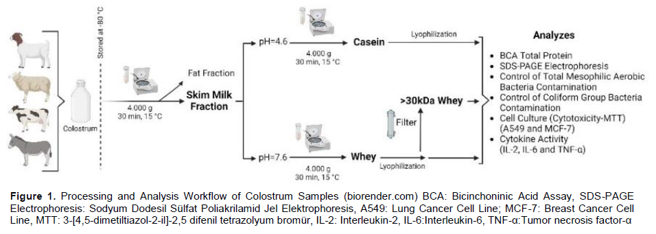
Collection of Colostrum Sample: The colostrum samples used in the study were obtained under hygienic conditions from 8 cows (Southeastern Anatolian Red), sheep (Akkaraman), goats (Hair goat), and donkeys (Anatolian donkey or Equus assinus) from four different farms in Diyarbakır province. In the study, colostrum was collected from cows, sheep, goats and donkeys on the first day after birth. The milk samples were transported to the laboratory under a cold chain. Colostrum samples were taken in different numbers from each species and combined. The samples were stored in a deep freezer at -80°C until the study was conducted.
Fractionation of Colostrum Samples: Defatted colostrum samples were adjusted to pH 7.6 by adding 1M NaOH and to pH 4.6 by adding 1N HCl. The samples were then centrifuged at 4000g for 30 minutes at 15°C to separate whey protein from the supernatant and casein protein from the pellet[6].
Determination of Total Mesophilic Aerobic Bacteria (TMAB), VRBLA Count: Before applying to cell culture, the bacterial contamination of the samples was assessed using the Total Mesophilic Aerobic Bacteria (TMAB) and Violet Red Bile Lactose Agar (VRBLA) methods. Plate Count Agar (PCA) (500 mL) was prepared according to the manufacturer's instructions (in a sterile environment in a safety cabinet) and then sterilized in an autoclave. Whey and casein proteins were passed through a 0.22 μm microfiltration membrane filter, and dilutions of 10-1 were prepared. From these dilutions, 1 mL was transferred to sterile Petri dishes. After incubation (24 hours at 37°C), the medium was checked for the development of col[15]. No growth was observed.
Lyophilization of Colostrum Samples: The samples were lyophilized using a Christ Freezone lyophilizer. Lyophilization, or freeze-drying, is a process that removes water from frozen samples by applying a pressure of 0.070 hPa (hectopascals) at -50°C. This technique preserves the integrity of the sample while converting it into a dry powder. After a 96-hour lyophilization process, the colostrum samples were completely converted into powder. These lyophilized samples were then stored at -80°C until needed for further analysis.
Electrophoresis of Whey and Casein Proteins: Before electrophoresis, the total protein concentrations in the samples were measured using the bicinchoninic acid (BCA, Bio-Rad, USA) colorimetric assay method. Absorbance was then measured at a wavelength of 562 nm using a Thermo Scientific Elisa Reader.
SDS-PAGE was conducted using the Bio-Rad System (Lot No. 161-0183). Colostrum samples were diluted at a ratio of 1:5 (v/v). After preparing the samples, SDS-PAGE gel was prepared by mixing with 30% glycerol and adding 5% β-mercaptoethanol. The mixture was then heated at 95°C for 5 minutes. A marker from the Bio-Rad kit was loaded into the first well. After loading, the gel was run at 200V for 40 minutes (Gel Electrophoresis Bio-Rad). Once the running process was completed, the gel was carefully removed and transferred to a gel imaging device (Bio-Rad) for visualization.
MTT Assay Cell Viability: To investigate the anticancer effects of whey and casein proteins at different doses and durations in vitro and to determine their mechanisms, A549 and MCF-7 cell lines were used. For this purpose, DMEM and RPMI media were utilized. The homogenized mixture consisted of 20% (50 mL) Fetal Bovine Serum (FBS) (Sigma-Aldrich, USA), 1% (5 mL) Penicillin/Streptomycin (Sigma-Aldrich, USA), Dulbecco's Modified Eagle Media (DMEM) medium containing 2.2 g/L Sodium Bicarbonate for the MCF-7 cell line, and Roswell Park Memorial Institute (RPMI) medium (pH 7) (Sigma-Aldrich, USA) for the A549 cell line. Here is the translation of the text from Turkish to English:
In our study, the MTT assay, which is one of the cell viability analyses, was used. The MTT assay was performed in 96-well plates to determine the cytotoxicity levels of whey, casein, and 30kDa concentrated whey proteins from different species, including cow, sheep, goat, and donkey. Cells exposed to trypsin were counted after centrifugation, and equal amounts of cells were seeded into each well: 90 μL containing 3x10³ cells for MCF cells and 90 μL containing 5x10³ cells for the A549 cell line. The sterile 96-well plates with the cell seeding were placed in an incubator at 37°C with 5% CO2 for 24 hours. After this period, adherence of the cells to the 96-well plates was checked using an inverted microscope. Whey and casein proteins from these species were weighed (0.064 g) and dissolved in 1 mL of 1% PBS. After complete homogenization, serial dilutions were prepared in sterile Eppendorf tubes at different concentrations under a laminar flow safety cabinet. The whey and casein proteins were added to the 96-well plates in serial dilutions at concentrations of 6.400 μg/mL, 3.200 μg/mL, 1.600 μg/mL, 800 μg/mL, and 400 μg/mL, with 10 μL added to each well. Before the experiment, the samples were filtered through a 0.2 μm filter. No sample was added to the control wells. Incubation was carried out for different durations: 24, 48, and 72 hours. After incubation, 10 μL of MTT solution (5 mg/mL) was added to the sterile wells and incubated at 37°C with 5% CO2 for 3 hours. To dissolve the formazan crystals, 100 μL of DMSO was added to the wells using a multi-channel pipette, and the plates were covered with aluminum foil and shaken for 10 minutes. Absorbance was measured at 570 nm using a microplate reader. The protocol was applied at 48 and 72 hours, and absorbance readings were obtained.
The first wells, which only contained the medium, served as the control group, with cell viability considered as 100%. The percentage of cell viability was calculated using the following formula:
%Cell Viability = (Absorbance of cells with different concentrations / Absorbance of control cells) × 100
Determination of IL-2, IL-6, and TNF-α Cytokine Levels: The determination of cytokine levels in whey proteins obtained from different animal species was performed using Sandwich ELISA (Kit No. 202011017) test kits. Samples were prepared in duplicate, and results were processed at 450-nm absorbance on the Thermo Scientific Multiskan Go Microplate Reader. BioTek ELx50 automated washer was used for plate washing. The concentrations were calculated based on standard curves. Serum concentrations of asprosin were expressed as nanograms per milliliter. The sensitivity and measuring range for IL-2, IL-6, and TNF-α as per the manufacturer's specifications were 2.51 ng/L and 5 to 2000 ng/L, 1.03 ng/L and 2 to 600 ng/L, 1.52 ng/L and 3 to 900 ng/L respectively.
Statistical Analysis: Quantitative data obtained in the study were expressed as an arithmetic mean ± standard deviation by post-hoc analysis. The results of the graphs related to the study were obtained with the GraphPad Prism 8 program (GraphPad Software, http://www.graphpad.com). In the MTT method, statistically significant differences between the samples were identified using the two-way ANOVA test followed by the post hoc Tukey HSD (Honestly Significant Difference) test in the SPSS 22 software program. The results of the repeated measures ANOVA revealed that the factors of time, type, and dose had a statistically significant effect on cell viability (p<0.001). Additionally, the interactions between time and type, time and dose, as well as the type × dose interaction were also found to be statistically significant (p<0.001). These findings indicate that the experimental conditions led to significant differences in cell viability both independently and through their interactions. As a result of the Tukey HSD (Honestly Significant Difference) test conducted for the type factor, statistically significant differences were identified among the types. Based on the analysis, the types were classified into five distinct homogeneous groups according to their mean cell viability levels. The significance level in each group was determined using p<0.05.
Bulgular
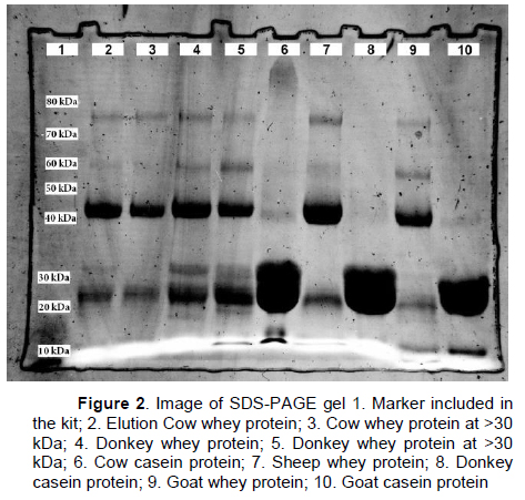
Whey and Casein SDS-PAGE (Sodium Dodecyl Sulphate–Polyacrylamide Gel Electrophoresis): SDS-PAGE electrophoresis was applied to determine the proteins in our sample. The marker included in the Bio-Rad kit was used as a marker. The obtained SDS-PAGE gel image is given in Figure 2.
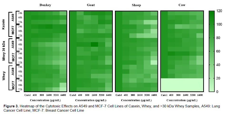
MTT Cell Viability: After lyophilization of whey and casein proteins obtained from colostrum samples, they were incubated with serial dilutions prepared from a high dose to a low dose of 6.400 μg/mL. A549 and MCF-7 cell lines were used in three duplicates, and the MTT test was used to examine the antiproliferative effects, viability changes, proliferation, and cytotoxicity determination methods. The control group (NT), which did not use whey or casein proteins, was considered to have 100% viability. The viability values of whey, whey 30 kda and casein samples were determined by proportional calculation by absorbance measurement in a microplate reader (570 nm) (Figure 3).
Viability percentages and standard deviations were determined from the data obtained from the MTT assay of cow, sheep, goat and donkey colostrum samples. In addition, IC50 value and R2 values, which is the growth inhibition concentration at 50% of tumor cells, were determined using the Excel program (Table 1).
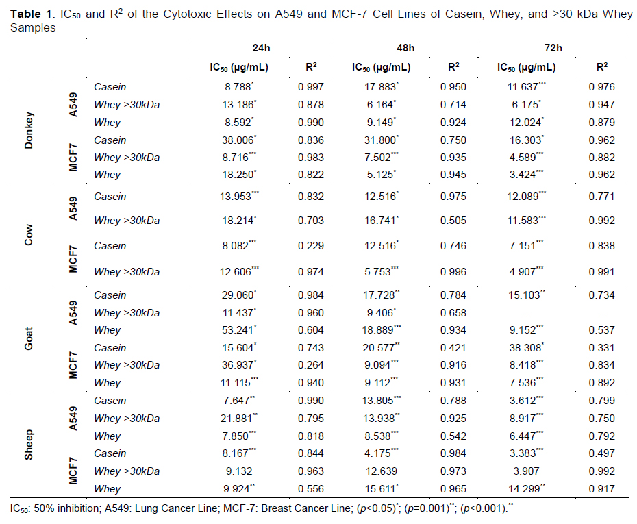
In this study, the cytotoxic effects of casein and whey proteins derived from different animal sources (donkey, cow, goat, and sheep) were evaluated on A549 (lung cancer) and MCF-7 (breast cancer) cell lines. IC₅₀ (half-maximal inhibitory concentration) values and their corresponding R² correlation coefficients were calculated over incubation periods of 24, 48, and 72 hours (Table 1).
In our study, IL-2 levels were found at concentrations of 3200 μg/mL and 6400 μg/mL, which showed a high cytotoxic effect among different animal samples. The highest IL-2 level was found at a concentration of 3200 μg/mL, which is >30 kDa in sheep whey, and R2=0.9949 (Figure 4).
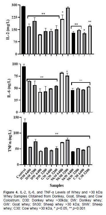
Among the cow, sheep, goat, and donkey samples we used in the study, the concentrations with high IL-6 levels were found to be between 3200 μg/mL and 6400 μg/mL. The highest IL-6 level was found at a concentration of 6400 μg/mL in sheep whey of >30 kDa type, with a R2=0.9861 In our investigation, samples with elevated levels of TNF-α had values ranging between 3200 and 6400 g/mL. The greatest TNF-α concentration was obtained at 3200 g/mL with sheep whey >30 kDa and R2= 0.9915 (Figure 4).
Tartışma
In cancer, cell division occurs in an uncontrolled and abnormal manner due to a variety of causes[16]. As a result of mutations that may occur during cell division, the failure to execute programmed cell death (apoptosis) can lead to the transformation of aging cells into cancerous cells. In general, chemotherapy, which is frequently preferred in cancer, has negative effects on healthy cells by showing toxicity in normal cells, which can sometimes cause lifelong irreversible side effects[17]. Therefore, the purpose of our study was to determine the anticancer role of colostrum components in different cancer cells (A549 and MCF-7) due to the many benefits they provide to human health and to be able to reveal a new approach in cancer treatment. In certain investigations on whey protein, it was revealed that these proteins include antioxidant, antibacterial, hypoglycaemic, antiviral, antifungal, anticancer, and antiaging properties[18].McIntosh et al.[19] demonstrated a protective role for dietary dairy proteins against tumour development, showing that dietary whey protein and casein were more protective against the development of intestinal cancers in rats than was red meat or soy bean protein. They concluded that dietary proteins differ in their ability to protect against cancer development and that the proteins in dairy foods, particularly the whey proteins, appear to play a significant role in cancer prevention. In this study, we suggest that the observed effect on tumor cells, which shows consistency between in vitro data and in vivo findings, may be attributed to whey proteins.
Bounous et al.[20] demonstrated that dietary whey protein concentrate (WPC) has potent anticancer properties against colon cancer induced by DMH in mice, and there is also some evidence from controlled clinical studies of the effectiveness of WPC in limiting metastasis during anticancer therapy. The authors have suggested that the protective efficacy of dietary WPC could be due to whey proteins enhancing tissue glutathione concentration, since whey protein is known to be rich in substrates for glutathione synthesis (Parodi, 21). The presence of high levels of glutathione in tissues has been suggested to suppress tumour development at various sites in the body, possibly by reducing free radical- and oxidant-induced damage to chromosomal DNA[22].
The majority of reports which have characterised milk-derived anticancer activity have come from in vitro studies using tumour cell lines, or in vivo studies using animal models of tumorigenesis. Although both approaches provide valuable evidence as to the potential anticancer actions of milk-derived molecules, caution should be taken when extrapolating results from such studies to statements on disease protection in humans. In the case of in vitro studies, the demonstration of an anticancer effect should be taken to imply that the component under test has the potential to regress tumour development (not initiation), and moreover any given biological effect of a component in vitro must be assessed in light of its perceived in vivo performance in the gastrointestinal tract. This is particularly important with respect to human intestinal physiology: Many potentially beneficial molecules in milk may be rendered inactive and/ or remain unabsorbed in the human digestive tract, following gastric processing. The same cautions apply to in vivo studies of tumorigenesis in animal models, where the rodent gastrointestinal system may well respond to anticancer factors in a different way to the human digestive system. Overall, there have been remarkably few well-designed clinical trials to determine conclusively the effects of milkderived anticancer agents. Although such studies are notoriously difficult to conduct and interpret, the proven efficacy of some milk-derived components (e.g. CLA and lactoferrin) in animal models should merit their trialling under controlled clinical conditions. In a similar fashion, several other minor constituents of milk, especially growth and inhibitory factors, have shown promising action in in vitro or laboratory animal studies, but remain untested in humans. Improvements in milk fractionation technology should facilitate the identification of further minor constituents of milk, particularly low-molecular-weight proteins and peptides, which may have important anticancer properties. Such technology has been successfully employed in identifying potent immunomodulatory molecules from milk[23].
Colostrum contains significant amounts of immunoglobulins, growth factors, antibodies, vitamins, minerals, enzymes, and amino acids in its composition. The structure of colostrum is more similar to that of blood. This also has physiological significance and provides nutritional compatibility for the new born puppy[24]. Milk composition is influenced by the animal's breed, age, nutrition, number of births, environmental factors, and health state[25]. Regional and environmental factors may differ in the amount of mammary secretion depending on intramammary pressure[26]. In addition to the amount of milk secretion, it also negatively affects the nutritional quality of the milk. The obtained whey contains vitamins, proteins, and a very small amount of milk fat.
In addition, the presence of high amounts of sulphur-containing amino acids in whey protein supports its antioxidant activity[27]. Whey contains several different types of proteins, including α-lactalbumin, β-lactaglobulin, lysozyme, lactoferrin, serum albumin, and immunoglobulins. According to the findings of many studies, a significant number of these proteins exhibit anticarcinogenic properties[28]. In addition to whey protein, milk also contains casein protein[
Laktoferrin It is considered an important host defence molecule and has been demonstrated to present several biological activities, such as antimicrobial, anti-oxidative, anti-inflammatory, anticancer, and immune regulatory in in vivo, in vitro and cell-line experiments (31-34). Historically, the journey toward the understanding of themolecular basis of the anticancer activity of milk components has started from the identification of a complex of α-LA and oleic acid (OA) in human milk[35-37]. Fragments of cow colostrum are casein, with the remaining 18% constituting whey proteins. In this investigation, the antiproliferative properties of caseins and whey proteins in the colostrum of several animals were examined. The strongest antiproliferative impact of whey protein against MCF-7 cells was identified in >30 kDa sheep whey, donkey whey protein, and cow whey protein, in that order. With the data obtained, it is possible to conclude that consumption of donkey whey protein can improve the nutrition of cancer patients undergoing chemotherapy by affecting the immune system in a positive manner due to the high nutritional content of lung cancer patients. Sheep casein exhibited a significant antiproliferative effect against MCF-7 cells among caseins. It is anticipated that the findings of this study will lead to the examination of cellular and molecular pathways that may contribute to the treatment of breast cancer. The findings obtained from the study revealed that the anticancer activity was high in whey protein and lower in casein, and this effect differed depending on animal species, dose, and time.
There are two main types of differentiation inducer that lead young tumour cells to maturation with normal physical functions. One can directly react with tumour cells, while the other type includes indirect inducers that activate the immune system to promote the secretion of cytokines with anti-tumour activity[38]. Splenocytes and macrophages play crucial roles in the immune system. Activated macrophages are considered as the pivotal immunocytes of host defence that inhibit tumour growth[39]. The tumoricidal activity of macrophages is mediated mainly through nitric oxide (NO) and cytokines of tumour necrosis factor-alpha (TNF-a), interleukin-6 (IL-6) and IL- 12[40].
Cow and donkey whey proteins and TNF-α, IL-2, and IL-6 cytokine levels of whey proteins with molecular masses less than 90 kDa show anti-proliferative effects against A549 and MCF-7 cells, and cow and donkey whey proteins produce cytokines. This suggests that it has a stimulating capacity and may support cytokine potentiation. Consumption of Laktoferrin-enriched food products may be a natural method to prevent or support treatment of MCF-7 tumor cells. The results of the present study showed that sheep whey could induce a marked increase in cytokine production of IL-2, IL-6, and TNF-a from lymphocytes and macrophages. These cytokines are contributory to an immune response and result in maturation, differentiation, and proliferation of immuno-competent cells for defence mechanisms of the host which explains the possible immuno-enhancing mechanism of the anti-tumour activity of sheep whey.
Conclusionly, colostrum structure contains different proteins. About 82% of the proteins in mammalian colostrum are casein, with the remaining 18% constituting whey proteins. In this investigation, the antiproliferative properties of caseins and whey proteins in the colostrum of several animals were examined. The strongest antiproliferative impact of whey protein against MCF-7 cells was identified in >30 kDa sheep whey, donkey whey protein, and cow whey protein, in that order. With the data obtained, it is possible to conclude that consumption of donkey whey protein can improve the nutrition of cancer patients undergoing chemotherapy by affecting the immune system in a positive manner due to the high nutritional content of lung cancer patients. Sheep casein exhibited a significant antiproliferative effect against MCF-7 cells among caseins. It is anticipated that the findings of this study will lead to the examination of cellular and molecular pathways that may contribute to the treatment of breast cancer. The findings obtained from the study revealed that the anticancer activity was high in whey protein and lower in casein, and this effect differed depending on animal species, dose, and time.
Cow and donkey whey proteins and TNF-α, IL-2, and IL-6 cytokine levels of whey proteins with molecular masses less than 90 kDa show anti-proliferative effects against A549 and MCF-7 cells, and cow and donkey whey proteins produce cytokines. This suggests that it has a stimulating capacity and may support cytokine potentiation. Consumption of LF-enriched food products may be a natural method to prevent or support treatment of relevant cancers.
References
- Yeni Doğan Buzağılarda Çeşitli Hematolojik ve Biyokimyasal Parametreler İle Kolostral İmmun Globulinler Arasındaki İlişkinin Belirlenmesi
- Origanum minutiflorum O. Schwarz et. P.H. Davis’in Terapötik Potansiyelinin Değerlendirilmesi
- Yerli Kara ve İsviçre Esmeri Irkı Sığırların Kolostrum Kalitesinin Karşılaştırılması
- Halep Keçisi ve Oğlaklarında Doğum Şekline Bağlı Olarak Kan ve Kolostrumda Bazı İz Element Seviyelerinin Araştırılması