
Kanatlı Hayvanlarda Nekropsi ile Tanımlanan Patolojik Değişikliklerin Bölgesel Analizi
Cilt 40, Sayı 2
Haziran 2026 · Sayfalar 183-187
Kanatlı Hayvanlarda Nekropsi ile Tanımlanan Patolojik Değişikliklerin Bölgesel Analizi
A Regional Analysis of Pathological Changes Identified by Necropsy in Poultry
Canan AKDENİZ İNCİLİ1, Zeynep YERLİKAYA2, Yeliz YALIKAYA KALPAKLIOĞLU1, Yesari ERÖKSÜZ1
2Fırat University, Faculty of Veterinary Medicine, Department of Microbiology, Elazığ, TÜRKİYE
Kanatlı hayvan nekropsisi, özellikle kümes hayvancılığı sektöründe potansiyel sağlık tehditlerinin belirlenmesinde veteriner hekimler tarafından kullanılan etkili tanı yöntemlerinden biridir. Bu çalışmada, Nisan 2025 ile Ocak 2026 tarihleri arasındaki 9 aylık dönemde nekropsisi yapılan toplam 35 kanatlı hayvan değerlendirildi. Olguların 16'sını hindi, 17'sini tavuk, 1'ini bıldırcın ve 1'ini leylek oluşturdu. Bu vakaların 16'sında perikardit (7), airsakkülit (7), fibrinöz pnömoni (6), koligranülom (1) ve hepatit (3) gibi bakteriyel etkenlerle ilişkili lezyonlar saptandı. Yedi vaka viral, dört vaka ise Histomonas meleagridis'in neden olduğu hepatit ve tiflit dahil olmak üzere paraziter kökenli olarak değerlendirildi. Bir vaka travmaya bağlıydı; yedi hayvanda ise makroskobik veya mikroskobik bulguya rastlanmadı. Bakteriyel solunum sistemi lezyonlarının en sık karşılaşılan bulgular arasında yer aldığını ve bu olguların özellikle ilkbahar ve yaz aylarında daha fazla görüldüğünü ortaya koydu. Bu sonuçlar, bakteriyel hastalıkların bölgesel dağılımının anlaşılmasında nekropsinin önemli bir tanısal araç olduğunu göstermektedir.
Avian necropsy is an important diagnostic tool for identifying health problems in poultry and other bird species. This study evaluated 35 avian necropsy cases submitted over a 9-month period between April 2025 and January 2026, including 16 turkeys, 17 chickens, 1 quail, and 1 stork. Among these cases, 16 showed lesions associated with bacterial infections, including pericarditis (1), airsacculitis (7), fibrinous pneumonia (6), coligranuloma (1), and hepatitis (3); 7 were associated with viral disease; 4 were associated with parasitic disease, including hepatitis and typhlitis caused by Histomonas meleagridis; 1 case was attributed to trauma; and 7 showed no significant macroscopic or microscopic findings. Bacterial respiratory lesions represented the largest diagnostic category and were observed most often in cases submitted during the spring and summer months. These findings highlight the diagnostic value of necropsy in avian disease investigation and provide regional data on the distribution of lesions in submitted cases.
Giriş
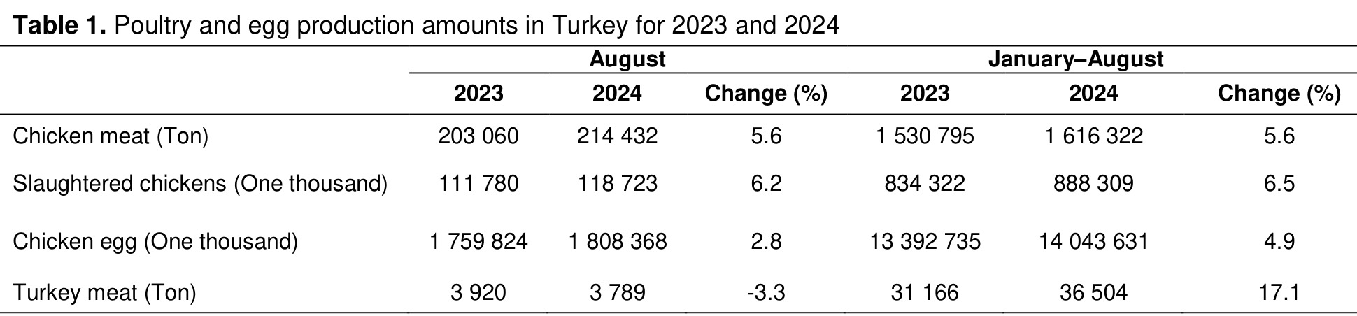
Poultry meat is an important source of animal protein for balanced and adequate human nutrition and is therefore one of the most widely consumed food products worldwide. It offers several advantages, including high nutritional quality, good palatability, relatively affordable cost compared with red meat, ease of production, and broad acceptance across different segments of society [1].Poultry products, including meat and eggs, represent an important means of meeting human needs for animal protein [2]. Population growth, increasing consumption, and rapid urbanization have contributed to a steady rise in the demand for poultry products. Poultry meat, particularly chicken meat, is the most popular type of white meat, and its consumption is expected to continue increasing over the next decade [3]. Annual poultry production is projected to exceed 37 billion animals by 2050 [4]. In Türkiye, poultry products supply a substantial share of basic nutritional needs and continue to expand within the livestock sector (Table 1) [5].
Among the most important factors threatening poultry production in Türkiye are flock-level poultry diseases, which can lead to substantial economic losses. Therefore, early diagnosis and the prevention of flock-related problems are of critical importance. In addition, the most frequently encountered bacterial agents causing disease in poultry include Salmonella spp., Escherichia spp., Clostridium spp., Staphylococcus spp., Campylobacter spp., Bacillus spp., and Listeria spp. These bacterial infectious agents may also cause disease in humans through poultry-derived food products. Thus, poultry diseases not only result in economic losses in poultry production but also pose serious public health concerns [6-9].
Bacterial, viral, fungal, parasitic, and nutritional diseases that significantly affect productivity in poultry can largely be identified through necropsy findings and the macroscopic and microscopic examination of lesions. This is highly important for preventing major potential losses. To date, numerous studies have been conducted, and continue to be conducted, to improve and increase poultry production; therefore, updating the current body of knowledge remains essential for future studies [10-12]. The aim of this study was to evaluate poultry diseases observed in both small-scale and industrial poultry enterprises in our region and to obtain up-to-date data.
Gereç ve Yöntem
Research and Publication Ethics: According to the Directive of the Local Ethics Committee for Animal Experiments of Fırat University, ethics committee approval was not required for this study, as it was considered within the scope of non-experimental clinical veterinary practice. Between 14.04.2025 and 01.01.2026, necropsies were performed on a total of 35 avian animals (16 turkeys, 17 chickens, 1 quail, and 1 stork) brought to the Department of Pathology, Faculty of Veterinary Medicine, Fırat University, from Elazığ and neighboring provinces (Table 2). Following necropsy, tissue samples were collected from lesioned areas for histopathological examination. These samples were processed routinely, serial sections of 3–5 µm thickness were obtained, and the prepared sections were stained with Hematoxylin and Eosin and examined and photographed under a light microscope.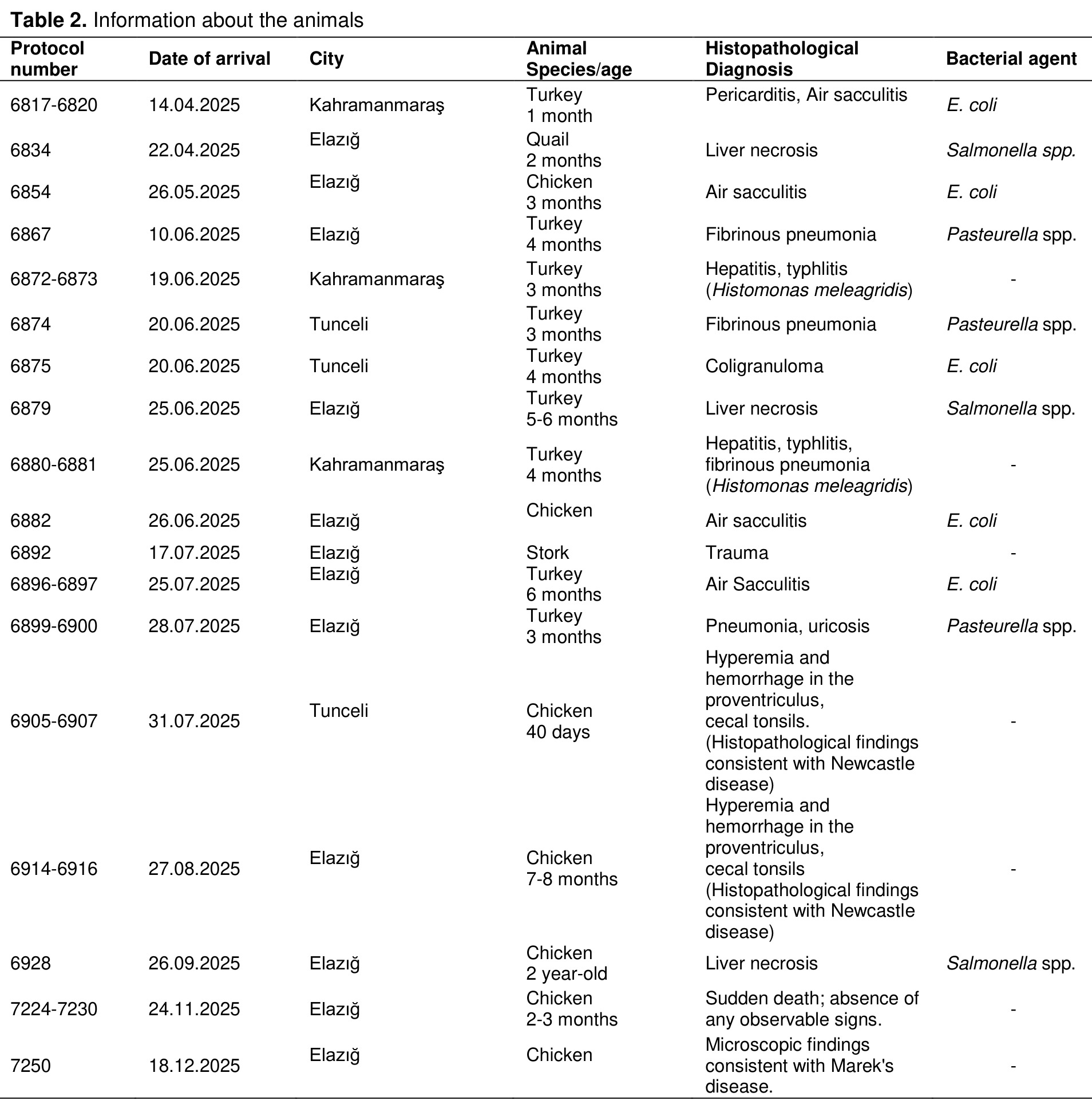
Bacteriological Isolation and Phenotypic Identification: Lung, liver, and air sac samples collected from the birds were aseptically inoculated onto 5% sheep blood agar (Oxoid, United Kingdom) and incubated aerobically at 37°C for 24 hours. Colonies showing growth were subcultured onto MacConkey agar and Eosin Methylene Blue (EMB) agar (Oxoid, United Kingdom) and re-incubated under the same conditions. Gram staining, catalase, and oxidase tests were used for the phenotypic examination of the isolates.
For the isolation of Salmonella spp., 1 g of liver sample was weighed and subjected to pre-enrichment in 9 mL of Buffered Peptone Water (Merck, Darmstadt, Germany) at 37°C for 24 hours. After pre-enrichment, 0.1 mL of this culture was transferred into Rappaport-Vassiliadis broth (Merck, Darmstadt, Germany) and selectively enriched at 42°C for 24 hours. Subsequently, 10 µL from the Rappaport-Vassiliadis broth culture was streaked onto XLT4 agar (Merck, Darmstadt, Germany), and the plates were incubated aerobically at 37°C for 24 hours. Suspected colonies obtained were purified, and MacConkey agar, EMB agar, Gram staining, oxidase testing, and Triple Sugar Iron (TSI) agar were used for phenotypic identification.
Statistical Analysis: The collected data were classified according to disease type. Statistical analyses were performed using the SPSS software package (IBM SPSS Statistics for Windows, Version 22.0), employing Fischer's Exact Chi-square Test. The main effects and interactions of the data are presented as mean ± standard error. Statistical significance was set at p<0.05.
Bulgular
Macroscopic Findings: Among the 35 avian animals examined, 16 exhibited lesions associated with bacterial agents, including pericarditis, airsacculitis, fibrinous pneumonia, coligranuloma, and hepatitis; 7 had lesions consistent with viral infection; 4 had parasitic lesions, including hepatitis and typhlitis caused by Histomonas meleagridis; 1 case was attributed to trauma; and 7 birds showed no macroscopic or microscopic abnormalities (Figures 1A–1H).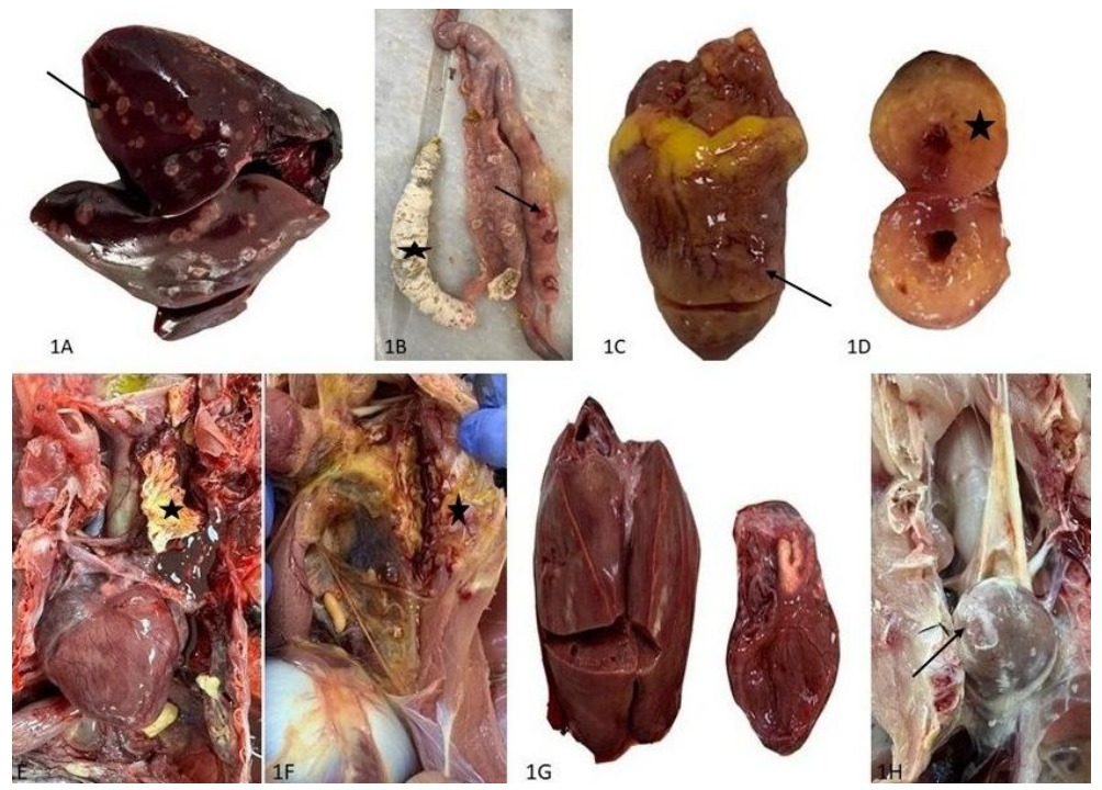
Histopathological Findings:
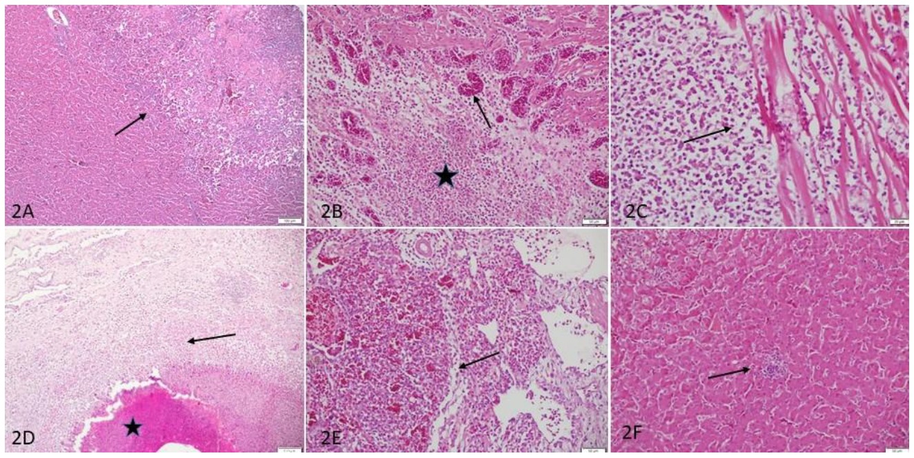
Liver; In the liver, pseudoglandular arrangement of hepatocytes, subcapsular hemorrhages, periportal inflammatory cell infiltration, and fibrosis (Figure 2A), multifocal typhoid nodules (Figure 2F) were observed.
Cecum; In the cecum, diffuse inflammatory cell infiltration and congestion were detected (Figure 2B), multifocal typhoid nodules and diffuse lymphohistiocytic infiltrations were observed.
Pericarditis; Widespread lymphocytic infiltration was observed in the heart.
Heart; In the heart, subepicardial lymphohistiocytic infiltrations were observed (Figure 2C).
Airsacculitis: In the air sacs, randomly distributed lymphohistiocytic infiltrations and foci of caseous necrosis were observed (Figure 2D).
Lungs; The lungs exhibited diffuse congestion, perivascular edema, widespread heterophilic infiltration, and fibrin accumulation within the interstitial tissues (Figure 2E).
Microbiological Findings: Microbiological examination revealed the isolation of E. coli from 9 air sac samples, Salmonella spp. from 3 liver samples, and Pasteurella spp. from 4 lung samples. The E. coli isolates produced pink to red colonies on MacConkey agar, consistent with lactose-positive characteristics, and exhibited colonies with dark centers and a greenish metallic sheen on EMB agar. On Gram staining, these isolates appeared as Gram-negative bacilli and were catalase-positive and oxidase-negative.
The Salmonella spp. isolates formed typical suspicious colonies with black centers on XLT4 agar and showed colorless colonies on MacConkey agar, consistent with lactose-negative characteristics. They were observed as Gram-negative bacilli on Gram staining and were catalase-positive and oxidase-negative. In addition, these isolates showed an alkaline slant/acid butt (K/A) reaction with H₂S production on TSI agar, consistent with glucose fermentation and the absence of lactose and sucrose fermentation.
The Pasteurella spp. isolates formed small, grayish-translucent, non-hemolytic colonies on blood agar and showed no growth on MacConkey agar. On Gram staining, they appeared as Gram-negative coccobacilli and yielded positive reactions for both catalase and oxidase tests.
Tartışma
The results of the present study demonstrated that bacterial infections were the diagnostic category among the avian cases accounting for 45.71%, this was followed by viral cases and individuals without lesions: 7 cases each (20.00%), parasitic cases were identified as 4 (11.43%), whilst traumatic cases were only 1 (2.86%). of all avian cases submitted from Elazığ and neighboring provinces to the Department of Pathology, Faculty of Veterinary Medicine, Fırat University, between 14 April 2025 and 18 December 2025 (Table 3). Of these bacterial cases, 13 involved the respiratory system and 3 involved the digestive system. In addition, disease occurrence increased during the spring and summer months. In the current study, E. coli was identified as the causative agent in nine cases of bacterial infections of the respiratory system. It is well known that colibacillosis, a prevalent bacterial infection, has been identified as a significant health concern affecting poultry worldwide. The disease has been documented to frequently result in air sacculitis (coligranuloma) and pericarditis, with its prevalence exhibiting regional variation. This variation has been attributed to climatic and farming practices [13].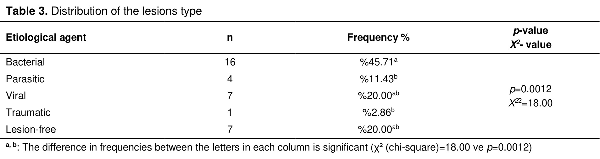
In the present research, Salmonella spp. was isolated from three cases of bacterial infections of the digestive system. The macroscopic and microscopic findings observed in these cases where Salmonella spp. was isolated are consistent with the literature. In this context, previous studies have also demonstrated that salmonellosis causes widespread areas of macroscopic necrosis in the liver and typhoid nodules at the microscopic level [14].
A macroscopic and microscopic examination revealed dilatation of the cecum, a significant quantity of necrotic material within the cecum, and necrotic pale areas were observed in the liver. These findings are consistent with those reported in other studies where histomoniasis (Histomonas meleagridis) on this subject [15]. Furthermore, in the case where Marek's disease was suspected, widespread pale areas were observed macroscopically in the heart, and widespread lymphocytic infiltration was observed microscopically; these findings are consistent with those reported in previous cases [16].
A previous study conducted using data obtained from the Turkish Ministry of Agriculture and Forestry and the Turkish Statistical Institute (TÜİK) similarly reported that bacterial and parasitic diseases were the predominant causes of disease and that their frequency increased during the spring and summer seasons. Bacterial diseases are commonly encountered in poultry flocks due to inadequate ventilation, poor hygiene conditions, overcrowding, and insufficient implementation of infection control measures [17]. Accordingly, it is essential to ensure adequate ventilation in poultry farms, avoid housing different animal species together, improve hygiene conditions, strengthen preventive measures against bacterial infections, and optimize nutritional management.
References
- Wahyono ND, Utami MMD. A Review of the Poultry meat production industry for food safety in Indonesia. J Phys Conf Ser 2018; 953(01): 21-25.rn
- Scholten MT, De Boer IJM. Gremmen B, Lokhorst C. Livestock farming with care: Towards sustainable production of animal-source food. NJAS: Wageningen J Life Sci 2013; 66(1): 3-5.rn
- Henchion M, Mc Carthy M, Resconi VCD. Troy meat consumption: Trends and quality matters. Meat Sci 2014; 98 (3): 561-568.rn
- Wu D, Cui D, Zhou M, Ying Y. Information perception in modern poultry farming. Comput Electron Agric 2022; 199: 107-131.rn
- Türkiye İstatistik Kurumu (TÜİK). ''Kümes hayvancılığı üretim miktarı, Ağustos 2024'' https://veriportali.tuik.gov.tr/tr/press/53569/14.10.2024.rn
- Eroğlu R, Çakıcı N. Kanatlı etlerinde gıda güvenliği: Bakteriyel tehlikeler. Environmental Toxicology and Ecology 2024; 4(1): 59-72.rn
- Özdemir Ö, Erer H. Tavukların önemli üst solunum yolu hastalıklarında patolojik değişiklikler. Adana Veteriner Kontrol Enstitüsü Müdürlüğü Dergisi 2012; 2: 29-38.rn
- Türkyılmaz S, Esenda ÖM. Kanatlı Hayvanlarda görülen solunum sistemi enfeksiyonları. Veteriner Hekimleri Mikrobiyoloji Dergisi 2002; 2(2): 43-49.rn
- Bıçakcıoğlu T, Müştak HK. Kümes hayvanlarında enteritise neden olan viral etkenler. Veteriner Hekimleri Derneği Dergisi 2022; 93(1): 67-75.rn
- Bett B, Kiunga P, Gachohi J, et al. Effects of climate change on the occurrence and distribution of livestock diseases. Prev Vet Med 2017; 137: 119-129.rn
- Brooks-Pollock E, de Jong MCM, Keeling MJ, Klinkenberg D, Wood JLN. Eight challenges in modelling infectious livestock diseases. Epidemics 2015; 10: 1-5.rn
- Valeria A, Sander Valeria A, Sander Edwin F, et al. Use of Veterinary vaccines for livestock as a strategy to control foodborne parasitic diseases. Front. Cell Infect Microbiol 2020; 10: 1-20.rn
- Elitok B, Bingüler N. Kanatlılarda Escherichia coli enfeksiyonları. Dicle Üniversitesi Veteriner Fakültesi Dergisi 2018; 11(1): 34-38.rn
- Hossain S, Chowdhury EH, Islam MM, Haider G, Hossain MM. Avian Salmonella infection: Isolation and identification of organısms and hıstopathologıcal study. Bang J Vet Med 2006; 4(1): 7-12.rn
- Esquent C, De Herdt P, De Bosschere H, et al. An outbreak of histomoniasis in free-range layer hens. Avian Pathol 2003; 32(3): 305-308.rn
- Dolka R, Sapierzyński WB, Malicka E, Żbikowski A, Szeleszczuk PS. Histopathology in diagnosis of broiler chicken and layer diseases – review of cases 1999-2010. Pol J Vet Sci 2012; 15(4): 773-779.rn
- Yeşilova ME. Türkiye'de Kanatlı hayvan hastalıkları ve nedenleri: İstatistiksel inceleme. Iğdır Üniversitesi Fen Bilimleri Enstitüsü Dergisi 2024; 3(1): 34-37.
- BROİLER PİLİÇLERDE PAMUK TOHUMU VE PAMUK TOHUMU KÜSPESİ TOKSİKASYONLARINDA (GOSSİPOL TOKSİKOSİS) PATOLOJİK İNCELEMELER
- ELAZIĞ ELET MEZBAHASINDA KESİLEN İNEKLERDE MASTİTİSLER ÜZERİNE PATOLOJİK İNCELEMELER
- BİR KOYUNDA BÖBREK KARSİNOMU
- BİR BROİLER CİVCİVDE NEFROBLASTOMA
- AVİAN ENCEPHALOMYELİTİS (AE) VİRUSU İLE ENFEKTE EDİLMİŞ KAZ (ANSER ANSER) EMBRİYOLARİNDA PATOLOJİK İNCELEMELER*